2830
Optimization of Compressed Sense accelerated 3D pelvis view for diagnosis of rectal cancer1Departments of Radiology, West China Hospital, Sichuan University, Cheng du, China, 2Departments of Radiology, West China Hospital, Sichuan University, Chengdu, China, 3Department of Clinical, West China Hospital, Sichuan University, Chengdu, China, 4Philips Healthcare, China, Chengdu, China
Synopsis
Although 3D T2W imaging can make up for deficiencies, such as thick layer thickness, low spatial resolution and no retrospective post-processing, of 2D acquisitions, the long imaging time and uncertain diagnostic benefits have limited its clinical application. We compared the quality of 2D, 3D and CS sense accelerated 3D T2W imaging in patients with rectal cancer and found that 3D T2W imaging resembled CS sense accelerated 3D T2W imaging, both superior to 2D acquisitions, while the latter scanning faster than the former. It’s conclude that CS sense accelerated 3D T2W imaging can substitute the 2D acquisitions.
Introduction
Neoadjuvant therapy combined with total mesorectal excision (TME) is the standard treatment for locally advanced rectal cancer (LARC). MRI plays an important role in TNM staging in the baseline and post-treatment settings, especially in evaluation of the depth of extramural invasion (T stage), and detection of extramural vascular invasion (EMVI) as well as involvement of mesorectal fascia (MRF). Although conventional two-dimensional (2D) T2W imaging has so far been one of the routine sequences for rectal MRI, it may lead to misdiagnosis or missed diagnosis due to its thick layer thickness, low spatial resolution and retrospective post-processing inavailability. Therefore, it’s necessary to explore a higher quality MR imaging method for precise diagnosis of LARC.Three-dimensional (3D) T2W imaging can make up for those deficiencies. However, the long scan and reconstruction time and uncertain diagnostic benefits have limited its clinical application. Compressed sense (CS sense) is a nonlinear iterative reconstruction technique, which can accelerate acquisition through k-space undersampling[1, 2]. Therefore, our study intends to investigate the value of CS sense accelerated 3D T2W imaging in improving diagnostic efficiency of rectal MRI.
Methods
Subjects: following internal review board approval and informed consent, 18 patients (13 males and 5 females) who had rectal cancer diagnosed by endoscopy with or without treatment have been included in the study.MRI exams: all exams were performed on a Philips 3.0T MRI scanner (Elition, Philips Healthcare, the Netherlands) and the detailed MRI protocol parameters are listed in Table 1.
Image quality analysis: compare image quality of the above three sequences in contrast resolution, edge sharpness, noise, and demonstration of anatomical structures such as tumor, rectal wall, mesorectal fascia, peritoneal reflex, extramural vessels and lymph nodes.
Results and discussion
Figure 1 and 2 show the 2D, 3D and CS sense accelerated 3D images of four male LARC patients. The overall image quality of 3D T2W imaging is better than 2D T2W imaging in contrast resolution, edge sharpness and the display of tumors, stratification of rectal wall, lymph nodes, extramural vessels and peritoneal reflection. The image quality of CS sense accelerated 3D is similar to that of 3D, and their image noise is more obvious than that of 2D. This is because the in-plane spatial resolution and z-axis resolution of 3D T2 weighted imaging are better than that of 2D T2 weighted imaging (Table 1). 3D T2 weighted imaging is limited by the small size of voxels, and higher isotropic high spatial resolution will reduce the signal-to-noise ratio. Fortunately, this does not affect diagnosis. Figure 3 displays the retroperitoneal invasion on oblique axial, sagittal and coronal planes of 2D, 3D and CS sense accelerated 3D sequences respectively, reflecting the advantages of 3D and CS sense accelerated 3D T2W imaging in retrospective post-processing over 2D T2W imaging.Conclusion
Our study preliminarily proved the feasibility of CS sense accelerated 3D T2W imaging for rectal cancer. The optimization of 3D T2 weighted imaging sequence parameters is still in progress. We believe that with the shortening of imaging time, 3D T2 weighted imaging will eventually replace 2D T2 weighted imaging.Acknowledgements
No acknowledgement found.References
[1] Lustig M, Donoho D, Pauly JM. Sparse MRI: The application of compressed sensing for rapid MR imaging. Magn Reson Med. 2007 Dec;58(6):1182-95.
[2] Altahawi FF, Blount KJ, Morley NP, Raithel E, Omar IM. Comparing an accelerated 3D fast spin-echo sequence (CS-SPACE) for knee 3-T magnetic resonance imaging with traditional 3D fast spin-echo (SPACE) and routine 2D sequences. Skeletal Radiol. 2017 Jan;46(1):7-15.
Figures
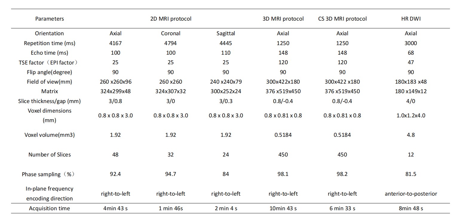
Table 1. Scan parameters of rectum MR imaging protocols
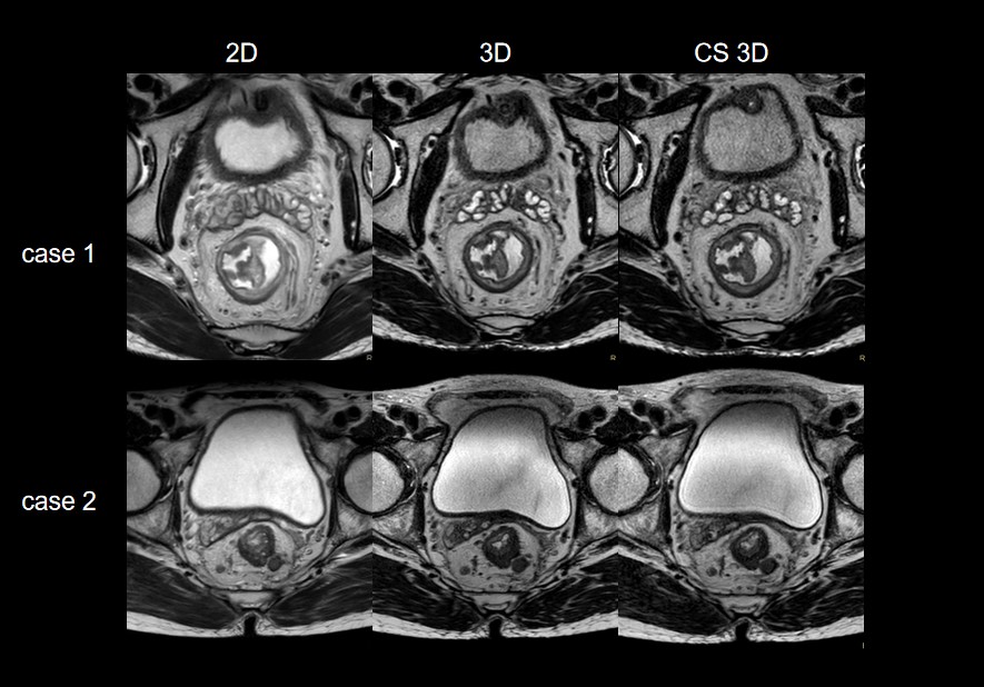
Figure 1. The display of tumors, stratification of rectal wall, lymph nodes, extramural vessels and peritoneal reflection in 2D, 3D and CS sense accelerated 3D T2W imaging.
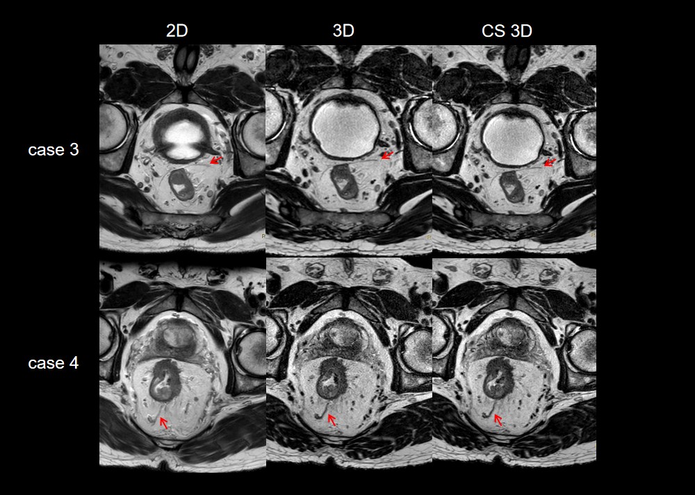
Figure 2. The display of peritoneal reflection and extramural vessels in 2D, 3D and CS sense accelerated 3D T2W imaging.

Figure 3. Contrast display of retroperitoneal invasion on oblique axial, sagittal and coronal planes of 2D, 3D and CS sense accelerated 3D T2 imaging.