2808
B0 shimming in human brain using rosette encoding at 7T1Radiology, University of Missouri Columbia, Columbia, MO, United States, 2Radiology, University of Pittsburgh, Pittsburgh, PA, United States, 3Resonance Research Inc., Billerica, MA, United States, 4University of Missouri Columbia, Columbia, MO, United States
Synopsis
With increasing use of ultra-high field, there is a need for fast and efficient field mapping. Towards this purpose, the non-Cartesian circular rosette trajectory is advantageous due to its low gradient demands and its efficiency due to the simultaneous acquisition of spatial and field encoding information. We describe the implementation of the circular rosette trajectory for field mapping at 7T in the human brain and compare this to the highly accurate although slow 5 echo Bolero field map.
Introduction
B0 field mapping is of increasing importance particularly with the increasing use and availability of ultra high field MR. The majority of methods for mapping the field are based on single time point measurements, which although very fast, commonly suffer from lower accuracy. This compares with the Bolero field map (1), which as acquired with 5 time points, is very accurate although commonly slow in acquisition. To achieve more accurate B0 field maps, we propose use of the non-Cartesian rosette trajectory (2). This trajectory is described as k= km*sin(w1t)*exp(iw2t) and as implemented with w1 = w2, executes a circular trajectory that repeatedly samples the origin. At low and moderate spatial resolution, its low gradient demands and smoothly continuous re-sampling makes the rosette advantageous for field mapping. The simultaneous use of time allocated to spatial encoding with off-resonance encoding also makes the rosette particularly efficient for mapping in comparison with conventional methods. We implement the circular rosette trajectory for B0 field mapping at 7T and as proof of principle, compare its performance to the 5 echo Bolero field map.Methods
A Siemens Magnetom 7T human system with a very high order shim insert and 8x2 transceiver array (Resonance Research Inc. Billerica MA) were used for all acquisitions, using RF shimming for the homogeneous B1 distribution for optimal whole brain coverage. The circular trajectory was implemented with a 2D GRE rosette acquisition, using w1=w2=1916 rad/s, and encoding dwell time of 1.6ms = pi/w1. With FOV = 216mm, 64x64 base resolution, 82 shots were used; the gradient strength and slew rates are constant in the circular trajectory here at 6.7mT/m and 25.3mT/m/ms. The reconstruction was based on methods described by Liu (ref. MRiLab, 3) and Schirda (4), with a 2D Kaiser Bessel window of 4 applied for regridding onto a 2-fold oversamled grid. In the time domain, the Tacq = 32ms and were binned according to the Bo encoding dwell time of 1.6ms. For initial demonstration of the acquisition, 33 slices (3mm thick), flip angle of 60 and TR = 1.2sec were used giving a total acquisition time of 100sec. Coil combination was performed using a sum of squares weighting performed over the initial time bin images. The first time bin data were also used to define subsequent phasing after the inclusion of a half-circle temporal shift of each shot to maintain each time bin centered on the k-space origin. To calculate the B0 values, temporal time bins 1, 2, 3, 5 and 9 were used, i.e., at 1.6ms encoding dwell times, delay times of 1.6, 3.2, 6.4, 12.8ms after the first acquisition were analyzed. For comparison to the Bolero acquisition (min), 1ms encoding dwell times (i.e., 1, 2, 4, and 8ms) were used as previously described (1). The field map acquisitions were acquired in n=3 healthy subjects acquired after targeted shimming through the temporal lobe.Results
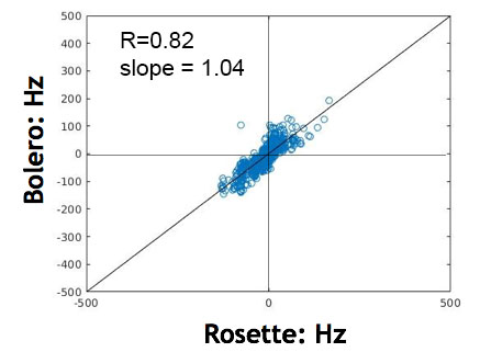
Fig. 1 shows the T1 co-registered images, single time bin reconstructed rosette images with calculated rosette and Bolero B0 maps from the temporal lobe. The rosette images (Fig. 1B) show some artifact due to the inter-shot crossing while the field maps are comparable between the GRE rosette and Bolero acquisitions. Fig. 2 shows a linear regression between the rosette and Bolero field values, R = 0.82 with a slope of 1.04, p<0.001.Conclusions
This proof of principle demonstration shows the feasibility of the circular rosette trajectory for robust field mapping. The approach is highly efficient for B0 encoding due to the repeated and simultaneous use of B0 encoding time with spatial sampling. With faster gradients, encoding bandwidths of +/-500Hz (temporal encoding time of 500us) can be achieved without need for temporal interleaves. As discussed by Noll (2), the accrual of phase due to off-resonance effects results in artifactual image appearance; however the phase consistency is maintained between the succeeding time bins with excellent fidelity in comparison to Bolero field mapping. With a sampling window of <15ms and resolution of ~3.5mm3, without any additional acceleration, the rosette field map should make whole brain measurements achievable with scan times of <45sec.Acknowledgements
This work is supported by NIH EB024408 and NS090417.References
1. Hetherington HP, Chu WJ, Gonen O, Pan JW.Magn Reson Med. 2006 Jul;56(1):26-33.
2. Noll D. Multishot rosette trajectories for spectrally selective MR imaging. IEEE Trans Med Imaging. 1997 Aug 16(4):372-7.
3. Liu F, Velikina JV, Block WF, Kijowski R, Samsonov AA. Fast Realistic MRI Simulations Based on Generalized Multi-Pool Exchange Tissue Model. IEEE Transactions on Medical Imaging. 2016. doi: 10.1109/TMI.2016.2620961
4. Schirda CV, Tanase C, Boada FE. Rosette spectroscopic imaging: optimal parameters for alias-free, high sensitivity spectroscopic imaging. J Magn Reson Imaging. 2009 Jun;29(6):1375-85.
Figures