2748
AI-driven markers of IDH1 mutational status using microstructure-based characterization of peritumoral region in gliomas1DiCIPHR (Diffusion and Connectomics in Precision Healthcare Research) Lab, University of Pennsylvania, Philadelphia, PA, United States, 2Department of Radiology, Department of Neurosurgery, University of Pennsylvania, Philadelphia, PA, United States
Synopsis
We developed a novel, microstructure-based, voxel-wise map of the peritumoral region in glioma brain tumors using DTI-based free water volume fraction map and deep-learning. This novel map captures the infiltrative heterogeneity of peritumoral region and can differentiate between gliomas with distinct IDH1 mutation status (IDH-mutant vs. IDH-wildtype). Thus, this new derived map that incorporates microstructure information can be used as a new diffusion based radiomic feature for various oncological investigations involving mutation status.
Introduction
The mutational status of isocitrate dehydrogenase (IDH1) is a defining feature of the World Health Organization (WHO) classification of gliomas. IDH-wildtype gliomas describe the most common malignant primary brain tumor in adults and have a worse prognosis than IDH-mutants1,2. There has been growing interest in developing computational methods to determine IDH1 mutation status using diverse MRI modalities3-6, taking into account the characteristics of the tumor, as well as the peritumoral region7. However, the use of diffusion tensor imaging (DTI) has been limited to using the standard diffusion measures like fractional anisotropy (FA) and mean diffusivity (MD)8,9. Using multi-compartment modeling on DTI, unique microstructural information can be derived about the tissue, particularly, free water volume fraction (FW-VF) that quantifies the amount of extracellular water at each voxel. We previously demonstrated that the FW-VF captures differences between infiltrative and vasogenic peritumoral regions based on differences in extracellular water content. These findings pave the way for further characterization of infiltrative heterogeneity in peritumoral tissue10,11 and using that information to determine the mutation status of the tumors. As the peritumoral infiltrative heterogeneity of high-grade gliomas is linked to their IDH gene mutational status12, and extracellular water content is linked to microstructural heterogeneity, we hypothesize that there is an association between the extracellular water content and IDH mutational status in glioma patients. We now introduce a novel heat map of the peritumoral region based on tissue microstructure extracted by deep-learning on the DTI-based free water volume fraction and use it to interrogate mutation-based differences.Method
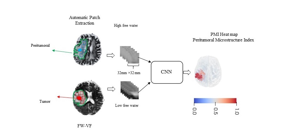
Figure 1 shows the pipeline of our approach. The dMRI data was acquired for 106 patients with brain tumors (66 glioblastomas and 40 metastases, ages 23-87 years, 51 females). Masks of the tumor and edema for each patient were created using DeepMedic13. FW-VF was extracted using FERNET 14, a multi-compartment modeling technique that extracts free water volume fractions (FW-VF) representative of extracellular water content from diffusion MR data. We leveraged the widely different peritumoral microenvironments of metastases and glioblastomas, the former consisting of vasogenic edema and the latter consisting of infiltrative tissue to provide reference labels. A set of (32mm ×32mm) patches was automatically extracted from peritumoral edema using random seed generators. We trained a Convolutional Neural Network (CNN) on patches in the peritumoral area from glioblastomas and metastases, labeled as either low free-water or high free-water, to extract a heat map containing a peritumoral microstructure index (PMI) for each voxel in the peritumoral region. PMI exploits the differential extracellular free-water in the voxel with and without infiltration and hence captures the infiltrative heterogeneity in the peritumoral region. An independent test set was used containing 261 gliomas (10 IDH-mutants versus 251 IDH-wildtypes, age 22-89, 105 females) and the voxel-wise PMI heat map was extracted for them. For each test subject, we averaged the CNN results among the three patches in the axial, sagittal, and coronal planes centered at each voxel to produce the voxel-wise PMI heat map. The average PMI of the peritumoral region was calculated for each patient. We performed a group comparison on average PMI between IDH-mutants and IDH-wildtypes using ordinary linear regression with sex and age as covariates.Results
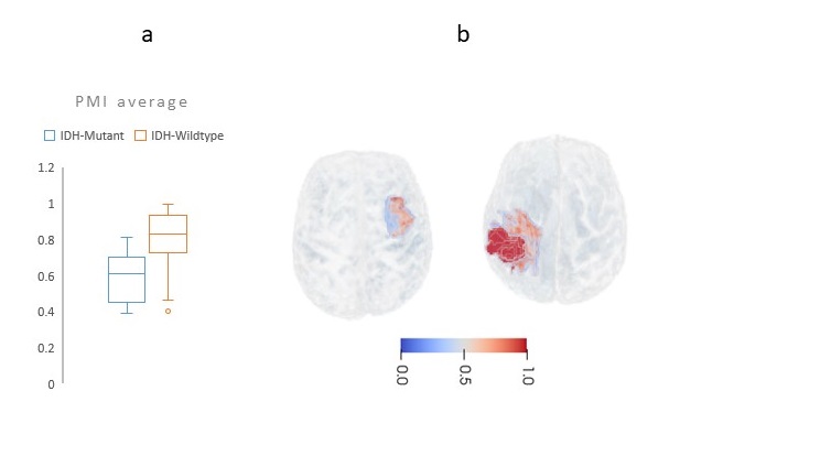
Figure 2 shows representative samples of PMI heat map for an IDH-mutant and IDH-Wildtype subject. We observed significant group difference between average PMI of IDH-mutants and IDH-wildtypes (t-statistics=4.4, p-value< 0.001) with higher values of PMI in IDH-wildtypes (Average: 0.82 ±Std: 0.12 for IDH-wildtypes vs. Average: 0.58 ±Std: 0.11 for IDH-mutants).Discussion
The microstructure-based characterization of infiltrative heterogeneity provided by the PMI heat map, showed significantly higher values in IDH-wildtypes. Our findings are consistent with previous literature describing that higher infiltration of glioma is associated with poorer prognosis15, and IDH-wildtypes have a worse prognosis than the IDH-mutant counterparts1. It further underlines the fact that the extracellular water-based characterization of peritumoral heterogeneity, the PMI, represents a powerful marker to discriminate mutation status, prior to biopsy.Conclusion
The proposed PMI heat map provides further information about the peritumoral microstructure that can be used to characterize the peritumoral area and discriminate IDH1 mutation status. This novel map potentially could complement other modalities and DTI-based measures like fractional anisotropy, to provide an integrated, biologically relevant characterization of the peritumoral microenvironment. Knowledge of IDH1 mutational status at initial presentation could highly influence clinical decision-making, especially, as mutant-IDH enzyme inhibitors and immunotherapy targeting IDH-mutant tumor cells are being developed.Acknowledgements
This research was supported by the National Institutes of Health (NIH) Grant R01NS096606 (PI: Ragini Verma).References
[1] Y.-Z. Chang, G.-Z. Li, B. Pang, K.-N. Zhang, X.-H. Zhang, Y.-Z. Wang, et al., "Transcriptional characteristics of IDH-wild type glioma subgroups highlight the biological processes underlying heterogeneity of IDH-wild type WHO grade IV gliomas," Frontiers in cell and developmental biology, vol. 8, p. 1132, 2020.
[2] D. N. Louis, A. Perry, P. Wesseling, D. J. Brat, I. A. Cree, D. Figarella-Branger, et al., "The 2021 WHO classification of tumors of the central nervous system: a summary," Neuro-oncology, vol. 23, pp. 1231-1251, 2021.
[3] S. Bakas, S. Rathore, M. Nasrallah, H. Akbari, Z. Binder, S. M. Ha, et al., "Nimg-40. Non-invasive in vivo signature of idh1 mutational status in high grade glioma, from clinically-acquired multi-parametric magnetic resonance imaging, using multivariate machine learning," Neuro-Oncology, vol. 20, pp. vi184-vi185, 2018.
[4] S. Nalawade, G. K. Murugesan, M. Vejdani-Jahromi, R. A. Fisicaro, C. G. B. Yogananda, B. Wagner, et al., "Classification of brain tumor isocitrate dehydrogenase status using MRI and deep learning," Journal of Medical Imaging, vol. 6, p. 046003, 2019.
[5] D. Alis, O. Bagcilar, Y. D. Senli, M. Yergin, C. Isler, N. Kocer, et al., "Machine learning-based quantitative texture analysis of conventional MRI combined with ADC maps for assessment of IDH1 mutation in high-grade gliomas," Japanese journal of radiology, vol. 38, pp. 135-143, 2020.
[6] C. De Looze, A. Beausang, J. Cryan, T. Loftus, P. G. Buckley, M. Farrell, et al., "Machine learning: a useful radiological adjunct in determination of a newly diagnosed glioma’s grade and IDH status," Journal of neuro-oncology, vol. 139, pp. 491-499, 2018.
[7] C. G. Bangalore Yogananda, B. R. Shah, M. Vejdani-Jahromi, S. S. Nalawade, G. K. Murugesan, F. F. Yu, et al., "A novel fully automated MRI-based deep-learning method for classification of IDH mutation status in brain gliomas," Neuro-oncology, vol. 22, pp. 402-411, 2020.
[8] S. Bisdas, J. Yuan, L. Mancini, and D. Roettger, "Can diffusion tensor MR imaging identify glioma IDH mutation status?," Annals of Oncology, vol. 29, p. viii129, 2018.
[9] Y. Park, K. Han, S. Ahn, Y. Choi, J. Chang, S. Kim, et al., "Whole-tumor histogram and texture analyses of DTI for evaluation of IDH1-mutation and 1p/19q-codeletion status in World Health Organization grade II gliomas," American Journal of Neuroradiology, vol. 39, pp. 693-698, 2018.
[10] Z. R. Samani, D. Parker, R. Wolf, W. Hodges, S. Brem, and R. Verma, "Distinct tumor signatures using deep learning-based characterization of the peritumoral microenvironment in glioblastomas and brain metastases," Scientific Reports, vol. 11, p. 14469, 2021/07/14 2021.
[11] Z. R. Samani, D. Parker, J. A. Alappatt, S. Brem, and R. Verma, "Deep learning-based peritumoral microstructure mapping in glioblastomas using free water volume fraction," in NEURO-ONCOLOGY, 2020, pp. 158-158.
[12] S. Lee, S. H. Choi, I. Ryoo, T. J. Yoon, T. M. Kim, S.-H. Lee, et al., "Evaluation of the microenvironmental heterogeneity in high-grade gliomas with IDH1/2 gene mutation using histogram analysis of diffusion-weighted imaging and dynamic-susceptibility contrast perfusion imaging," Journal of neuro-oncology, vol. 121, pp. 141-150, 2015.
[13] K. Kamnitsas, C. Ledig, V. F. Newcombe, J. P. Simpson, A. D. Kane, D. K. Menon, et al., "Efficient multi-scale 3D CNN with fully connected CRF for accurate brain lesion segmentation," Medical image analysis, vol. 36, pp. 61-78, 2017.
[14] D. Parker, A. A. Ould Ismail, R. Wolf, S. Brem, S. Alexander, W. Hodges, et al., "Freewater estimatoR using iNtErpolated iniTialization (FERNET): Characterizing peritumoral edema using clinically feasible diffusion MRI data," Plos one, vol. 15, p. e0233645, 2020.
[15] S. L. Di Jia, D. Li, H. Xue, D. Yang, and Y. Liu, "Mining TCGA database for genes of prognostic value in glioblastoma microenvironment," Aging (Albany NY), vol. 10, p. 592, 2018.
Figures