2648
Diagnosing Parkinson’s disease by Combining Neuromelanin and Iron Image Features Using Automatic SN Subregions Detection Approach1Ruijin Hospital, Shanghai Jiao Tong University School of Medicine, Shanghai, China, 2Magnetic Resonance Innovations, Inc., Bingham Farms, MI, United States, 3Radiology, Wayne State University, Detroit, MI, United States, 4Biomedical Engineering, Wayne State University, Detroit, MI, United States
Synopsis
A total of 100 Parkinson’s disease (PD) patients and 100 age- and sex-matched healthy controls (HCs) were scanned using a single 3D gradient echo magnetization transfer sequence. We developed an automatic substantia nigra (SN) subregions segmentation approach to get neuromelanin (NM) and iron measurements in the SN. These measures along with their overlap region volume and the nigrosome-1 (N1) sign showed reliable results indicative of promising diagnostic biomarkers to differentiate PD patients from HCs.
Methods: A total of 100 PD patients (age range 62.6 ± 8.6 years old) and 100 age- and sex-matched healthy controls (HC) (age range 63.2 ± 5.9 years old) were imaged on a 3T Philips scanner using a 3D multi-echo gradient echo SWI sequence with an activated MTC pulse. The imaging parameters included: seven echoes with TE1 = 7.5ms, ΔTE = 7.5ms, with TR = 62ms, flip angle = 30˚, pixel bandwidth = 174Hz/pixel, matrix size = 384 × 384, slice thickness = 2mm, and an original spatial in-plane resolution = 0.67 × 1.34mm2 then interpolated to 0.67 × 0.67mm2. The first echo of the MTC-SWI magnitude image (TE = 7.5ms) was used to delineate the NM content since that provided the key MT contrast. The second echo (TE = 15ms) was used for QSM reconstruction. The midbrain template was then created and the boundaries of each midbrain nucleus in template space were obtained. We mapped these boundaries back to the original space and then fine-tuned the boundaries in the original space using a dynamic programming algorithm to match the details of each individual’s NM and iron features.4,5 To evaluate the overlap volume between putative NM content and iron deposition in the SN, the NM regions-of-interest (ROIs) were superimposed on those of the SN from the corresponding MTC-QSM maps and normalized to the SN volume. Thresholds of 50 ppb on the QSM data and a normalized NM contrast threshold of 0.15 were found to be optimum for separating the two cohorts. Also the nigrosome 1 (N1) was assessed manually by two raters who reached a consensus for all the 200 cases in this study. Furthermore, in order to assess the sensitivity and specificity of the proposed models to distinguish between the HC and PD cases, binary logistic regression, a receiver operating curve (ROC) and area under the curve (AUC) analysis were performed on different parameters using IBM SPSS Statistics Version 22.0.
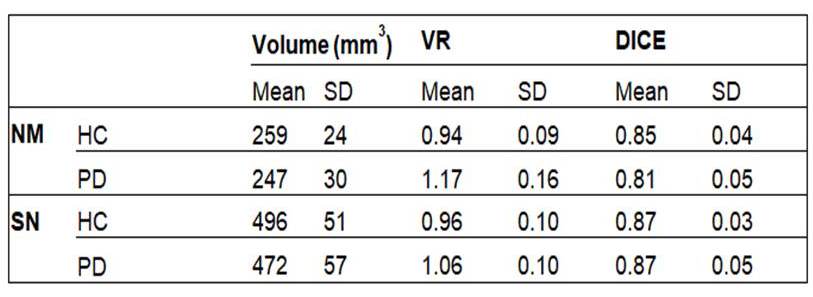
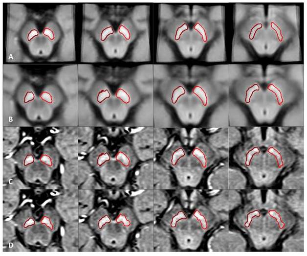
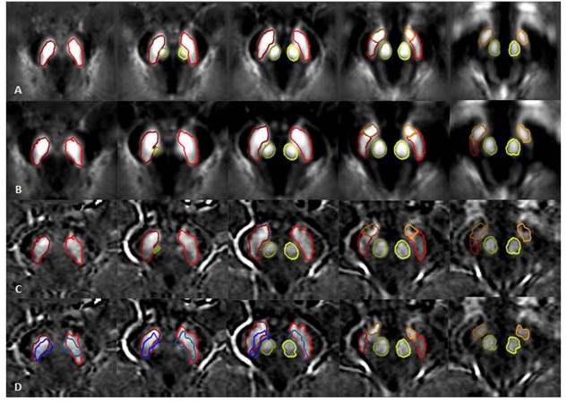
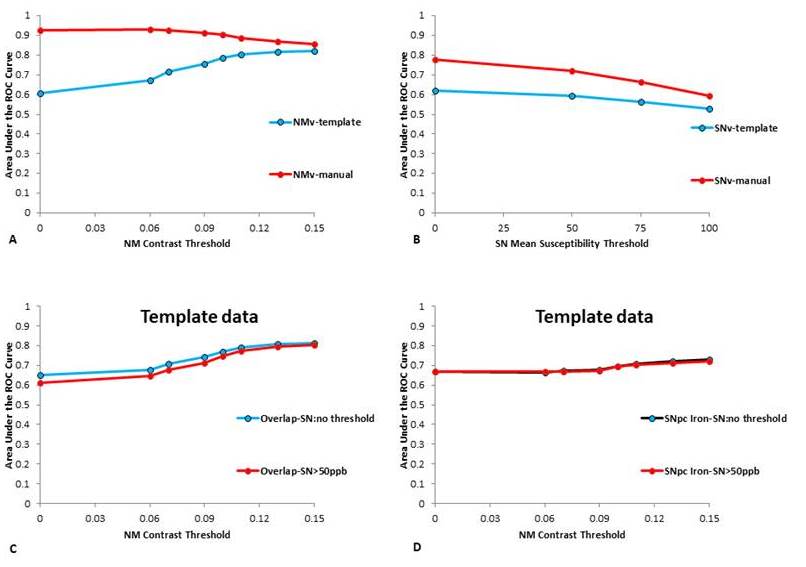
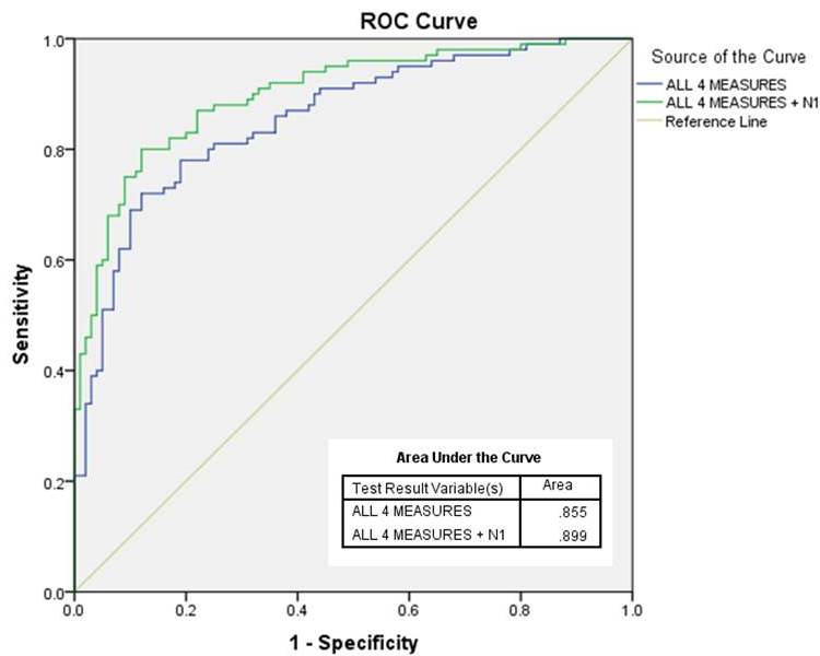
Results: Table 1 summarizes the results associated with the estimated template volumes, volume ratio (VR) values of the template boundaries over the manual ROIs and DICE similarity coefficients for the NM and SN, confirming the satisfactory performance of the template used in this study. The mean and standard deviation values are shown for the HC and PD cohorts. The slices from the original and transformed space as well as the boundaries associated with the MTC and QSM data are shown in Figure 1 and 2, respectively. Figure 3 shows the change in the AUC at different NM and SN thresholds for the individual measures, which are NM volume, SN volume, the NM/SN overlap region volume, and substantia nigra pars compacta (SNpc) iron content. From this data the optimum threshold for the NM contrast (normalized by NM background) and QSM data were selected as 0.15 and 50 ppb, respectively. Figure 4 shows the ROC curve for the combined measures at the thresholds of 0.15 and 50ppb for the normalized NM contrast and SN, respectively. The combination of the four measures (NM volume, SN volume, NM/SN overlap volume and SNpc iron) yielded an AUC of 0.855. However, the AUC improved to 0.899 when we combined these four measures with the N1 sign.
Discussion and conclusion: In this work, we have introduced an automatic SN subregions segmentation approach to measure the iron and NM contents and their overlap volume using a single multi-echo sequence in an attempt to develop a comprehensive pathophysiological biomarker that could be used clinically to diagnose PD. The combination of NM and SN volume, NM/iron overlap volume and iron content as well as the N1 sign provided strong measures to differentiate PD patients from HCs. We have demonstrated that this diagnostic biomarker can be determined using a fully automatic approach with clinically useful diagnostic accuracy (with an AUC as high as 90%).
Acknowledgements
We thank Mr. Emil Pacurar for handling all the data issues and automatically processing the data. This work was supported, in part, by a grant from the National Natural Science Foundation of China and the Innovative Research Team of High-level Local Universities in Shanghai.References
1. Group GBDNDC. Global, regional, and national burden of neurological disorders during 1990-2015: a systematic analysis for the Global Burden of Disease Study 2015. Lancet Neurol 2017; 16(11): 877-97.
2. Kowal, S.L., Dall, T.M., Chakrabarti, R., Storm, M.V., and Jain, A. (2013). The current and projected economic burden of Parkinson's disease in the United States. Mov Disord 28, 311-318.
3. Dexter, D.T., Carayon, A., Javoy-Agid, F., Agid, Y., Wells, F.R., Daniel, S.E., Lees, A.J., Jenner, P., and Marsden, C.D. (1991). Alterations in the levels of iron, ferritin and other trace metals in Parkinson's disease and other neurodegenerative diseases affecting the basal ganglia. Brain 114 ( Pt 4), 1953-1975.
4. Jiang, J., Haacke, E.M., Dong, M., 2007. Dependence of vessel area accuracy and precision as a function of MR imaging parameters and boundary detection algorithm. J Magn Reson Imaging 25, 1226-1234. 5. Zhang, T.Y., Suen, C., 1984. A fast parallel algorithm for thinning digital patterns. Comm. of the ACM 27.
Figures