2641
Bullying is associated with reduced cognitive scores and altered brain morphometry over time in preadolescent children.1Diagnostic Radiology and Nuclear Medicine, University of Maryland School of Medicine, Baltimore, MD, United States, 2Otorhinolaryngology—Head and Neck Surgery, University of Maryland School of Medicine, Baltimore, MD, United States, 3Pediatrics, University of Maryland School of Medicine, Baltimore, MD, United States, 4Neurology, University of Maryland School of Medicine, Baltimore, MD, United States, 5Neurology, Johns Hopkins University School of Medicine, Baltimore, MD, United States
Synopsis
Few studies identified brain morphometric changes associated with peer-victimization (“being bullied”), and no study examined how these changes can mediate the relationship between bullying and cognition. Using T1-weighted MRI scans and cognitive test scores from the Adolescent Brain Cognitive Development longitudinal dataset, we found that bullying is associated with reduced cognitive performance over time. Bullied children had smaller putamen volumes and right insula surface areas, thinner left precentral and banks of superior temporal sulcus cortices, but larger left entorhinal and right pars orbitalis surface areas. Importantly, these altered brain measures partially mediated the relationship between bullying and cognitive scores.
Introduction
Bullying victimization affects 20% of adolescents across the US,1 and was identified as a risk factor for suicide attempts2 and anxiety disorders3 into adulthood. Additionally, bullying by age 6 was associated with reduced executive function two years later,4 and 8-9-year-old children who were bullied were 6-9 months behind their peers in reading, writing, and grammar.5Rodent studies of “social-victimization”6 defined several vulnerable brain regions, including the prefrontal cortex, amygdala, and hippocampus.7,8 One preadolescent human imaging study identified thinner fusiform cortices at age ten in bullied children.9 Another longitudinal adolescent study reported that greater peer victimization was associated with generalized anxiety through decreases in left putamen volume.10
This study aims to use the longitudinal Adolescent Brain Cognitive Development (ABCD) dataset to test the hypotheses that children who were bullied would have brain changes and reduced cognition, and that the brain morphometric changes will mediate the relationships between bullying and cognitive outcomes.
Methods
The ABCD study is an ongoing, 10-year longitudinal study of children, starting at ages 9-10 years old, and includes caregiver-reported data regarding their children at 21 sites across the US. Baseline (Year 1) and second annual follow-up (Year 3) ABCD datasets (NDA release v.3.0.1) from 6,571 participants were assessed. The caregivers were asked whether their child had bullying problems at school or in their neighborhood.11 Bullied participants (N=323) were matched to non-bullied participants for sex, age, race/ethnicity, caregiver education level, and family income level (Table 1). The NIH Cognition Battery Toolbox®12 uncorrected scores in five domains and one composite area were included. T1-weighted MP-RAGE scans13 were acquired at each timepoint on 3T scanners (1-mm isotropic resolution). Cortical area and thickness and subcortical volumes were measured using FreeSurfer.14 We used linear mixed effects models and emmeans in R (version 4.1.1) to determine whether being bullied was related to changes in cognitive scores or brain structures over time and whether the changes in brain morphometry mediated the relationships between being bullied and changes in cognitive scores. Covariates included age, sex, visit, intracranial volume, and scanner site.Results
At Year 1, the children were 9.9±0.6 years of age, 60% were boys and 50% were racial minorities, with similar distributions between bullied and non-bullied groups (Table 1). Overall, bullied children had lower scores in the domains of reading, processing speed, inhibitory control and attention, and crystallized composite (reading and episodic memory) (Figure 1A; P=0.001-0.040). Bullying and visit showed an interaction, with bullied children showing greater increases in reading scores between visits than the non-bullied group (Figure 1B; interaction-P=0.037). Boys had lower episodic memory (Picture Sequence Memory) scores than girls in the non-bullied group only (Figure 1C; interaction-P=0.002).Bullied children had smaller putamen volumes than non-bullied children at both timepoints (Figure 2A; P=0.003-0.040), but both groups showed similar increases in putamen volumes over time (Figure 2B-2C). Compared to the non-bullied children, the bullied children also had smaller cortical areas in the right insula (Figure 3A-C; P=0.038), but larger cortical areas in four other regions (Figure 3A-C; P=0.008-0.038). Importantly, the right pars orbitalis was larger in the bullied group at Year 3, but not at Year 1 (Figure 3C; interaction-P=0.012). Furthermore, left entorhinal and right pars orbitalis areas mediated the relationship between bullying and reading scores (P=0.002), and the right pars orbitalis mediated between bullying and crystallized composite scores (P=0.023) (Figure 3D). Bullied children had thinner cortices in four additional regions at both timepoints (Figure 4A-C; P=0.008-0.037). Cortical thickness in the left banks of the superior temporal sulcus (STS) and precentral gyrus mediated the relationships between bullying and processing speed scores (Figure 4D; P=0.001), and the left precentral gyrus also mediated between bullying and inhibitory control and attention scores (Figure 4D; P=0.037).
Discussion
We identified novel findings that bullied children had poorer processing speed, inhibitory control and attention, and crystallized composite cognition than non-bullied children at both timepoints. Additionally, we found that bullied children had larger surface areas and thinner cortices in several cortical regions. Importantly, the relationships between bullying and four cognitive domain scores were mediated by brain morphometry.The insula was previously hypothesized to play a role in emotional processing and regulation in children who were bullied;9 hence our finding of reduced surface area in the right insula of bullied children suggests that they may develop problems with emotional processing. Crystallized intelligence was associated with larger surface areas in language-related regions,15 which is consistent with our finding that the pars orbitalis, one of the brain regions involved in language and emotion processing,16 partially mediated the relationships between bullying, reading, and crystallized composite scores. Interestingly, we found a larger surface area in the pars orbitalis by Year 3 in the bullied group. This may indicate that bullied children experience a slower dendritic pruning process, similar to those with lower IQ.17
Additional follow-up over a longer period will validate whether bullying is associated with the altered morphometric and cognitive changes and how the frequency and intensity of bullying might impact these outcomes.
Conclusions
Our findings support our hypotheses that bullying is associated with brain morphometric alterations and poorer cognitive development, and that these brain changes may mediate the relationships between bullying and cognitive outcomes.Acknowledgements
We thank all the ABCD Study participants. The ABCD Study® is supported by the National Institutes of Health and additional federal partners under award numbers U01DA041048, U01DA050989, U01DA051016, U01DA041022, U01DA051018, U01DA051037, U01DA050987, U01DA041174, U01DA041106, U01DA041117, U01DA041028, U01DA041134, U01DA050988, U01DA051039, U01DA041156, U01DA041025, U01DA041120, U01DA051038, U01DA041148, U01DA041093, U01DA041089, U24DA041123, U24DA041147. A full list of supporters is available at https://abcdstudy.org/federal-partners.html.References
1. Wang, K., Chen, Y., Zhang, J. & Oudekerk, B. Indicators of School Crime and Safety: 2019. https://nces.ed.gov/pubsearch/pubsinfo.asp?pubid=2020063 (2020).
2. Meltzer, H., Vostanis, P., Ford, T., Bebbington, P. & Dennis, M. S. Victims of bullying in childhood and suicide attempts in adulthood. Eur. Psychiatry J. Assoc. Eur. Psychiatr. 26, 498–503 (2011).
3. Copeland, W. E., Wolke, D., Angold, A. & Costello, E. J. Adult Psychiatric Outcomes of Bullying and Being Bullied by Peers in Childhood and Adolescence. JAMA Psychiatry 70, 419–426 (2013).
4. Holmes, C. J., Kim-Spoon, J. & Deater-Deckard, K. Linking Executive Function and Peer Problems from Early Childhood Through Middle Adolescence. J. Abnorm. Child Psychol. 44, 31–42 (2016).
5. Mundy, L. K. et al. Peer Victimization and Academic Performance in Primary School Children. Acad. Pediatr. 17, 830–836 (2017).
6. Golden, S. A., Covington, H. E., Berton, O. & Russo, S. J. A standardized protocol for repeated social defeat stress in mice. Nat. Protoc. 6, 1183–1191 (2011).
7. Colyn, L., Venzala, E., Marco, S., Perez-Otaño, I. & Tordera, R. M. Chronic social defeat stress induces sustained synaptic structural changes in the prefrontal cortex and amygdala. Behav. Brain Res. 373, 112079 (2019).
8. Zhang, T. R. et al. Negative Memory Engrams in the Hippocampus Enhance the Susceptibility to Chronic Social Defeat Stress. J. Neurosci. Off. J. Soc. Neurosci. 39, 7576–7590 (2019).
9. Muetzel, R. L. et al. Frequent Bullying Involvement and Brain Morphology in Children. Front. Psychiatry 10, 696 (2019).
10. Quinlan, E. B. et al. Peer victimization and its impact on adolescent brain development and psychopathology. Mol. Psychiatry 25, 3066–3076 (2020).
11. Kaufman, J., Birmaher, B., Brent, D. A., Ryan, N. D. & Rao, U. K-SADS-PL. J. Am. Acad. Child Adolesc. Psychiatry 39, 1208 (2000).
12. Luciana, M. et al. Adolescent neurocognitive development and impacts of substance use: Overview of the adolescent brain cognitive development (ABCD) baseline neurocognition battery. Dev. Cogn. Neurosci. 32, 67–79 (2018).
13. Casey, B. J. et al. The Adolescent Brain Cognitive Development (ABCD) study: Imaging acquisition across 21 sites. Dev. Cogn. Neurosci. 32, 43–54 (2018).
14. Hagler, D. J. et al. Image processing and analysis methods for the Adolescent Brain Cognitive Development Study. NeuroImage 202, 116091 (2019).
15. Tadayon, E., Pascual-Leone, A. & Santarnecchi, E. Differential Contribution of Cortical Thickness, Surface Area, and Gyrification to Fluid and Crystallized Intelligence. Cereb. Cortex N. Y. N 1991 30, 215–225 (2020).
16. Belyk, M., Brown, S., Lim, J. & Kotz, S. A. Convergence of semantics and emotional expression within the IFG pars orbitalis. NeuroImage 156, 240–248 (2017).
17. Schnack, H. G. et al. Changes in thickness and surface area of the human cortex and their relationship with intelligence. Cereb. Cortex N. Y. N 1991 25, 1608–1617 (2015).
Figures
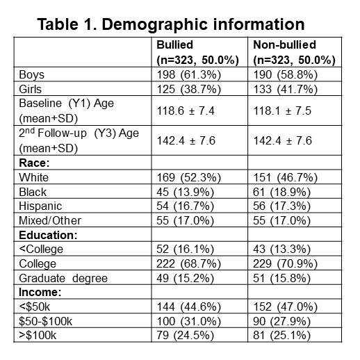
Table 1 Legend. Bullied children with MRI data from Year 1 (Baseline, Y1) and Year 3 (2nd follow-up, Y3) visits were matched with non-bullied children for sex, age, race, education, and income.
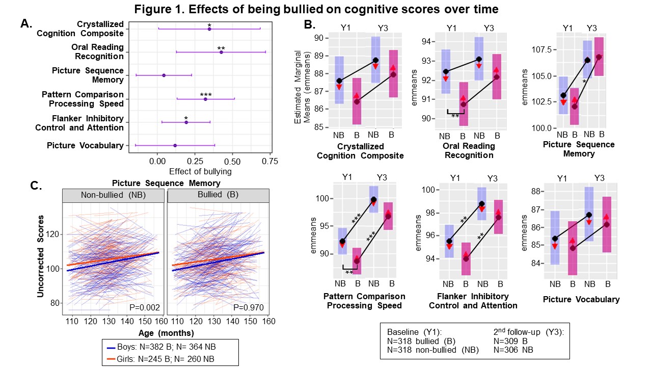
Figure 1 Legend. A-B display the main effects of bullying (A) and interactive effects of bullying and visit (B) on cognitive scores, calculated using emmeans and linear mixed effects models (*P<0.05; **P<0.01; ***P<0.001; adjusted for multiple comparisons). C plots individual and mean Picture Sequence Memory scores by age; with a sex difference in the non-bullied group only. Picture Vocabulary (P=0.024), Pattern Comparison Processing Speed (P=0.003), and Picture Sequence Memory (P=0.007; not shown) had significant sex differences overall.
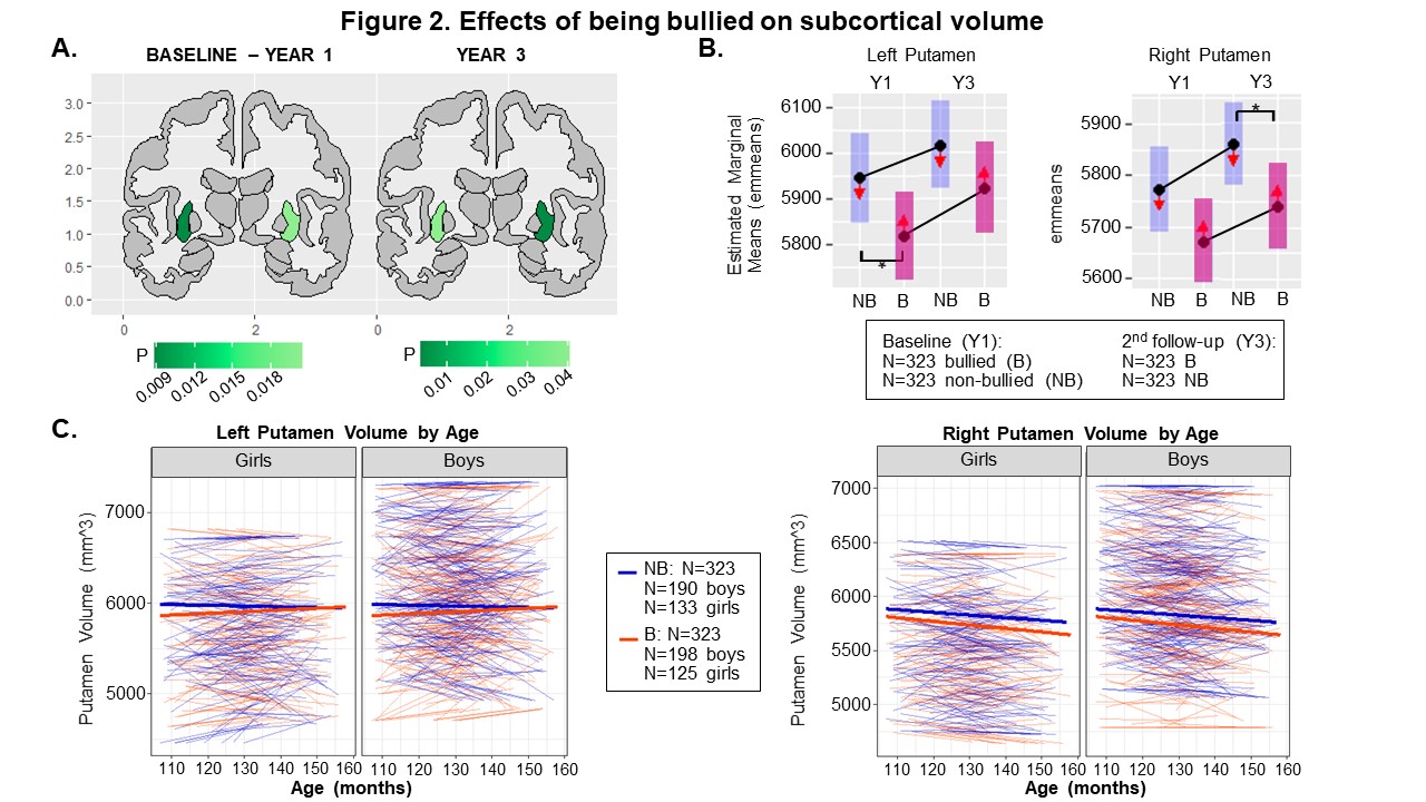
Figure 2 Legend. A-B display the main effects of bullying (A) and interactive effects of bullying and visit (B) on subcortical putamen volumes, calculated using emmeans and linear mixed effects models (*P<0.05; **P<0.01; ***P<0.001; adjusted for multiple comparisons). C plots individual and mean putamen volumes by age. Although left (P<0.001) and right (P<0.001) putamen volumes showed sex-differences, no sex-specific effects are observed (left: interaction-P=0.26; right: interaction-P=0.57).
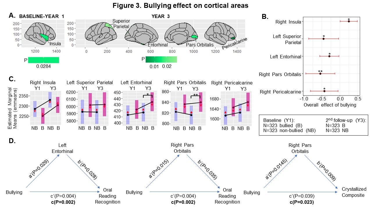
Figure 3 Legend. A-C display the effects of bullying (A-B) and interactive effects of bullying and visit (C) on cortical areas, calculated using emmeans and linear mixed effects models (LMEM). (*P<0.05; **P<0.01; ***P<0.001; adjusted for multiple comparisons). While girls have smaller areas in all regions except the pericalcarine (P=<0.001-0.003), there are no interactions between sex and bullying (P=0.08-0.95). D displays LMEM p-values to show the relationship between bullying and cognition, and how cortical area acts as a partial mediator.
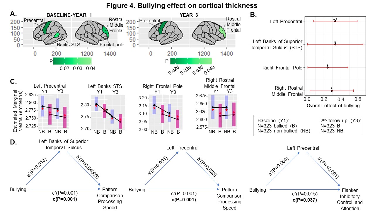
Figure 4 Legend. A-C display the effects of bullying (A-B) and interactive effects of bullying and visit (C) on cortical thickness, calculated using emmeans and linear mixed effects models (LMEM). (*P<0.05; **P<0.01; ***P<0.001; adjusted for multiple comparisons). Girls have smaller precentral (P<0.001) and banks of STS (P<0.001) cortices than boys but there are no interactions between sex and bullying (P=0.46-0.82). D displays LMEM p-values to show the relationship of bullying and cognition, and how cortical thickness acts as a partial mediator.