2637
On the basis of sex: Preclinical 23Na MRI at 21.1 T Delineates Sodium Distribution Differences in Females Following Central Sensitization1National High Magnetic Field Laboratory, Florida State University, Tallahassee, FL, United States, 2Chemical & Biomedical Engineering, FAMU-FSU College of Engineering, Tallahassee, FL, United States, 3Neurology, University of Southern California, Los Angeles, CA, United States
Synopsis
23Na FID-based 3D CSI was implemented in female rats at 21.1 T to examine sex differences in sodium response to NTG-triggered central sensitization. The female brainstem was resilient to NTG-triggered increases in sodium seen with males, despite sodium increases in the female cisterna magna and fourth ventricle. Lateral ventricles and thalamus sodium in females decreased with NTG trigger. Female resilience in brainstem and thalamus, both connected to trigeminal first order neuron, holds interesting implications for migraine.
Introduction
Traditionally in preclinical examination of migraine, the male Sprague-Dawley rat model is implemented, despite the vast majority of human migraineurs being female. This choice in animal model is commonly implemented to avoid potential complications from estradiol. While the estrus cycle may add variance within results, concerns of interference with the fundamental mechanisms of migraine may be unwarranted, e.g. the Sprague-Dawley estrus cycle has been shown not to influence CGRP release in the trigeminal nerve1. Previously, preclinical 23Na MRI at 21.1 T demonstrated sodium increases within the male Sprague-Dawley model as a result of nitroglycerin (NTG)-triggered central sensitization, a well-established model of migraine2. In this study, preclinical 23Na 3D chemical shift imaging (CSI) was performed at 21.1 T in female Sprague-Dawley rats to elucidate potential sex differences in the sodium response to NTG-triggered central sensitization.Methods
Animal Model: Prior to scanning, animals (108-220 g) were anesthetized with 5% isoflurane in oxygen to implant an intraperitoneal (IP) line for in situ NTG delivery while in the vertical magnet. During scanning, rodents were maintained at 2-3% isoflurane. Once baseline values were acquired, the NTG dose was administered IP. Female animals were injected with 2-mg/kg bodyweight NTG (N=14), 10-mg/kg bodyweight NTG (N=14) or an equivalent volume of saline (N=14).MR Acquisitions: For 23Na FID-based 3D CSI, Hamming-filtered data were acquired to maximize SNR within the preferred temporal resolution. Scans were performed interdigitated, with 10-min CSI acquisitions repeated every 20 min, out to 2 h post-injection. Reported times correspond to the end of the scan. The acquired image resolution was 1x1x3 mm.
Data Processing & Analysis: Manual image reconstruction and segmentation were performed in MATLAB. An exponential filter was applied to each FID; during Fourier transform, the image was zero-padded to a final resolution of 0.25x0.25x1 mm (Figure 1). Segmentation was performed directly on the sodium images, based on the Waxholm SD rat brain atlas. Regions segmented were the brainstem, thalamus, cisterna magna and ventricular aqueduct as well as the lateral, third and fourth ventricles. Statistical analyses were performed in JMP Pro 15. A mixed model with repeated measures with a Toeplitz covariance matrix was performed; post-hoc Tukey HSD tests were performed with p<0.05 for all significances.
Results & Discussion
Both doses of NTG (2 and 10 mg/kg) showed similar trends in sodium signal for all segmented regions, with 10 mg/kg displaying more drastic changes. Male Sprague-Dawley rats previously had demonstrated increases in neural sodium with a 10-mg/kg NTG trigger of central sensitization, displaying sodium impacts from baseline in the brainstem, third and fourth ventricles and cisterna magna2. In the female model, sodium response to NTG mirrors the male in the fourth ventricle and cisterna magna (Figure 2), with sharp increases in sodium signal. In both of these CSF regions, the 10-mg/kg NTG group reached significance to baseline and saline at a majority of all times. Adjacent neural tissue structure showed the first divergence between the female and male rat models.Upon NTG-triggered central sensitization, the female brainstem displays no significant changes from baseline sodium (Figure 3), albeit with high variance within each group. Female resistance to increases in brainstem sodium seen with the male model holds interesting implications for the study of migraine, as the brainstem contains direct connections to the trigeminal first order neuron and is critical for the onset and progression of central sensitization. It is possible that the lack of estradiol control is in part responsible for the wide deviations seen in the female brainstem data. However, both NTG doses follow the same brainstem signal trend as saline animals, implying this resilience is a sex difference independent of cyclical estradiol levels.
The third ventricle (data not shown) is marked by the same large deviations seen within the brainstem, although a few significances are evident: 10-mg/kg NTG induces a significant increase from baseline values at 40 min, followed by a slow decrease that reaches significance to saline at 120 min.
Sex differences are most apparent in the lateral ventricles and thalamus. In females, both regions show sustained statistically significant decreases in sodium signal upon central sensitization (Figures 4-5). These decreases contrast sharply to the male model, in which thalamic and lateral ventricular sodium significantly increased with acute 10-mg/kg NTG3. As differences were most pronounced in the rostral ventricular system and brain regions innervated by the trigeminal ganglia (thalamus and brainstem), this data suggests that the established lower sensory threshold in females may arise from a sex-based difference in sodium homeostatic regulation.
Conclusion
23Na FID-based 3D CSI was performed at 21.1 T to examine the impacts of central sensitization on the preclinical female Sprague-Dawley rat model. Two doses of NTG were examined, although dose did not significantly impact the directionality of sodium changes, only their severity.The female rat brainstem is resilient to the NTG-based changes in sodium evident for males; female lateral ventricle and thalamus sodium signals see sustained decreases throughout central sensitization in sharp contrast to the male model.
Acknowledgements
This work is supported by the NIH through R01-NS072497. Part of this work was performed at the US National High Magnetic Field Laboratory (NHMFL), which is supported by the State of Florida and the National Science Foundation Cooperative Agreement No. DMR-1644779.References
1. Moussaoui, S., Duval, P., Lenoir, V., Garret, C. & Kerdelhue, B. CGRP in the trigeminal nucleus, spinal cord and hypothalamus: Effect of gonadal steroids. Neuropeptides 30, 546–550 (1996).
2. Abad, N., Rosenberg, J. T., Hike, D. C., Harrington, M. G. & Grant, S. C. Dynamic sodium imaging at ultra-high field reveals progression in a preclinical migraine model. Pain 159, 2058–2065 (2018).
3. Abad, N. Acute NTG Triggered Migraine: Evaluating the Holy Trinity in Neurological Disorders. Florida State University, (2019)
Figures
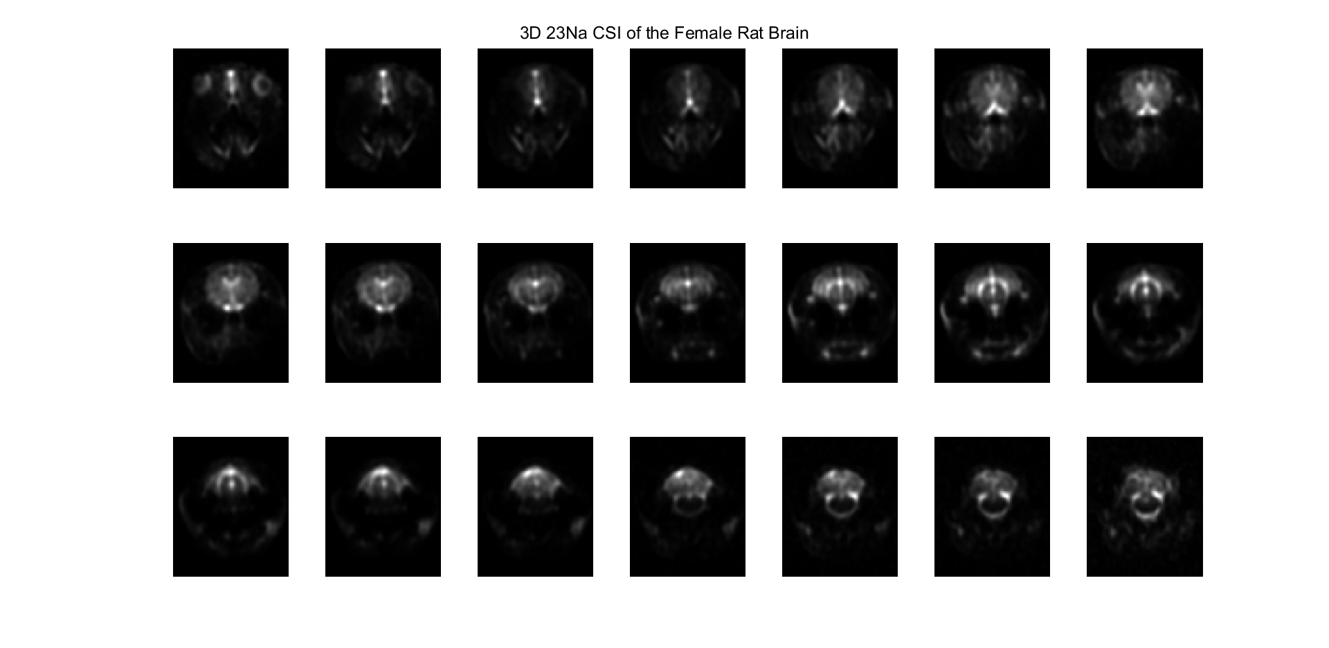
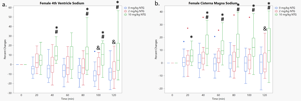
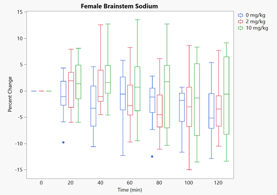
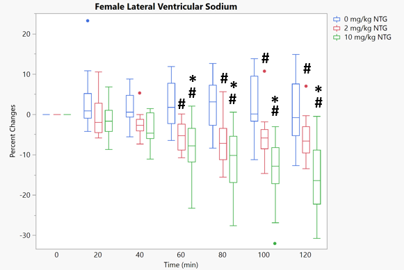
Figure 4. Percentage changes in 23Na signal in the female Sprague-Dawley lateral ventricles post-NTG administration. N=14 for all three doses. * denotes significance to baseline values, # denotes significance to saline. p<0.05 for all significances.
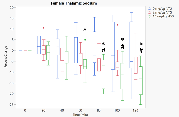
Figure 5. Percentage changes in 23Na signal in the female Sprague-Dawley thalamus post-NTG administration. N=14 for all three doses.* denotes significance to baseline values, # denotes significance to saline. p<0.05 for all significances.