2627
Continuous monitoring of MRI staff exposure to stray magnetic field at 1.5 T and 3 T1Consiglio Nazionale Ricerche - Istituto SPIN, L'Aquila, Italy, 2Istituto Nazionale Fisica Nucleare - Laboratori Nazionali del Gran Sasso, L'Aquila, Italy, 3Università dell'Aquila, L'Aquila, Italy, 4Tecnorad SrL, Verona, Italy, 5Azienda Sanitaria Locale 1 - Abruzzo, L'Aquila, Italy
Synopsis
We show how to monitor the MRI staff exposure to the $$$B_0$$$ stray-field. Monitoring is extended to several body districts without interferences with normal hospital operative procedures. The Weighted Peak index is calculated, for different Action Levels limits as from the current EU regulations, to consider the non-sinusoidal nature of exposure. Results show that exposure limits are exceeded even at 1.5 T and their occurrences significantly increase at 3.0 T scanners.
Introduction
MRI staff personnel is exposed to non-sinusoidal time-variable magnetic fields and induced electric fields, during their movements inside the scanner room. EU regulations [1] establish limits on worker’s exposure which are expressed in terms of non-measurable E field in the body or by means of the Action Levels (ALs), parameters defined from the measurable magnetic field in the specific position occupied by the body. In this work we report a novel method suitable to test ALs compliance and monitor a plurality of body districts by means of easily available and cheap hardware. To show the effectiveness of the method, we present the results of several monitoring days, in a standard hospital setting, with 1.5 and 3 T scanners.Methods
Measurements were performed in the Radiology Department of the San Salvatore Hospital, L’Aquila (Italy), equipped with a 1.5 T Siemens Magneton and a 3.0 T GE Optima MR750w. We used five wearable gaussmeters (Talete©, Technorad SrL, Verona) [2] with 0-3 T range, 1 mT sensitivity, and built-in data storage capacity. Sensors were first calibrated using a THM1176 Three-axis Hall Magnetometers (Metrolab Instruments SA, Geneva, Switzerland). The TALETE firmware was modified to perform a B modulus measurement every 2 ms (250 Hz bandwidth). We stored the B values if B $$$\ge$$$ 10 mT, thus considering only the time windows with the operator inside the MRI scanner room. For each work sheet (approx. 3 h) a single operator was monitored, and a sensor was firmly fixed on each wrist, the belt (abdomen), the shirt pocket (heart) and the temple of plastic glasses (head). Recorded data were processed offline to calculate the Weighted Peak (WP) as a function of time [1,3], using the frequency-domain approach based on the Fourier decomposition of the recorded signal:$$WP(t) = \left| \sum_i \frac{B(f_i)}{B_L (f_i)\sqrt{2}} \cos{(2\pi f_i t+\theta (f_i) + \phi (f_i))} \right|$$
where $$$B(f_i), \theta (f_i)$$$ are the measured signal spectral amplitudes and phases at frequency $$$f_i$$$, respectively, while $$$B_L (f_i), \phi (f_i)$$$ are the frequency-dependent limits as reported in Table 1.
| Table 1 | ||||
| | BL (uT) | $$$\phi$$$ (deg) | ||
| Frequency range (Hz) | Low ALs | High ALs | Limb ALs | |
| $$$1 \le f < 8$$$ | 2.0 105/f2 | 3.0 105/f | 9.0 105/f | +180 |
| $$$8 \le f < 25$$$ | 2.5 104/f | 3.0 105/f | 9.0 105/f | + 90 |
| $$$25 \le f < 250$$$ | 1.0 103 | 3.0 105/f | 9.0 105/f | 0 |
WP(t) was calculated each 10 ms for the three ALs. The total number of time points with WP(t) > 1, a condition corresponding to an exposure above the AL threshold, was counted.
Results
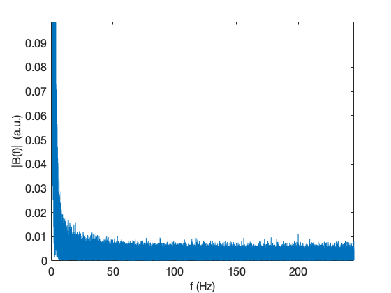
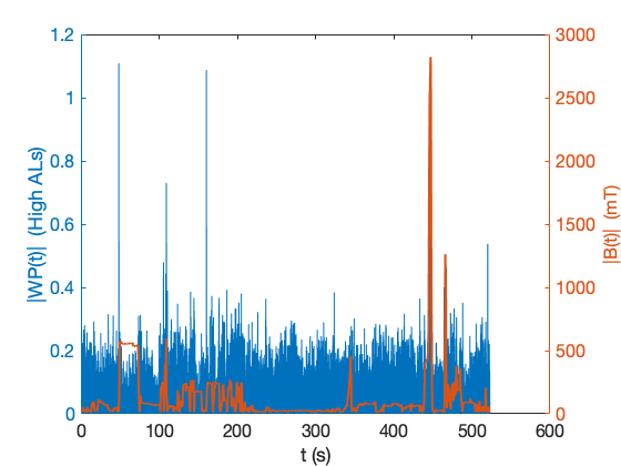
As reported in Fig. 1, a typical amplitude spectrum of B(t) is peaked at f=0 Hz and, as expected, decreases at increasing frequencies, with amplitude smaller that the detector noise for frequencies higher than 100 Hz. Based on this, to avoid spurious contribution from noise to the computed WP(t), the analysis was restricted to the $$$1 \le f \le 100$$$ Hz range.Figure 2 shows the measured B(t) and the calculated WP(t) for the High ALs threshold over a total of 523 s, corresponding to the total time spent by an MRI worker within the 3 T scanner room in a 3 h period. As expected, the observed WP(t) peaks are related to rapid changes of the magnetic field due to fast worker movements close the bore entrance. Two values above 1, exceeding the High AL exposure limit, are present.
In Table 2 we summarize the WP(t) > 1 observed occurrences for the 1.5 T and 3 T scanners mesurements.
| Table 2 | |||||
| 1.5 T (n=8 MRI operators) | |||||
| | Wrist Right | Wrist Left | Abdomen | Hearth | Head |
| Low ALs | NA | NA | 983 | 107 | 89 |
| High ALs | NA | NA | 6 | 0 | 0 |
| Limb ALs | 0 | 0 | NA | NA | NA |
| 3.0 T (n=5 MRI operators) | |||||
| Low ALs | NA | NA | 2856 | 243 | 205 |
| High ALs | NA | NA | 84 | 6 | 0 |
| Limb ALs | 0 | 0 | NA | NA | NA |
Discussion
The results in Table 2 show that, increasing the static magnetic field, the number of above-the-threshold events increases. Even if the hands are probably the fastest moving body part, their position, often close to the main magnet axis, and the higher limits established for limbs, prevent them to exceed the threshold. The worst case applies to the abdomen, probably due to its close position to the highest $$$B_0$$$ gradient region when the operator is in the proximity of the bore access.Conclusions
To the best of our knowledge, we report for the first time a multi body-districs monitoring of exposure limits, according to EU regulations, over a standard working shift of MRI operators. We observed that both Low ALs and High Als limits are exceeded during standard imaging protocols, suggesting that continuous monitoring of the full body districts [5,6] is necessary. Our main conclusion is that specific training sessions for the MRI workers should be adopted, to instruct them on how to properly move within the scanner room to stay below the EU limits.Acknowledgements
We acknowledge Prof. C. Masciocchi and E. Di Cesare of the San Salvatore Hospital in L’Aquila for allowing access to the MRI scanners.
References
[1] Directive 2013/35/EU of the European Parliament and of the Council of 26 June 2013, “On the minimum health and safety requirements regarding the exposure of workers to the risks arising from physical agents (electromagnetic fields)”, Official Journal of the European Union L179, 1-21 (2013).
[2] https://tecnorad.it/product/talete/
[3] ICNIRP (International Commission on Non-Ionizing Radiation Protection). “Guidelines for limiting exposure to time-varying electric and magnetic fields (1 Hz to 100 kHz)”. Health Phys. (2010) 99:818–836.
[4] D. Andreuccetti, L. Biagi, G. Burriesci, V. Cannatà, G.M. Contessa, R. Falsaperla, E. Genovese, R. Lodato, V. Lopresto, C. Merla, A. Napolitano, R. Pinto, G. Tiberi, M. Tosetti, N. Zoppetti, “Occupational exposure in MR facilities due to movements in the static magnetic field”, Med. Phys. (2017) 44:5988.
[5] A. Galante, M. Alecci, M. Fantasia, C. Rinaldi, F. Federici, F. Franchi, “Real-time monitoring and training for professionals exposed to static magnetic fields”, Proc. IEEE Medical Measurements Applications (2018) :255-259.
[6] A. Galante, G. Placidi, “Method and apparatus for monitoring the personal exposure to static or quasi-static magnetic fields”, EP 14716012.1, (2019).
Figures