2576
Quantitative susceptibility mapping in the dynamic evaluation of renal ischemia-reperfusion injury in rabbits: a feasibility study1Third Affiliated Hospital of Soochow University, changzhou, China, 2MR Research China, GE Healthcare, Shanghai, China
Synopsis
The purpose was to explore whether quantitative susceptibility mapping (QSM) can assess the dynamic process of renal ischemia-reperfusion injury (IRI) in rabbits. We found that the magnetic susceptibility values of the outer medulla were statistically significant among the IRI group. Additionally, the magnetic susceptibility values of the outer medulla was highly correlated with the pathological score of renal injury. With these findings, QSM could serve as a quantitative biomarker to assess the dynamic changes of the outer medulla in the early stage of renal IRI.
Introduction
Renal ischemia-reperfusion injury (IRI) often occurs in the process of major cardiovascular surgery, renal transplantation, shock and severe trauma, which is one of the main causes of acute renal injury and even renal failure[1]. Therefore, early diagnosis and treatment of renal IRI has become the focus of clinical attention. At present, biochemical and hematological tests are commonly used to evaluate renal IRI, but those indicators cannot reflect unilateral renal function and have a delaying effect. Renal biopsy is the gold standard for the evaluation of renal injury, but its clinical application is limited by the associated invasiveness and complications[2]. Quantitative susceptibility mapping (QSM) is a relatively novel magnetic resonance imaging technique for quantitatively measuring tissue magnetic susceptibility[3]. This technique has been widely used in the disease diagnosis and treatment effect evaluation such as Neurodegenerative diseases, hepatic iron overload, and prostate cancer et al[4, 5]. Therefore, the main goal of this study was to firstly explore if QSM has potential in assessing the dynamic process of renal IRI in rabbits and to secondly evaluate the relationship between the magnetic susceptibility and the pathological score of renal injury.Methods
Animal model In this animal care committee–approved study, thirty-six rabbits were divided into the IRI group(n=30) and the sham group(n=6). In the IRI group, rabbits underwent the left kidney surgery by clamping the left renal artery and vein for 60 minutes and then releasing the clamp to establish renal IRI model. In the sham group, rabbits underwent the same operation, but without clamping the left renal artery and vein. MRI experiment MRI experiments were performed five times (IRI-pre, IRI-1h, IRI-12h, IRI-24h and IRI-48h) for all rabbits on an 3.0-Telsa MR (750w, GE Healthcare, USA) with a sixteen-channel phase array body coil employed. Multi-echo fast-gradient-echo sequence was used to acquire QSM imaging. The corresponding scan parameters were of field-of-view = 15cm×12cm, matrix size =448×176, repetition time (TR)= 36.7ms, echo of time (TE): 4.4ms/ 9.2ms/ 14.1ms/ 18.9ms/ 23.8ms/ 28.6ms, flip angle = 10°. The scan time was 23 seconds. Data analysis All data were analyzed with a vendor-provided QSM fitting software on ADW 4.6 workstation (GE Healthcare). Dynamic Renal the magnetic susceptibility values were measured in the outer medulla at five time points. In addition, all specimens of left kidneys were stained with hematoxylin-eosin (HE). The pathological score of renal injury were calculated[6]. All statistical analyses were performed in SPSS software. The repeated measurement analysis of variance was used to compare the differences in the magnetic susceptibility among IRI group at different time points and sham group. Differences in the pathological score of renal injury were analyzed by the nonparametric Kruskal-Wallis test. Spearman correlation analysis was used to evaluate the relationship between the magnetic susceptibility of outer medulla and the pathological score of renal injury in the IRI group. Significance threshold was set as P<0.05.Results
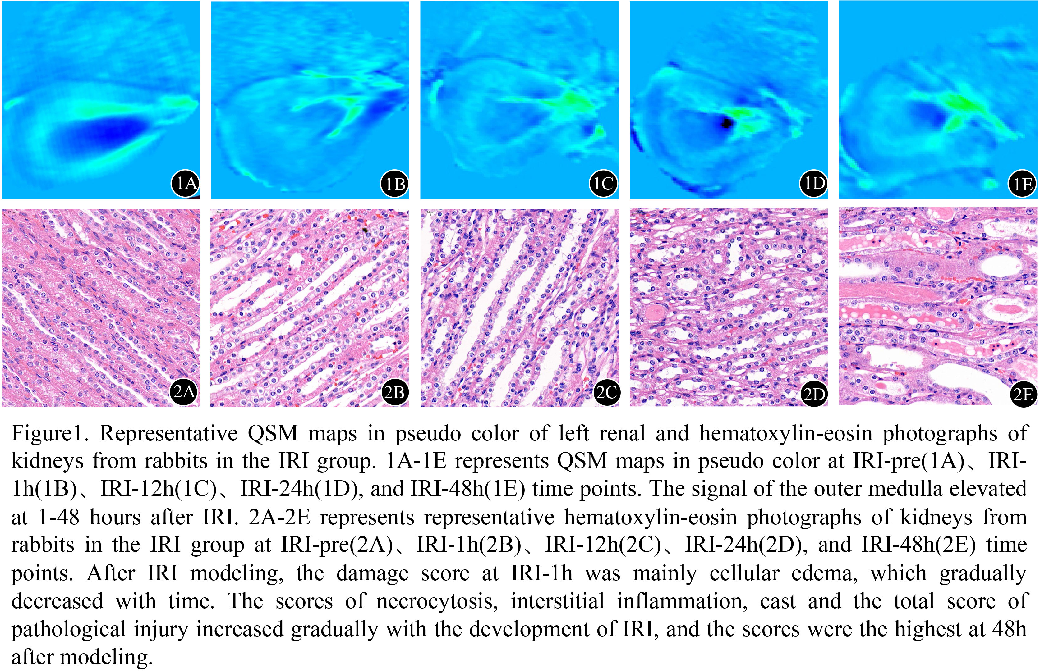
In IRI group, the magnetic susceptibility values of the outer medulla at IRI-pre、IRI-1h、IRI-12h、IRI-24h, and IRI-48h time points were (43.23 ± 3.06)×10-3ppm, (﹣5.78 ± 1.82)×10-3ppm, (6.87 ± 3.85)×10-3ppm, (10.56 ± 2.28)×10-3ppm, (19.93 ± 2.18)×10-3ppm, respectively, and showed significant differences between each two of different time points (P<0.05;Fig.1). In comparison, for the sham group, comparable magnetic susceptibility values of the outer medulla were revealed between each two of different time points (P>0.05). In the IRI group, the magnetic susceptibility values of the outer medulla had negative correlation with the scores of cell edema (r=﹣0.70, P<0.05), and showed positive correlation with the scores of necrocytosis, interstitial inflammation, cast and the total pathological score of renal injury (r=0.71, 0.60, 0.76, 0.53, P<0.05, respectively).Discussion
This study investigated the dynamic relationship between the magnetic susceptibility values of the outer medulla and the early pathological characteristics of renal IRI. The results demonstrated that the magnetic susceptibility values could reflect the dynamic changes of renal IRI. The magnetic susceptibility values of the renal outer medulla decreased decreased at 1 hour after IRI and decreased from 1 to 48 hours. The magnetic susceptibility values of the renal outer medulla was positive correlated with the scores of necrocytosis, interstitial inflammation, cast and the total pathological score of renal injury, and negative correlated with the scores of cell edema.Conclusion
QSM has been demonstrated to assess the dynamic changes of the outer medulla in the early stage of renal IRI of rabbits, and might thus be used as an effective method to evaluate the early stage of renal IRI.Acknowledgements
No acknowledgement found.References
[1] Munshi R, Hsu C, Himmelfarb J. Advances in understanding ischemic acute kidney injury [J]. BMC Med, 2011, 9: 11.
[2] Basile D P, Anderson M D, Sutton T A. Pathophysiology of acute kidney injury [J]. Compr Physiol, 2012, 2(2): 1303-53.
[3] Deistung A, Schweser F, Reichenbach J R. Overview of quantitative susceptibility mapping [J]. NMR Biomed, 2017, 30(4): e3569.
[4] Liu C, Li W, Tong K A, et al. Susceptibility-weighted imaging and quantitative susceptibility mapping in the brain [J]. J Magn Reson Imaging, 2015, 42(1): 23-41.
[5] Wang Y, Liu T. Quantitative susceptibility mapping (QSM): Decoding MRI data for a tissue magnetic biomarker [J]. Magn Reson Med, 2015, 73(1): 82-101.
[6] de Carvalho A L, Vital R B, Kakuda C M, et al. Dexmedetomidine on renal ischemia-reperfusion injury in rats: assessment by means of NGAL and histology [J]. Ren Fail, 2015, 37(3): 526-30.
Figures