2572
Comparison of the reproducibility of human renal amide proton transfer weighted imaging in axial and coronal orientations1Xi'an Gaoxin Hospital, Xi'an, China, 2Philips Healthcare, Beijing, China
Synopsis
Three-dimensional (3D) amide proton transfer (APT)-weighted (APTw) techniques have been applied in clinical work, However, image quality and feasibility of APTw imaging in normal kidneys at different scanning orientations have not been well confirmed. This study conducted on 27 healthy adult volunteers and all volunteers underwent intermittent end-respiratory breath-holding of axial and coronal 3D-APTw imaging. The preliminary results of this study show that the image quality of the image quality of renal APTw axial plane is better than that of coronal plane, and the reproducibility of APTw measurement values in axial plane is superior to that of coronal plane.
Synopsis
Three-dimensional (3D) amide proton transfer (APT)-weighted (APTw) techniques have been applied in clinical work, However, image quality and feasibility of APTw imaging in normal kidneys at different scanning orientations have not been well confirmed. This study conducted on 27 healthy adult volunteers and all volunteers underwent intermittent end-respiratory breath-holding of axial and coronal 3D-APTw imaging. The preliminary results of this study show that the image quality of the image quality of renal APTw axial plane is better than that of coronal plane, and the reproducibility of APTw measurement values in axial plane is superior to that of coronal plane.Introduction
The amide proton transfer (APT) imaging technology is a kind of chemical exchange saturation transfer (CEST) imaging technology [1] which can indirectly reflect the proliferation and protein metabolism levels of tissues and lesions. The application of APT-weighted (APTw) imaging technology in craniocerebral diseases is relatively mature and has been affirmed [2]. APTw imaging has been initially applied in the urinary system such as the prostate, cervix and rectum [3-5]. At present, APTw imaging technology is rarely reported in kidney research. The purpose of this study was to compare the reproducibility of renal APTw imaging in axial and coronal plane.Methods
From September 2021 to November 2021, 27 healthy volunteers (age range from 24 to 46 years, 30±8.9, 10 males) underwent intermittent end-respiratory breath-holding of renal axial and coronal 3D-APTw imaging on a 3.0T MR scanner (Ingenia CX, Philips Healthcare). APTw imaging was performed using a saturation pulse with duration of 2s and strength of 2uT, other parameters were shown in Table 1. According to the Likert Scale scoring method for APTw image quality evaluation (Table 1), lower scores (1 or 2) were not included in the measurement of APTw values. APTw images were analyzed independently by three radiologists with 3-, 6- and 10-years’experience,blinded to each other’s measurements.A region of interest (ROI) was manually placed by each observer on the location of the anterior, middle and posterior parts of axial APTw image, and upper, middle and lower parts of coronal plane. Three ROIs were placed on every part (Table 5) in right kidney, and the averaged APTw value in each part was measured. All ROIs avoid blood vessels and artifacts. The intraclass correlation coefficient (ICC) was used to evaluate the consistency of the three observers. One-way analysis of variance was used to analyze the data between parts of APTw imaging in each plane. The two independents sample t-tests were used to assess the difference between the axial and coronal APTw values.Results
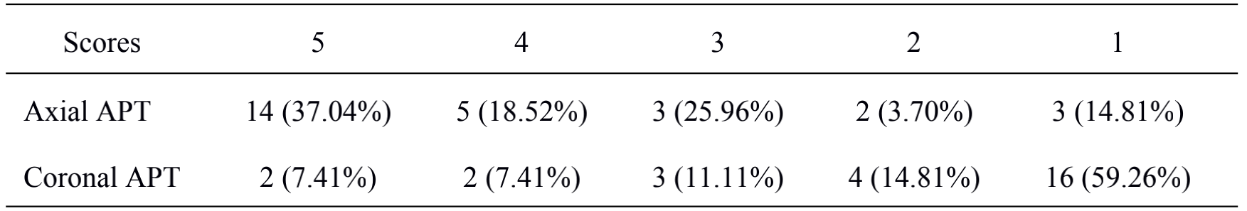
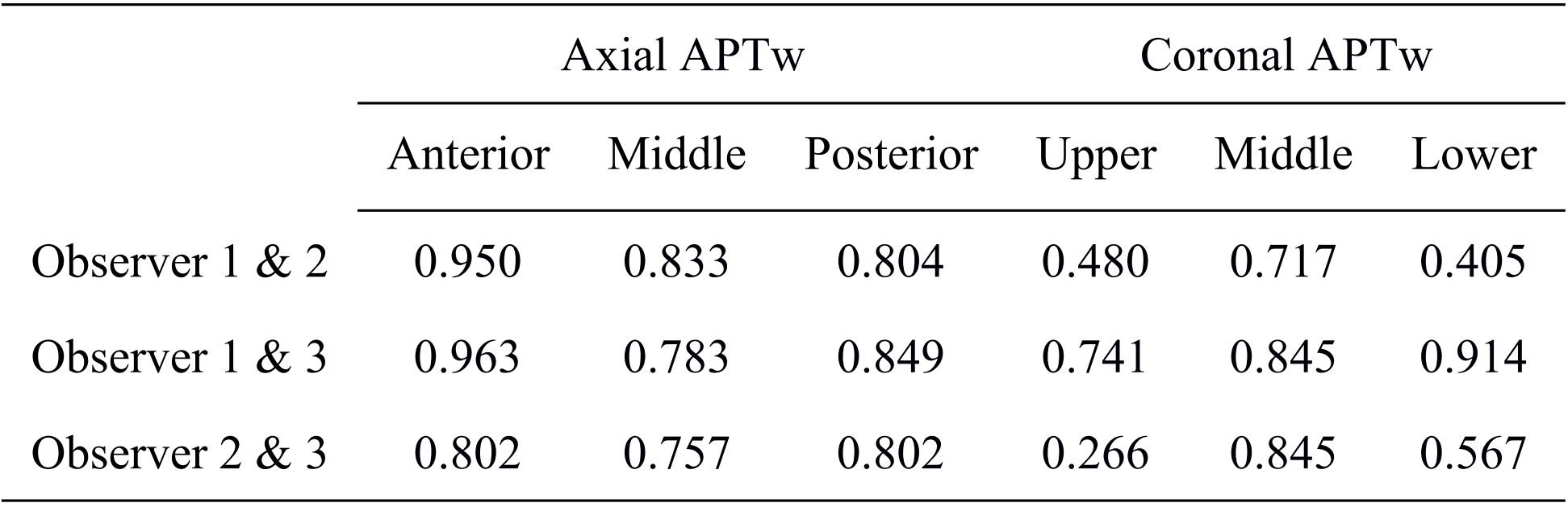
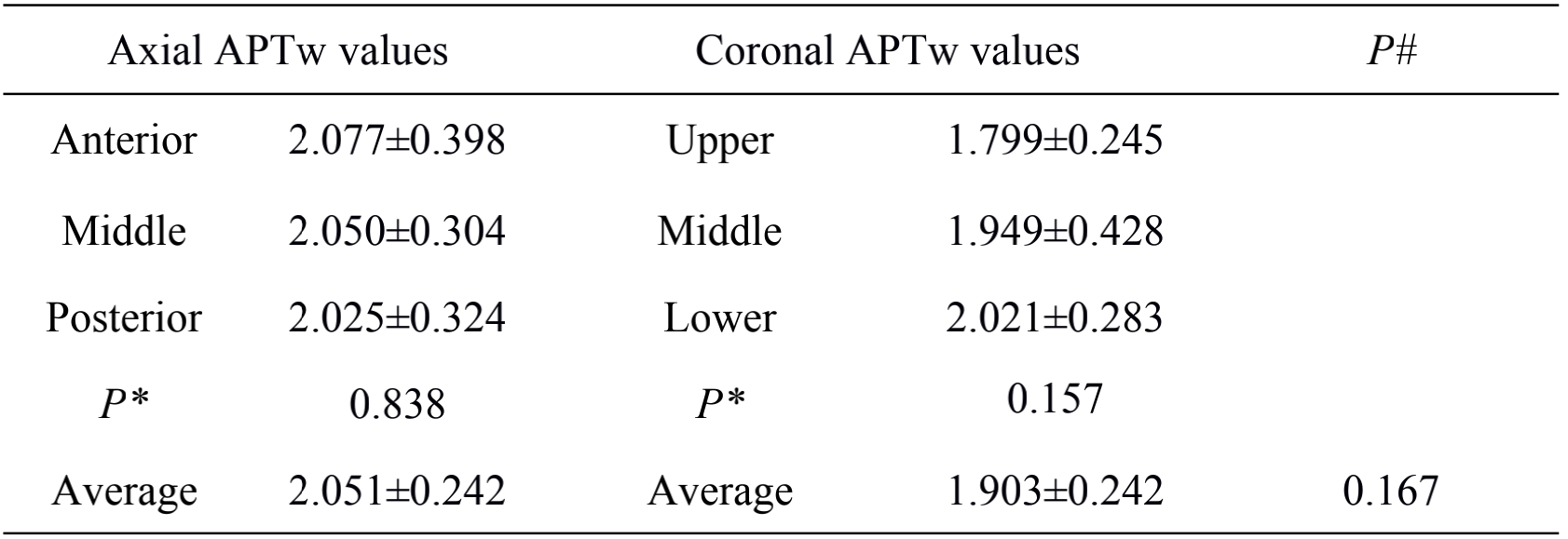
The image quality scoring results (Table 2 and Figure 1) showed that the renal axial APTw image quality is better than that of coronal APTw image. APTw ROI measurements were completed in 22 cases (22/27, 81.48%) in axial plane, and only 7 cases in coronal plane (7/27, 25.92%) (Table3). APTw values measured by three observers were well consistent in axial plane, (ICC>0.75), and unstable in coronal plane (ICC between 0.266~0.914, most of them are less than 0.75) (Table 4). The reproducibility of the APTw measured value of the axial plane is better than that of the coronal plane. There was no statistical difference between the APTw values in 3 parts in each plane. The difference between APTw values in the axial (2.051±0.284%) and coronal (1.903±0.242%) planes was not statistically significant (P = 0.167) (Table 5).Discussion and Conclusion:
The preliminary results of this study show that the image quality of intermittent breath-hold 3D-APT imaging of the normal kidney in axial plane is better than that of coronal plane, and the reproducibility of APTw measurement values in axial plane is superior to that of coronal plane. The axial plane of 3D-APTw imaging has potential clinical value.The study of APT scan orientation and image quality in normal kidneys can provide a basis for the detection of various pathological changes in the kidney. The current results of APT studies on the kidney show that APT can be used as a non-invasive biological marker in the diagnosis and treatment of a variety of renal pathologies, such as CKD, preoperative diagnosis and grading of renal tumors and assessment of efficacy and prognosis, and allows controlled studies to be performed previously by various research platforms and teams. Therefore, this technology has a very important clinical value. On the other hand, the use of APT scan parameters as non-invasive biological markers requires a high reproducibility of the parameter measurements.
Acknowledgements
NOReferences
[1] Choi SH. Can amide proton transfer MRI distinguish benign and malignant head and neck tumors?[J]. Radiology, 2018, 288: 791-792.
[2] Suh CH, Park JE, Jung SC, et al. Amide proton transfer-weighted MRI in distinguishing high-and low-grade gliomas:a systematic review and meta-analysis[J]. Neuroradiology, 2019, 61(5):525-534.
[3] Jia G, Abaza R, Williams JD, et al. Amide proton transfer MR imaging of prostate cancer: a preliminary study[J]. J Magn Reson Imaging, 2011, 33(3): 647-654. DOI:10.1002/jmri.22480.
[4] Meng N, Wang X, Sun J, et al. Application of the amide proton transfer-weighted imaging and diffusion kurtosis imaging in the study of cervical cancer[J]. Eur Radiol, 2020, 30(10): 5758-5767.
[5] Ishimatsu K, Nishie A, Takayama Y, et al. Amide proton transfer imaging for differentiating benign ovarian cystic lesions: Potential of first time right[J]. European Journal of Radiology, 2019, 120: 108656.
Figures

Table 1. Detailed scan parameters of APTw images
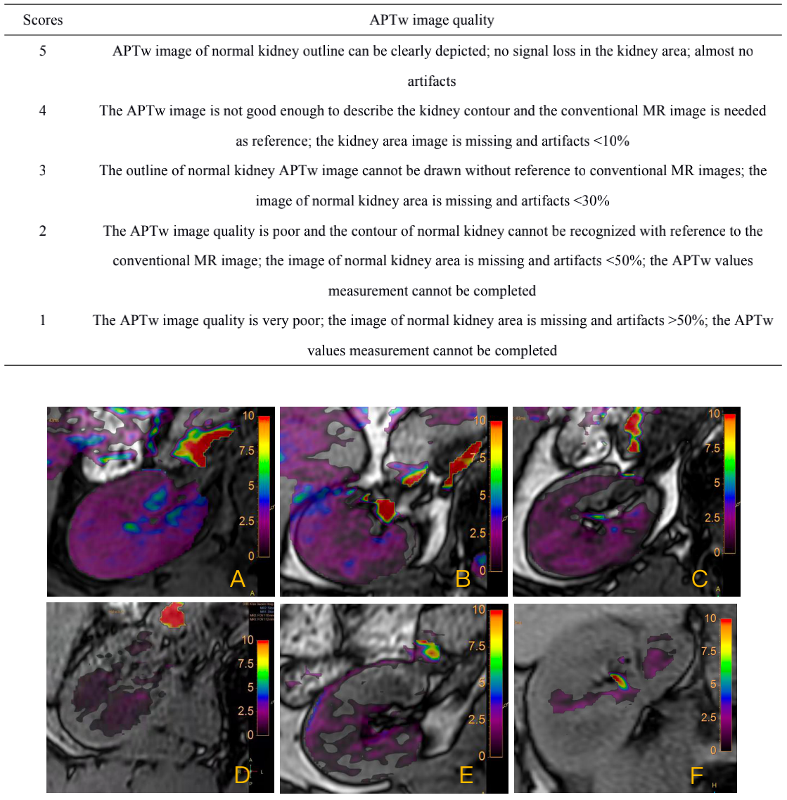
Table 2. Likert scale scoring of APTw image. Figure 1. Likert scale scores of right normal kidney of APTw images (A to E were evaluated as 5 to 1 point in order),F for coronal APTw 1point image.