2571
Abnormal stability of dynamic functional architecture in amyotrophic lateral sclerosis: a preliminary resting-state fMRI study1Radiology, Fujian Medical University Union Hospital, Fuzhou, China, 2Fujian Medical University Union Hospital, Fuzhou, China
Synopsis
Static and dynamic analyses for identifying functional connectivity(FC)have demonstrated brain dysfunctions in amyotrophic lateral sclerosis(ALS).We are to make the first attempt to perform studies about the stability of dynamic FC analysis based on resting-state fMRI data, in order to provide the new insight into the mechanism in ALS and investigate its correlation with disease severity. Our observation revealed that the abnormal pattern of FC stability involved sensorimotor regions(i.e.left precentral and postcentral gyrus)and non-sensorimotor areas(i.e.right temporal pole and middle/inferior frontal gyrus)in ALS patients, suggesting the potential of functional stability serving as the biomarker for monitoring disease progression in ALS.
Introduction
Amyotrophic lateral sclerosis (ALS) is an adult-onset neurodegenerative motor neuron disorder characterized by progressive upper and lower motor neuron 1. Resting-state functional magnetic resonance imaging (fMRI) which is based on blood oxygen level-dependent effect has been widely used in recording the signals of spontaneous brain activity2. Functional connectivity (FC), which is measured based on resting-state fMRI signal, has shown promise as a potential biomarker for its association with neurological disorders3. Although the functional architecture of the brain is dynamic in nature, its functional stability in a continuous state is also critical for maintaining normal brain functions4. Currently, the functional stability property of brain has attracted more and more attention4. For instance, based on resting-state fMRI data, a recent study has characterized the stability of the brain's functional architecture by measuring the concordance of dynamical functional connections over time in the form of voxel-wise4. Of note, the recent resting-state fMRI study has shown that the analysis of functional stability contributes to explore the mechanisms underlying various neuropsychiatric disorders5. Static and dynamic analyses for identifying functional connectivity (FC) have demonstrated brain dysfunctions in amyotrophic lateral sclerosis (ALS)6; 7. In this study, we are to make the first attempt to perform functional stability analysis based on resting-state fMRI data, in order to provide the new insight into the mechanism in ALS and investigate its correlation with disease severity.Methods
Resting-state functional magnetic resonance data were collected from ALS patients (n=20) and healthy controls (n=22). The severity of the disease was assessed using the Revised ALS Functional Rating Scale (ALSFRS-R). Sliding window correlation approach was employed to calculate dynamic FC. The functional stability of each voxel was calculated by measuring the concordance of dynamic FC over time. The between-group difference in functional stability was assessed by two-sample t test voxel-wisely. The correlation between functional stability index and ALSFRS-R was evaluated by Spearman’s correlation analysis among ALS patients.Results
Compared with HC, ALS patients had significantly increased functional stability in the left precentral and postcentral gyrus and right temporal pole while decreased functional stability in the right middle and inferior frontal gyrus. A significant correlation was observed between ALSFRS-R and mean functional stability in the right temporal pole (r = -0.452 and P = 0.046) in ALS group.Conclusion
ALS patients have abnormal stability of brain functional architecture, which is associated with the severity of the disease.Acknowledgements
The National Natural Science Foundation of China (No. 82071900) and Fujian Province Joint Funds for the Innovation of Science and Technology (No. 2019Y9067) supported this study.References
1. Turner MR, Hardiman O, Benatar M et al (2013) Controversies and priorities in amyotrophic lateral sclerosis. The Lancet Neurology 12:310-322
2. Ogawa S (2012) Finding the BOLD effect in brain images. Neuroimage 62:608-609
3. Hohenfeld C, Werner CJ, Reetz K (2018) Resting-state connectivity in neurodegenerative disorders: Is there potential for an imaging biomarker? Neuroimage Clin 18:849-870
4. Li L, Lu B, Yan CG (2020) Stability of dynamic functional architecture differs between brain networks and states. Neuroimage 216:116230
5. Zhu J, Zhang S, Cai H, Wang C, Yu Y (2020) Common and distinct functional stability abnormalities across three major psychiatric disorders. Neuroimage Clin 27:102352
6. Chenji S, Jha S, Lee D et al (2016) Investigating Default Mode and Sensorimotor Network Connectivity in Amyotrophic Lateral Sclerosis. PLoS One 11:e0157443
7. Chen HJ, Zou ZY, Zhang XH, Shi JY, Huang NX, Lin YJ (2021) Dynamic Changes in Functional Network Connectivity Involving Amyotrophic Lateral Sclerosis and Its Correlation With Disease Severity. J Magn Reson Imaging. 10.1002/jmri.27521
Figures
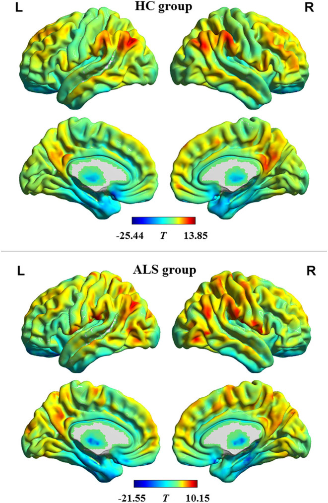
The profile of intrinsic functional stability within each group. The brain maps of T values show the results of one-sample t tests on functional stability in resting-state. High and low stability is indicated by red and blue color, respectively.
HC, healthy control; ALS, amyotrophic lateral sclerosis; L, left; R, right.
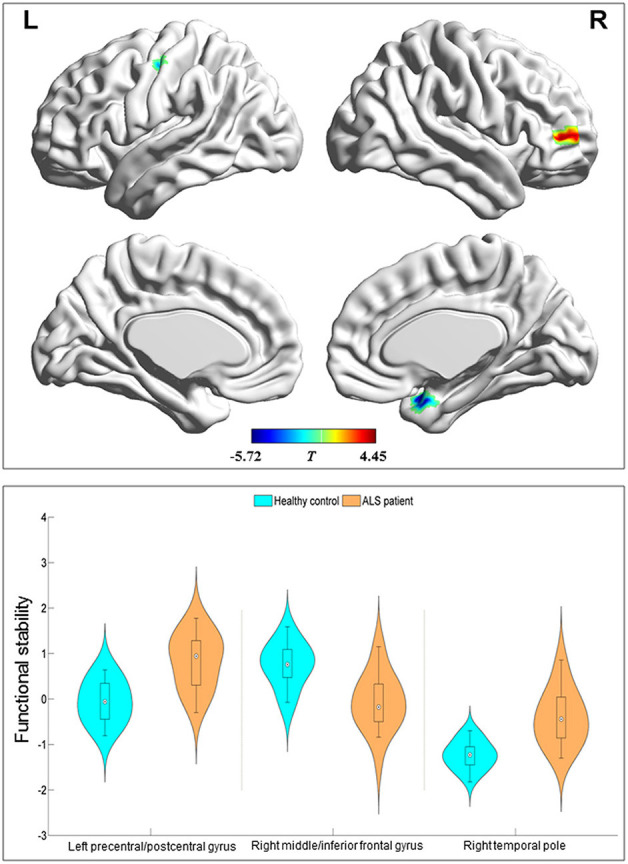
Regions with between-group difference in functional stability. The red and blue color indicate the regions with the decreased and increased functional stability in the patients with amyotrophic lateral sclerosis, respectively. The combination of violin and box plots shows the distribution and between-group differences (all P< 0.0001) of mean value of functional stability in these regions.
L, left; R, right.
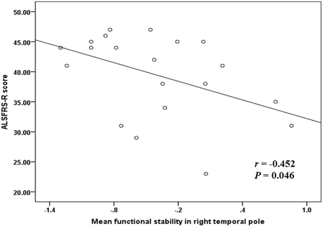
The correlation between functional stability and disease severity reflected by ALSFRS-R score.
ALSFRS-R, revised ALS Functional Rating Scale.