2544
Versatile Open source B0 shimtool1Advance Imaging Research Center, University of Texas Southwestern Medical Center, Dallas, TX, United States, 2Department of Bioengineering, University of Texas at Arlington, Arlington, TX, United States, 3Medical Metrology, Physikalisch Technische Bundesanstalt, Berlin, Germany, 4Max Planck Institute for Biological Cybernetics, Tuebingen, Germany
Synopsis
In this work, we compare the spectroscopy results and metabolite maps for different shimming routine. We use vendor default shimming technique and our own shimming technique. We tried to do shimming in different regions of brain (i.e. prefrontal cortex, occipital, and insula), also we tried to do multivoxel shimming. We compared the frequency shift maps between vendor’s implemented shim routine and our own shim algorithm. Also, we compared metabolite maps we got from different shimmed region after shimming using vendors and our own shimming routine.
Introduction
MR image quality can vary across sites due to differences in experimental conditions, sequence implementation, and image reconstruction and preprocessing choices. Significant progress has been made to making data acquisition and post processing more consistent across sites and vendor platform [1-2]. Another aspect that directly affects the image quality in functional, diffusion and perfusion MRI as well as magnetic resonance spectroscopy (MRS) is B0 inhomogeneity. In fact one of the most important challenges to obtain good MRS data quality is the inhomogeneity of the static magnetic B0 field in different regions of the human brain and body [3]. Poor B0 inhomogeneity renders adjacent spectral peaks separated by only few hertz indistinguishable and reduces the accuracy and precision of metabolite concentration estimates. In this work, we have developed a versatile open source B0 shimtool that combines several recent algorithmic developments [3-8] and considers recommendations of a recent consensus paper [3]. We have compared the B0 shimming performance of the open source B0 shimtool against the vendor implemented autoshim algorithm for different shim problems (i.e. single voxel, multi voxel, ROI) and evaluated the resulting spectroscopy data quality.Methods
Software tool: This open source B0 shimtool is generally implemented in MATLAB (Natick, MA, USA). In addition, we have incorporated FSL packages like ‘prelude’ and ‘bet’ for phase unwrapping and Brain skull stripping respectively. Real shim fields of all shim coils are considered by decomposition of respective acquired B0 calibration maps into a linear combination of spherical harmonic functions yielding respective spherical harmonic coefficients for each shim channel [6]. Since previous work reported that an iterative pseudo inverse method to solve the subject specific shim problem shows the most robust convergence for most B0 shimming problems the B0 shimtool returns optimized shim values using the respective ConsTru (constrained TSVD inversion method) algorithm [3]. This approach considers maximum available shim field strength by truncating the smallest singular value of spherical harmonic function and reinvert the new matrix in an iterative manner unless all the shim values fit inside our hardware constrains [5]. With the shimtool we can import square or rectangular spectroscopy voxels, draw random shim ROI shapes in any specific region or we can select whole brain shimming based on automatic skull stripping. Also we have implemented the region of less interest (ROLI) approach to consider shimming needs outside of the shim ROI for better shimming and artefact control in some regions [5]. Finally, automatized calculations of slice specific optimal shim values as input for dynamic shim experiments [7]. In principle the B0 shimtool is vendor and field strength independent.Experimental validation: We evaluated the shim tool at a 3T MRI scanner equipped with 2nd order shim coils (Siemens Prisma). First, we obtained real shim field calibration data of the B0 fields produced by gradient coils and second order shim coils using a 3D GRE sequence. This calibration process needs to be performed once and it is unique for each system. Subsequently, we performed different phantom and in vivo spectroscopy experiments in four subjects after IRB approval and signed consent to test the B0 shimtool performance: (i) 2nd order single voxel shimming in the frontal and occipital cortex of the human brain in vivo and (II) 2nd order shimming for 1H MRSI in the human brain using a global brain shim ROI versus a ROI matching the MRSI FOV. To evaluate respective B0 shimming performance single voxel 1H MRS data were acquired using a PRESS sequence (FOV = 20x20x20 mm3, TE = 30 ms, TR = 2sec, Angulation = 0) and 1H MRSI data were recorded using a semi laser sequence chemical shift imaging (CSI) sequence (FOV = 95x125x20 mm3, TE = 40 ms, TR = 1.5sec, Angulation = 0) respectively. We cross-validated our B0 shimtool against the vendor implemented default shim routine We have compared the metabolite spectrum and metabolite maps we are getting from different brain regions for the vender’s implemented algorithm and our shimtool.
Results & Discussion:
Figure 1 shows the user interface of the B0 shimtool and Figure 2 the tools B0 shim analysis capabilities. Figure 3 A and B shows the shimming performance of the B0 shim tool versus the vendors shim method in single voxel spectroscopy. Figure 4 A and B shows a comparison of 1H MRSI spectra after B0 shimming with the vendors implemented shim method versus our B0 shimtool, while in Figure 5 the comparison of respective NAA maps are shown .These results show that in single voxel spectroscopy applications the vendors shimming method shows similar performance as the B0 shimtool in the OCC (easy to shim), while the B0 shimtool clearly outperforms the vendors shimming method in the frontal cortex. Similarly the B0 shimtool leads to improved spectral quality in 1H MRSI. NAA maps look more homogenous if we do shimming using our custom made B0 shimtool in comparison to the vendor implemented method.
Conclusion
We have introduced a versatile open source GUI based B0 shimtool that provides better B0 shimming combining real shim field calibration with the ConsTru algorithm for different shimming problems compared to vendor’s pre implemented shim in different human brain areas which are difficult to shim.Acknowledgements
This work was performed at Advance Imaging Research Center At University OF Texas Southwestern Medical Center Dallas. This work was supported by Cancer Prevention and Research Institute of Texas (CPRIT) grant / RR180056.References
1 Nielsen, JF, Noll, DC: TOPPE: A framework for rapid prototyping of MR pulse sequences. Magn Reson Med. 2018; 79:3128-3134.
2 Ravi, KS, Potdar, S., Poojar, P, Reddy, AK, Kroboth, S., Nielsen, JF., Zaitsev, M., Venkatesan, R., and Geethanath, S.: Pulseq-Graphical Programming Interface: Open source visual environment for prototyping pulse sequences and integrated magnetic resonance imaging algorithm development. Magn Reson Imaging 2018; 52:9-15.
3 Juchem, C., Cudalbu, C., de Graaf, R., Gruetter, R., Henning, A., Hetherington, H., Boer, V.: B0 Shimming for in Vivo Magnetic Resonance Spectroscopy: Experts' Consensus Recommendations. NMR in Biomedicine 34 (5), pp. 1 - 20 (2021)
4 Gast, L., Henning, A., Hensel, B., Uder, M., Nagel, A., Localized B0 shimming based on 23Na MRI at 7 T. Magnetic Resonance in Medicine 83 (4), pp. 1339 - 1347 (2020)
5 Nassirpour, S., Chang, P., Fillmer, A., Henning, A., A comparison of optimization algorithms for localized in vivo B0 shimming. Magnetic Resonance in Medicine 79 (2), pp. 1145 - 1156 (2018)
6 Chang, P., Nassirpour, S., Henning, A.: Modeling Real Shim Fields for very High Degree (and order) B0 shimming of the Human Brain at 9.4T. pp. 529 - 540 (2018)
7 Fillmer, A., Vannesjö, S.; Pavan, M., Scheidegger, M., Pruessmann, K., Henning, A.: Fast Iterative Pre-Emphasis Calibration Method Enabling Third-Order Dynamic Shim Updated fMRI. Magnetic Resonance in Medicine 75 (3), pp. 1119 - 1131 (2016)
8 Fillmer, A., Kirchner, T., Cameron, D., Henning, A.: Constrained image-based B0 shimming accounting for "local minimum traps" in the optimization and field inhomogeneities outside the region of interest. Magnetic Resonance in Medicine 73 (4), pp. 1370 - 1380 (2015)
Figures
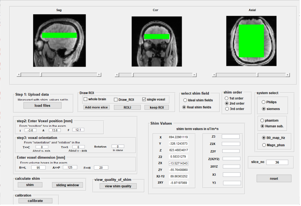
Figure 1: GUI of the open source B0 shimtool.
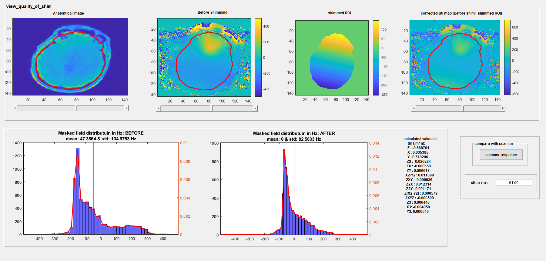
Figure 2: B0 shim performance analysis prediction and analysis capabilities of the B0 shimtool
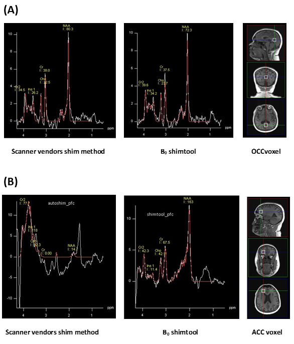
Figure 3: Performance of B0 shimtool versus scanner implemented B0 shimming method for single voxel spectroscopy (short TE PRESS) at 3T in (A) the occipital lobe and (B) in the frontal cortex. While spectral appearance and line width in the occipital lobe are comparable between the two shim approaches, the B0 shimtool clearly outperforms the vendors method in the more challenging frontal cortex location.
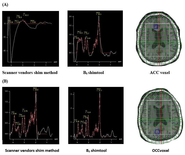
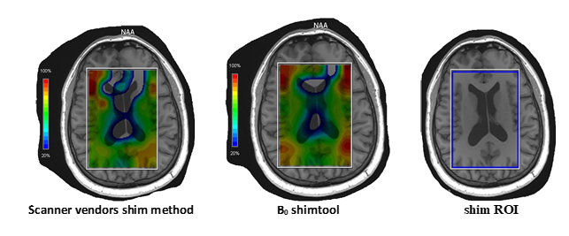
Figure 5 NAA maps from 1H MRSI at 3T after B0 shimming with the vendors method (left) versus the B0 shimtool (mid). The shim ROI corresponded to the semiLASER localization volume of the 1H MRSI sequence (right).