2506
Value of diffusion and intravoxel incoherent motion virtual MR elastography in differentiating benign and malignant focal liver lesions1the First Affiliated Hospital of Xi’an Jiaotong University, xi'an, China, 2Xi’an Jiaotong University, xi'an, China
Synopsis
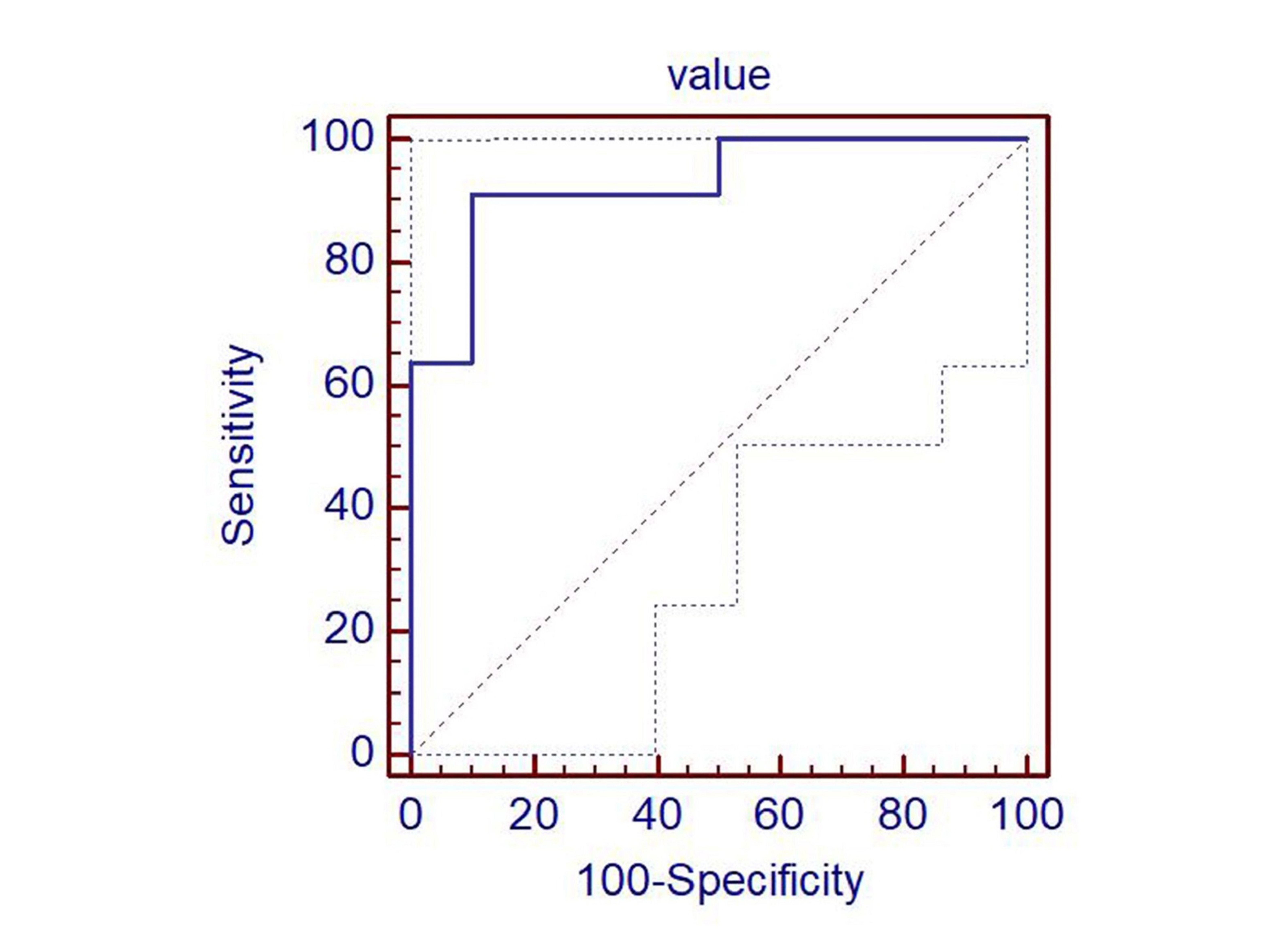
Diffusion and IVIM virtual MR elastography (dMRE) provides quantitative estimates of tissue stiffness without using mechanical vibrations. It rarely applied to identify focal liver lesions (FLL). This retrospective study included 21 subjects (11 malignant lesions and 10 benign lesions). DWI were obtained to calculating dMRE. The kPa values of malignant FLLs (7.93 ± 2.37) was significantly higher than that of benign FLLs (-0.28±5.37) (P < 0.01). ROC showed an AUC of 0.93 for dMRE in differentiating maglinant FLLs and benign FLLs. The sensitivity and specificity were 91% and 90%, respectively. Malignant lesions could be distinguished from benign FLLs by dMRE.
Background
It is important to differentiate benign and malignant focal liver lesions (FLL) accurately. Diffusion and intravoxel incoherent motion (IVIM) virtual MR elastography (dMRE) provides quantitative estimates of tissue stiffness without using mechanical vibrations. Hypothesizing that there might be a strong relationship between tissue water diffusivity and tissue elastic properties. As a new technology, its contribution to focal liver lesions is still uncertain.Aim
To explore the value of dMRE in differential diagnosis of benign and malignant FLL.Methods
This retrospective study included 21 subjects (13 men and 8 women; mean age± standard deviation(SD), 59 years ± 10),and diffusion weighted images(DWI) were obtained with a 3T MRI. The DWI of the lower b-value (Slow, b value = 200 s/mm2) and that of the higher b-value (Shigh, b value = 800 s/mm2) were used to estimate the virtual shear stiffness1,2: virtual shear stiffness = a·ln (Slow/Shigh) + b. The scaling (a) and the shift (b) factors were separately set to −9.8 and 14 according to the previous calibration studies1,2. Three kPa values were acquired after 3 consecutive measurements for each FLL, and the mean value was recorded. The kPa values of FLL were reported as mean ± SD. The kPa values of FLL was compared using Wilcoxon rank test. Receiver operating characteristic curve was performed to analyze the effectiveness of virtual elastography in distinguishing malignant and benign FLLs.Results
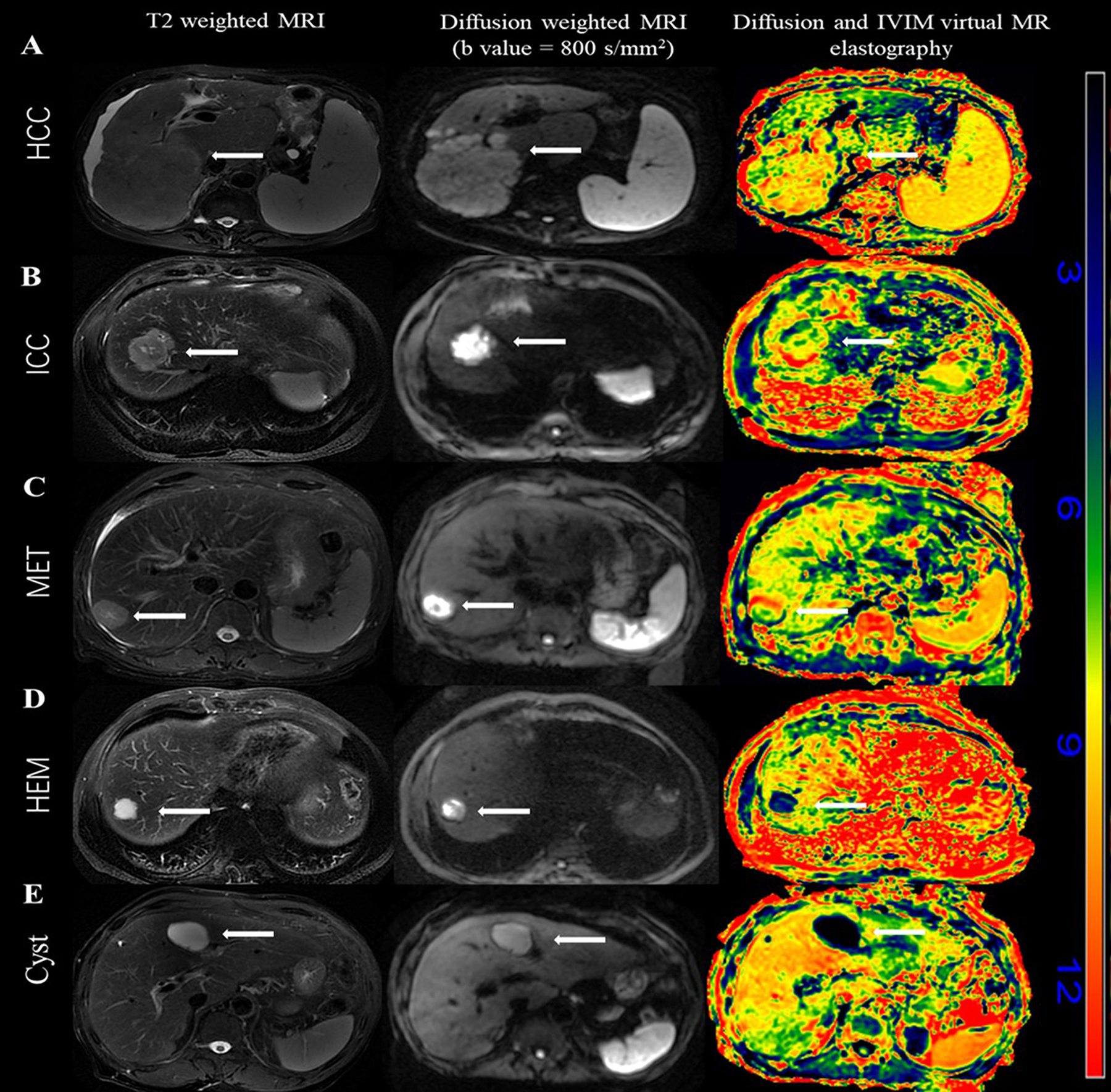
There were 11 malignant lesions (5 cases of hepatocellular carcinoma, 2 cases of cholangiocarcinoma and 4 cases of metastases) and 10 benign lesions (5 cases of cavernous hemangioma and 5 cases of cyst) in this study. The kPa values of hepatocellular carcinoma, cholangiocarcinoma and metastases were 7.30 ± 2.85, 8.25±2.47 and 8.58±2.13, respectively (P>0.05). The kPa values of cavernous hemangioma and cyst were 4.70 ± 1.15 and -5.27±1.24 (P<0.01).The kPa values of malignant FLLs (7.93 ± 2.37) was significantly higher than that of benign FLLs (-0.28±5.37)(P < 0.01). Receiver operating characteristic analysis showed an AUC of 0.93 (95% CI:0.73-0.99) for dMRE in differentiating maglinant FLLs and benign FLLs. The sensitivity and specificity were 91%(95% CI: 59–100) and 90%(95% CI: 56–100).Discussion
The dMRE provides quantitative estimates of tissue stiffness without using mechanical vibrations, and there are few studies currently applied to the differential diagnosis of focal liver lesions. With standard MRE propagating shear waves through tissues lead to microscopic tissue displacements resulting in phase shifts in the presence of magnetic gradient pulses, which can be exploited to estimate tissue shear stiffness. Mechanically-induced phase shifts induced by standard MRE are spatially distributed and results in a phase dispersion in each voxel and an amplitude signal attenuation, that is an IVIM effect 3,4. There was a strong correlation between liver tissue shear stiffness and tissue microstructure, as shown with diffusion MR imaging1. Elastography has a high sensitivity and specificity in differentiation for benign and malignant liver lesions, including shear wave elastography and tomoelastography5,6. Our results showed that dMRE could distinguish malignant from benign lesions, which was consistent with the previous literature about elastography. Furthermore, malignant tumors can accumulate hydrophobic and disorganized proteins, which increase tissue stiffness5. Cavernous hemangioma is a tumor of vascular origin composed of abnormal vascular clusters. Cyst is mostly caused by abnormal bile duct development during the embryonic period, and contain a large amount of fluid. Their stiffness was lower than that of malignant tumors5,6. However, the b values we selected were different from that of the previous study1, further researches are needed for the difference in diagnostic efficacy and image quality evaluation. Besides, the study revealed that the difference in kPa values between malignant lesions was not statistically significant. Considering the limitation of sample size, the further relevant research is still needed.Conclusion
Diffusion and IVIM virtual MR elastography could differentiate benign and malignant focal liver lesions, and the further research is needed for the discrimination between malignant lesions.Acknowledgements
This work was supported by the Key Research and Development Program of Shaanxi (Program No.2019SF-007)References
[1] Le Bihan D, Ichikawa S, Motosugi U. Diffusion and intravoxel incoherent motion mr imaging-based virtual elastography: a hypothesis-generating study in the liver. Radiology, 2017, 285(2):609–619.
[2] Lagerstrand K, Gaedes N, Eriksson S, et al. Virtual magnetic resonance elastography has the feasibility to evaluate preoperative pituitary adenoma consistency. Pituitary, 2021, 24:530–541.
[3] Glaser KJ, Felmlee JP, Manduca A, et al. Shear stiffness estimation using intravoxel phase dispersion in magnetic resonance elastography. Magn Reson Med. 2003 Dec;50(6):1256-65.
[4] Le Bihan D. What can we see with IVIM MRI? Neuroimage. 2019 Feb 15; 187:56-67.
[5] Shahryari M, Tzschätzsch H, Guo J, et.al. Tomoelastography Distinguishes Noninvasively between Benign and Malignant Liver Lesions.Cancer Res. 2019 Nov 15;79(22):5704-5710.
[6] Zhang HP, Gu JY, Bai M, et.al. Value of shear wave elastography with maximal elasticity in differentiating benign and malignant solid focal liver lesions.World J Gastroenterol. 2020 Dec 14;26(46):7416-7424.
Figures