2490
Dixon-MRI to quantitatively evaluate the correlation between abnormal fat deposition and chronic kidney disease with diabetic complications1Tianjin First Center Hospital, Tianjin medical university, Tianjin, China, 2Tianjin medical university, Tianjin, China, 3Tianjin First Center Hospital, Tianjin, China, 4Siemens Healthcare, Beijing, China
Synopsis
The study use Dixon and DKI sequence to quantitative analysis the ectopic fat deposition in chronic kidney disease and diabetic nephropathy. Compare the degree of fat infiltration in different organs and tissues including Liver, pancreas, kidney and muscles, combined with clinical laboratory experiments, to study the relationship between fat deposition and the development of CKD or diabetic nephropathy. Aim to point out the significance of treatment to fat deposition for CKD or diabetic nephropathy patient.
Introduction
Chronic kidney disease (CKD) has become a major global health problem]. Current studies have determined that the decline of renal function is related to the occurrence of insulin resistance, which is mainly related to the impaired glucose utilization of peripheral target tissues induced by insulin. A variety of mechanisms play a role, including inflammatory factors, adipokines, lipotoxicity and uremic toxins[1,2]. Lipotoxicity plays an important role in insulin resistances. Long term insulin resistance will eventually develop into type 2 diabetes, which leads to the development of diabetic nephropathy. It has high cardiovascular risk and will infect the quality of life and long-term survival of patients.[3]Therefore, it is necessary to clarify the correlation between ectopic fat deposition and clinical insulin resistance in CKD patients and diabetic nephropathy patients.Method
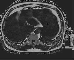
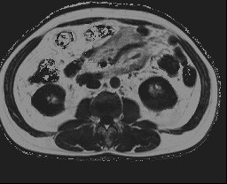
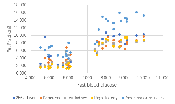
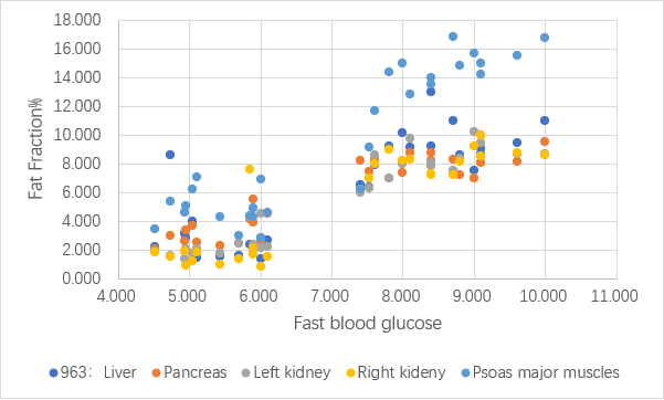
The current experiment included 35 patients, 20 patients with chronic kidney disease, 15 patients with diabetic nephropathy or nephropathy accompanied diabetes. All patients underwent magnetic resonance imaging. The scanning machine was Prisma 3.0T MR scanner (Siemens Healthcare, Erlangen, Germany). The inclusion criteria: 1. Age: 20-90 years old, unlimited gender, easy to move; 2. clinical diagnosis of chronic kidney disease 3, 4, 5, diabetic nephropathy or nephropathy with diabetes mellitus; 3. Complete clinical biochemical indexes, including biochemical items and fasting insulin. Exclusion criteria: 1. Receiving dialysis maintenance treatment or transplantation treatment; 2. Have a history of alcoholism; 3. There is a history of primary liver disease and malignant tumor. 3. Those with MRI contraindications. The main Dixon sequences included: Abdomen_qdx_tra(Slice thickness: 3.5mm, TR: 9.3ms, TE: 1.3ms, Matrix:256×156),T2star_qdx_963_tra, dewpi_DKI+IVIM. The achieved image data were analyzed in INFINITT software, and the ROI was put on the Fat Fraction (FF) image. The ROI was placed in the liver (avoiding large blood vessels and bile ducts, except for S1), paravertebral muscles (bilateral psoas major muscle in L2 and 3 segment), pancreas (ROIs were placed in the head, body and tail), kidney (ROIs were placed in the upper, middle and lower parts of each side), the fat fraction of each organ and tissue was obtained respectively, and the results were expressed as mean value ± standard deviation. SPSS 21.0 software was used for statistical analysis.Results
According to the creatinine, glomerular filtration rate, fasting blood glucose and other indicators, patients were divided into simple chronic kidney disease group and diabetic nephropathy (or nephropathy with diabetes) group. Two different Dixon sequences were used to assess the degree of fat infiltration in each tissue and organ. The results showed that the two sequences had high consistency. Comparing the degree of fat deposition between different tissues and organs of the two groups, we find that the degree of fat deposition in the tissues and organs of patients with diabetic nephropathy (or nephropathy with diabetes) is different from those of patients with chronic kidney disease, and in the two groups, the degree of fat infiltration of skeletal muscle is higher than that of other organs (liver, pancreas and kidney). No special distribution difference was found in the degree of fat infiltration in liver, pancreas and both kidneys. According to the glomerular filtration rate, the chronic kidney disease group was divided into three subgroups: stage 3, 4 and 5 of chronic kidney disease. There was no significant difference between the three subgroups in the degree of fat infiltration in various tissues and organsDiscussion and conclusion
In patients with diabetic nephropathy (or nephropathy with diabetes), the degree of fat infiltration in the organs is higher than that in the simple chronic kidney disease group, indicating that the distribution of fat in the body plays an important role in the development of diabetic nephropathy and diabetic nephropathy. The higher fat deposition in skeletal muscle may be due to skeletal muscle as an important insulin receptor in human body. The Lipotoxicity caused by fat deposition in it blocks the normal reception of insulin signal by skeletal muscle insulin receptor. Even if patients receive insulin injection treatment every day, skeletal muscle cannot normally play aerobic fermentation because it cannot receive insulin signal, resulting in poor effect to reduce blood glucose.Acknowledgements
We thank Professor. Wen Shen and Professor.Wenxiu Chang for the guidance to the experiment, and Dr. Jinxia Zhu for the technology support.References
[1] Witasp A, Carrero JJ, Heimbürger O, et al. Increased expression of pro-inflammatory genes in abdominal subcutaneous fat in advanced chronic kidney disease patients, J. Intern. Med., 2011, vol. 269 (pg. 410-419).
[2] Roubicek T, Bartlova M, Krajickova J, et al. Increased production of proinflammatory cytokines in adipose tissue of patients with end-stage renal disease, Nutrition, 2009, vol. 25 (pg. 762-768)
[3] B.J. Arsenault, E.P. Beaumont, J.-P. Després, E. Larose. Mapping body fat distribution: a key step towards the identification of the vulnerable patient? Ann. Med., 44 (2012), pp. 758-772
Figures