2474
Topological organization effects of different theta burst stimulation protocols over precuneus in healthy adults1College of Physics, Sichuan University, Chengdu, China, 2Huaxi MR Research Center (HMRRC), Functional and molecular imaging Key Laboratory of Sichuan Province, Department of Radiology, West China Hospital, Sichuan University,, Chengdu, China, 3Research Unit of Psychoradiology, Chinese Academy of Medical Sciences, Chengdu, China
Synopsis
This study explored the seed-based functional connectivity (FC) and topological organization alteration applied different TBS protocols over precuneus in healthy adults. Increased FC were found in sensorimotor network and insula after application of iTBS or cTBS, decreased FC in left thalamus was found after cTBS. Only cTBS induced significant topological alterations in whole-brain network characterized by small-worldness on FC and functional topology organization, The findings suggested distal effects were complicated and could not been simply summarized as inhibition/facilitation like as proximal rTMS effects. This study may help to elucidate the underlying mechanisms of information transmission through brain function connectivity.
Purpose
It is widely accepted that low-frequency repetitive transcranial magnetic stimulation (rTMS) (≤1Hz) decreases neuronal excitability and high-frequency rTMS (≥5Hz) increases neuronal excitability at the target site (Rossi et al., 2009). Regarding effects on the other regions functionally connected the target site, previous studies have yield heterogeneous results(Beynel et al., 2020). Theta-burst stimulation (TBS) is a form of rTMS involving the use of three pulse bursts at a high-frequency (50 Hz or at 30 Hz) repeated five times per second, which is thought to induce more rapid and longer-lasting effects on synaptic plasticity than with conventional rTMS protocols (Huang et al., 2005). Huang and his colleagues had demonstrated continuous theta burst stimulation (cTBS) increases cortical excitability and intermittent protocol (iTBS) decreases cortical excitability (Huang et al., 2011). Here, we combined resting state functional MRI (rs-fMRI) with TBS to investigate the seed-based functional connectivity and topological organization alteration after application of the two TBS protocols over precuneus in healthy adults.Materials and methods
Participants and study designTwenty-five, right-handed subjects (mean age: 21.7 years, range: 20-26 years, 9 males) participated in this study. All participants underwent one baseline scan session (S0) and two TBS sessions (iTBS and cTBS), 7 days apart.
TBS intervention
The TBS was applied using a figure-of-eight coil. The target stimulation sites were selected in the system by transformation of the MNI coordinates to participant’s native brain. The precuneus located at MNI coordinates x=6, y=-70, z=44. The motor threshold (MT) was defined as the lowest TMS intensity delivered over the motor cortex necessary to elicit visible twitches of the right index finger in at least 5 out of 10 consecutive pulses. The stimulations were applied to the target following protocols: stimulation intensity: 100% MT, total number of pulses: 1,200.
MRI data acquisition and preprocessing
Resting-state fMRI was performed before and after TBS sessions (three scans per subject in total) via a 3-Tesla United Imaging MRI scanner with a 16-channel phase-array head coil. The following scanning parameters were used: number of slice=34, time repetition (TR) =2000ms, time echo=30ms, flip angle=90◦, slice thickness = 3.5mm with no slice gap, field of view =224×224, and 240 volumes in each run. The rs-fMRI data were preprocessed using the fMRIPrep-20.2.1 pipeline. In brief, slice timing, normalization, spatial smoothing with a Gaussian-smoothing kernel of 6-mm FWHM, motion correction using ICA-AROMA(Pruim et al., 2015), regressing the confounds (mean white matter and cerebrospinal fluid signals), detrending and bandpass filtered (0.01–0.1 Hz).
Seed based functional connectivity analysis
The seed region was focused on precuneus, 10mm spheres around x=6, y=-70, z=44. The FC analysis was performed by computing the correlation between the mean BOLD signals from seed region to all other voxels in the brain. Paired t test was used to compare between the pre-stimulation and post-stimulation scans. the significant results were reported when they survived a statistical threshold (p<0.001, p-FWE corrected <0.01 at cluster level).
Topological organization analysis
We extracted 800 regions of interest (ROI) from a prior functional atlas (Schaefer et al., 2018) and calculated an 800×800 correlation matrix for each scan. We selected a sparsity range of 0.1-0.4 to construct a binary undirected graph and calculated the small-world parameters(small-worldness, σ ; normalized clustering coefficient, γ ; normalized characteristic path length, λ ) and global efficiency(Eg). Paired t test was used to compare the differences and p values were corrected using False Discovery Rate (FDR), p<0.05.
Results
The mean MT was 33.6% (SD: 4.9%) of the system’s maximum output. The mean interval between the last TBS pulse and the start of rs-fMRI scan was 771.6 seconds (SD: 135.6 s) for iTBS and 753.6 seconds (SD: 87.4 s). No adverse events occurred.The alteration of Seed-based FC
As shown in Figure 1 and Table 1, the precuneus-based FC results applied two TBS protocols were largely consistent., Increased FC were found almost in sensorimotor network and insula. Particularly, decreased FC in left thalamus was found only after cTBS.
The alteration of topological structure
All brain networks exhibited small-woldness. Relative to the baseline, no significant differences were observed after iTBS. The subjects showed statistically lower gamma, sigma and global efficiency after cTBS.
Discussion
TMS combined fMRI has provided a non-invasive method to investigate the human brain. In this study, we explored the alteration of precuneus-based FC and topological organization after application of two TBS protocols. Unlike the opposing effects of TBS induced on the activity of the simulation area, both iTBS and cTBS increased functional connectivity between most brain regions with simulation site. As for the topological structure effects, only cTBS induced significant topological alterations in whole-brain network characterized by small-worldness and global efficiency. These findings indicated that active TBS induced significant changes on FC and functional topology organization, the distal effects were complicated and could not been simply summarized as inhibition/facilitation like as proximal rTMS effects. This study may provide evidence to select the appropriate TBS protocol and help to elucidate the underlying mechanisms of information transmission through brain functional connectivity.Acknowledgements
This study was supported by the National Key Technologies R&D Program of China (Grant No. 82027808), the Key Research Project of Sichuan Science and Technology Department (Grant No. 2020YFS0048) and the Science Specialty Program of Sichuan University (Grand. No. 2020SCUNL210).
References
Beynel, L. et al. (2020). Effects of repetitive transcranial magnetic stimulation on resting-state connectivity: A systematic review. Neuroimage, 211, 116596. doi:10.1016/j.neuroimage.2020.116596
Huang, Y. Z. et al. (2005). Theta burst stimulation of the human motor cortex. Neuron, 45(2), 201-206. doi:10.1016/j.neuron.2004.12.033
Huang, Y. Z. et al. (2011). The theoretical model of theta burst form of repetitive transcranial magnetic stimulation. Clin Neurophysiol, 122(5), 1011-1018. doi:10.1016/j.clinph.2010.08.016
Pruim, R. H. R. et al. (2015). ICA-AROMA: A robust ICA-based strategy for removing motion artifacts from fMRI data. Neuroimage, 112, 267-277. doi:10.1016/j.neuroimage.2015.02.064
Rossi, S. et al. (2009). Safety, ethical considerations, and application guidelines for the use of transcranial magnetic stimulation in clinical practice and research. Clin Neurophysiol, 120(12), 2008-2039. doi:10.1016/j.clinph.2009.08.016
Schaefer, A. et al. (2018). Local-Global Parcellation of the Human Cerebral Cortex from Intrinsic Functional Connectivity MRI. Cereb Cortex, 28(9), 3095-3114. doi:10.1093/cercor/bhx179
Figures
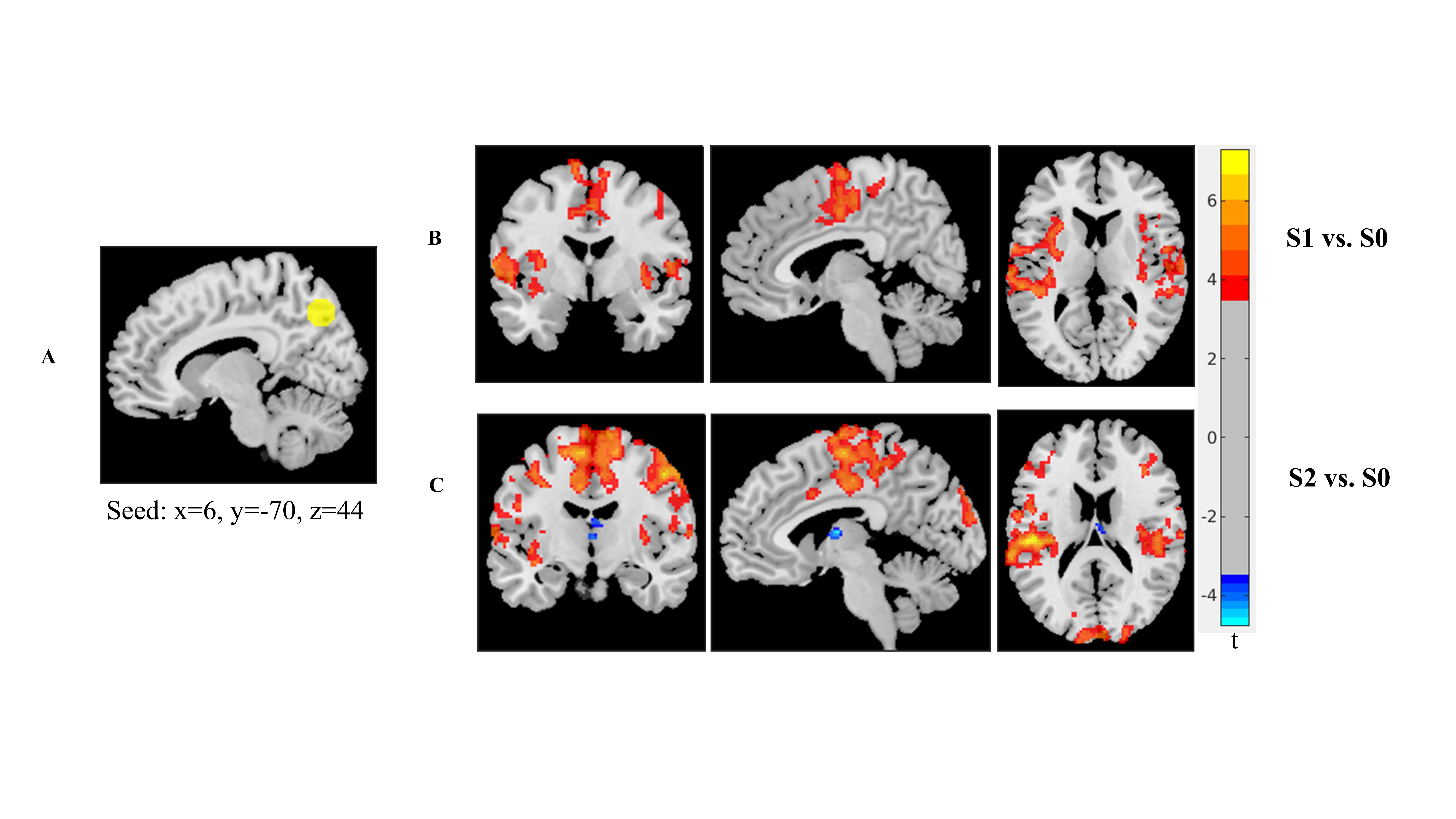
Figure 1. The results of seed-based functional connectivity (A) right precuneus seed (B) the differences between baseline scan and post-stimulation scan after iTBS (C) the differences between baseline scan and post-stimulation scan after cTBS. S0, baseline; S1, iTBS; S2, cTBS

Figure 2. The alteration of topological organization (A) the differences between pre-stimulation and post-iTBS on small-woldness parameters and global efficiency (B) the differences between pre-stimulation and post-cTBS on small-woldness parameters and global efficiency. γ, normalized clustering coefficient; λ, normalized characteristic path length; σ, normalized small-world; Eg, global efficiency. * P<0.05, FDR corrected.
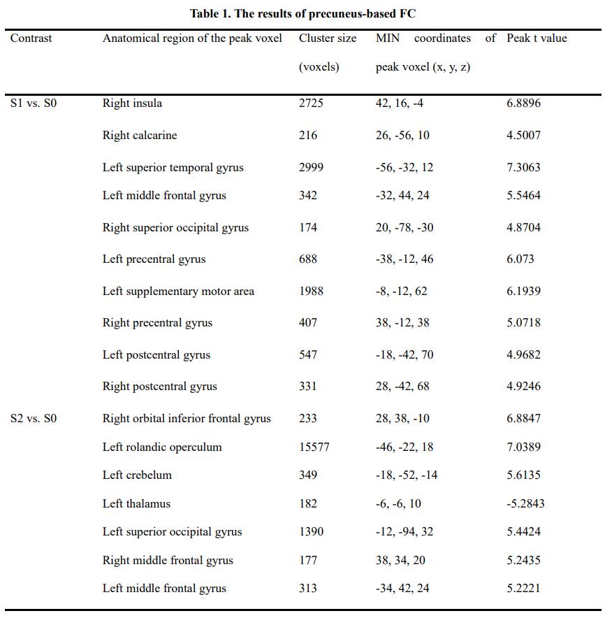
Table 1. The results of precuneus-based FC
S0, baseline scan; S1, post-iTBS scan; S2, post-cTBS scan