2464
Chi-sepnet: Susceptibility source separation using deep neural network1Seoul National University, Seoul, Korea, Republic of, 2Seoul St Mary’s Hospital, Seoul, Korea, Republic of, 3Harvard Medical School, Boston, MA, United States
Synopsis
The separation of positive and negative susceptibility source distributions (e.g., iron and myelin distributions) has important meanings in neuroscience and clinic. In this study, a deep learning-based χ-separation method is proposed to generate high-quality susceptibility source maps. For network training, multi-orientation head data are utilized, providing artifact-free label data. For the input data, either R2’ or R2* maps are utilized in addition to local field and QSM maps, producing two neural networks, χ-sepnet-R2’ and χ-sepnet-R2* (the latter requires no T2). The results of χ-sepnets outperformed the conventional method, revealing details of brain structures both in healthy volunteers and patients.
Introduction
Recently, approaches (e.g., χ-separation) have been proposed to separate positive and negative susceptibility source distributions.1-3 These methods require to resolve the dipole-inversion problem, which is ill-posed and creates steaking artifacts. Hence, the results of χ-separation reveal image degradation, hampering accurate estimation of susceptibility contribution from positive and negative sources. The artifacts can be resolved using multiple head orientation datasets as demonstrated in COSMOS QSM4 or a neural network that is trained using COSMOS dataset as shown in QSMnet.5 The latter is advantageous because a COSMOS-quality QSM map can be inferred from single orientation data once the network is fully trained. In this study, we developed a neural network, χ-sepnet, that generates COSMOS-quality χ-separation results by training the network using multi-orientation χ-separation data. Additionally, another χ-sepnet that only inputs multi-echo GRE data is designed to test the feasibility of using a dataset without T2 maps. The two χ-sepnets are evaluated in healthy volunteers and patients.Methods
Multi-echo GRE and multi-echo SE data were acquired from six subjects (see REF.1 for scan parameters; 4:1:1 subjects for train:validation:test; IRB-approved). Each subject GRE dataset consisted of six head orientations. These multi-orientation GRE data and multi-echo SE data were processed using a COSMOS-like χ-separation algorithm that created artifact-free positive (χpos) and negative (χneg) susceptibility maps (χ-sep-COSMOS; Fig. 1a). These maps were utilized as labels to train the two networks (χ-sepnet-R2’ that requires R2 and χ-sepnet-R2* that is free from R2; Fig. 1a). The original χ-separation algorithm using the MEDI prior is also utilized (χ-sepnet-MEDI; Fig. 1a).The networks had the same 3D U-net6 structure as QSMnet. For the input, not only local field and R2’ (or R2*) but also a QSM map generated from QSMnet was concatenated, improving the results. The loss function had three losses: The first loss is the L1 loss with
$$\left\|f({R_2}'|{R_2}^*,\Delta f,g(\Delta f))_{pos}-\chi _{pos}\right \|_1+\left \|f({R_2}'|{R_2}^*,\Delta f,g(\Delta f))_{neg}-\chi_{neg}\right\|_1$$
where $$$f$$$ denotes χ-sepnet, for which a R2’ or R2* map $$$({R_2}'|{R_2}^*)$$$ is used as the input. $$$\Delta f$$$ is local field map. $$$g(\Delta f)$$$ is the output of QSMnet. The second loss is the gradient loss with
$$\left\||\triangledown f({R_2}'|{R_2}^*,\Delta f,g(\Delta f))|_{x}-|\triangledown\chi _{pos}|_{x}\right \|_1+\left \||\triangledown f({R_2}'|{R_2}^*,\Delta f,g(\Delta f))|_{y}-|\triangledown\chi_{pos}|_{y}\right \|_1+\left \||\triangledown f({R_2}'|{R_2}^*,\Delta f,g(\Delta f))|_{z}-|\triangledown\chi_{pos}|_{z}\right \|_1$$.
The last loss is model loss that enforces to learn information from local field maps and R2’ maps and is written as
$$\left\||(d*(f({R_2}'|{R_2}^*,\Delta f,g(\Delta f))_{pos}+f({R_2}'|{R_2}^*,\Delta f,g(\Delta f))_{neg}))-\triangle f\right \|_1+\left \|(|f({R_2}',\Delta f,g(\Delta f))_{neg}|)-{R_2}' \right \|_1$$
where $$$d$$$ denotes a dipole kernel. Data augmentation was performed by rotating images by random angles (-30˚ - 30˚) relative to $$$B_0$$$ direction, resulting in a total of 48 datasets for training.
For quantitative evaluation, the test subject results were compared for normalized root-mean-squared error (NRMSE), peak signal-to-noise (PSNR), and structural similarity (SSIM) with χ-sep-COSMOS results as a reference. An ROI analysis was performed in 11 ROIs (Fig. 4), reporting the mean and standard deviation of susceptibility values across head orientation.
Two additional subjects, one multiple sclerosis (MS) patient and the other subject with calcification (IRB-approved), were utilized for the evaluation of the networks in clinical practice.
Results
The positive and the negative susceptibility maps from all four methods are summarized in Fig. 2. The χpos and χneg maps from the two χ-sepnets show high-quality results that are comparable or even cleaner than the reference maps from χ-sep-COSMOS. The improvements may be from the denoising nature of neural networks and registration issues in the χ-sep-COSMOS data. Streaking artifacts are observed in the χ-sep-MEDI maps (yellow arrows) but are less noticeable in the other maps. Quantitative metrics report χ-sepnet-R2’ generates the best results while χ-sep net-R2* provides good outcomes. On the other hand, χ-sep-MEDI results reveal poor metrics against the reference results.The three zoomed-in regions reveal well-known structures of the brain, which are the deep gray matters (CN: caudate nucleus, Put: putamen, GP: globus pallidus, Pul: thalamus including pulvinar, ND: nucleus dorsomedialis) and the brainstem regions in Fig. 3. In particular, the hand knob region (HK, orange circles) reveals that the positive susceptibility maps report higher values in the motor cortex (MC) than in the sensory cortex (SC), which is a well-known iron distribution from histology7. This finding is most clearly observed in the χ-sepnet-R2’ and χ-sepnet-R2* results and can be confirmed in the χ-sepnet-COSMOS map, which may suffer from a registration issue when processing multi-orientation data. Since the χ-sepnets utilize only single orientation input, they do not experience the registration issue.
The ROI analysis results demonstrate consistency in the χ-sepnet measurements, reporting much smaller standard deviation than that of χ-sepnet-MEDI (Fig. 4).
Lastly, MS lesions and calcifications are delineated clearly in the results from χ-sepnets (Fig. 5).
Discussion and Conclusions
In this study, we proposed the neural network, χ-sepnet, to reconstruct artifact-free susceptibility source distribution maps. The methods only require single head orientation data. Furthermore, χ-sepnet-R2* requires only multi-echo GRE data, reducing the requirement of R2 maps in χ-separation at the cost of slightly decreased accuracy. This has a significant impact, considering increasing availability in clinical situations. Specifically, x-sepnet-R2* had better quality than x-sep-MEDI in the limited clinical dataset. It demonstrated MS lesion characterization and complex and dense co-localized iron and calcification in globus pallidus more clearly without R2 map.Acknowledgements
This work has been supported by the National Research Foundation of Korea (NRF) grant funded by the Korea government (MSIT) (No. 2021M3E5D2A01024795) and NRF-2021R1A2B5B03002783.References
1. Shin, H. G., Lee, J., Yun, Y. H., Yoo, S. H., Jang, J., Oh, S. H., ... & Lee, J. (2021). 𝜒-separation: Magnetic susceptibility source separation toward iron and myelin mapping in the brain. NeuroImage, 240, 118371.
2. Chen, J., Gong, N. J., Chaim, K. T., Otaduy, M. C. G., & Liu, C. (2021). Decompose quantitative susceptibility mapping (QSM) to sub-voxel diamagnetic and paramagnetic components based on gradient-echo MRI data. NeuroImage, 242, 118477.
3. Emmerich, J., Bachert, P., Ladd, M. E., & Straub, S. (2021). On the separation of susceptibility sources in quantitative susceptibility mapping: Theory and phantom validation with an in vivo application to multiple sclerosis lesions of different age. Journal of Magnetic Resonance, 330, 107033.
4. Liu, T., Spincemaille, P., De Rochefort, L., Kressler, B., & Wang, Y. (2009). Calculation of susceptibility through multiple orientation sampling (COSMOS): a method for conditioning the inverse problem from measured magnetic field map to susceptibility source image in MRI. Magnetic Resonance in Medicine: An Official Journal of the International Society for Magnetic Resonance in Medicine, 61(1), 196-204.
5. Yoon, J., Gong, E., Chatnuntawech, I., Bilgic, B., Lee, J., Jung, W., ... & Lee, J. (2018). Quantitative susceptibility mapping using deep neural network: QSMnet. Neuroimage, 179, 199-206.
6. Ronneberger, O., Fischer, P., & Brox, T. (2015, October). U-net: Convolutional networks for biomedical image segmentation. In International Conference on Medical image computing and computer-assisted intervention (pp. 234-241). Springer, Cham.
7. Costagli, M., Donatelli, G., Biagi, L., Ienco, E. C., Siciliano, G., Tosetti, M., & Cosottini, M. (2016). Magnetic susceptibility in the deep layers of the primary motor cortex in amyotrophic lateral sclerosis. NeuroImage: Clinical, 12, 965-969.
Figures
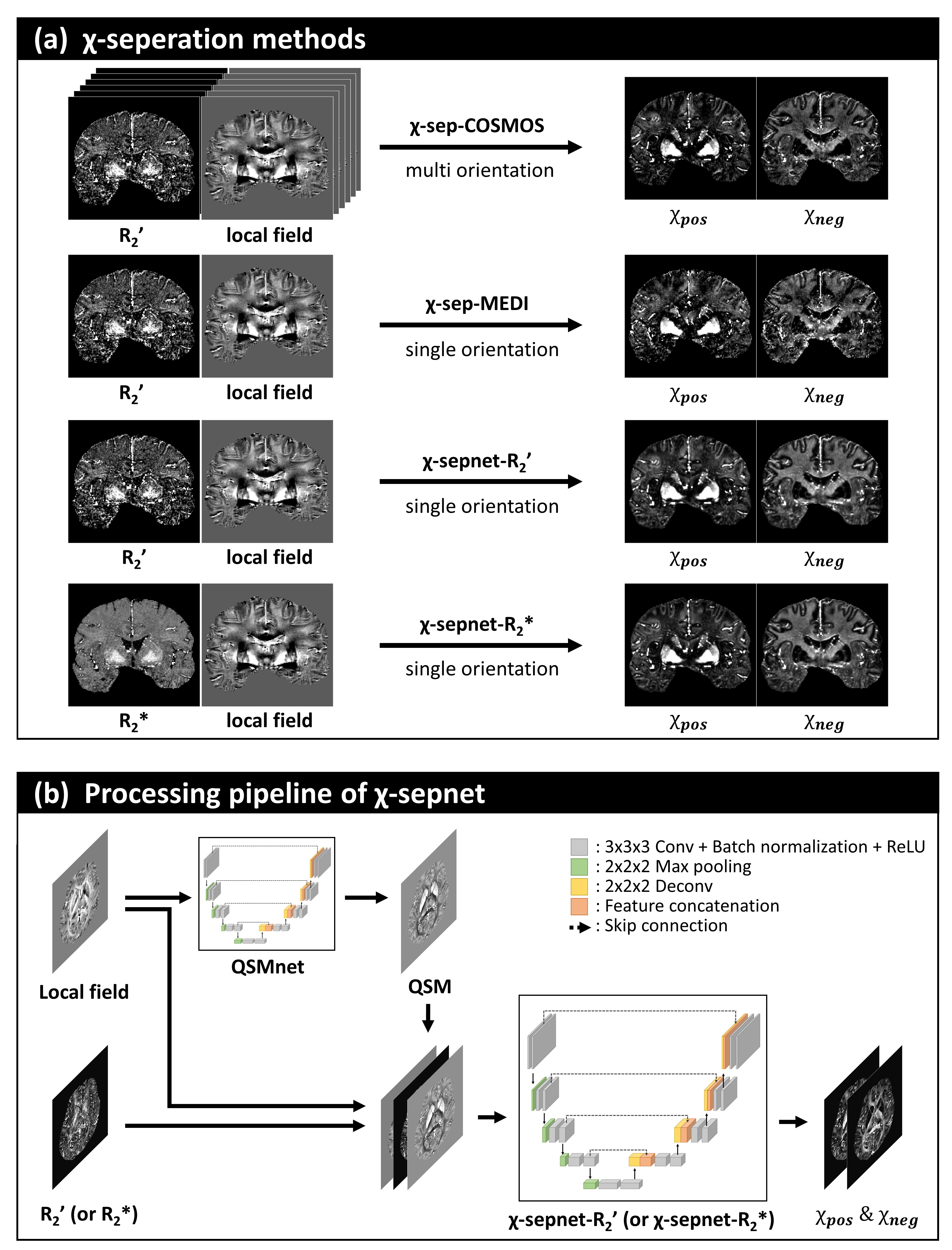
Fig. 1 (a) Overview of the four χ-separation methods applied (χ-sep-COSMOS and χ-sep-MEDI) or developed (χ-sepnet-R2’ and χ-sep net-R2*) in this study. (b) Processing pipeline of χ-sepnet. The network architecture of χ-sepnet had the same 3D U-net structure as QSMnet. From a local field map, QSM is reconstructed using QSMnet. Then R2’ (or R2*), local field and QSM maps are concatenated for the input of χ-sepnet, generating positive and negative susceptibility maps.
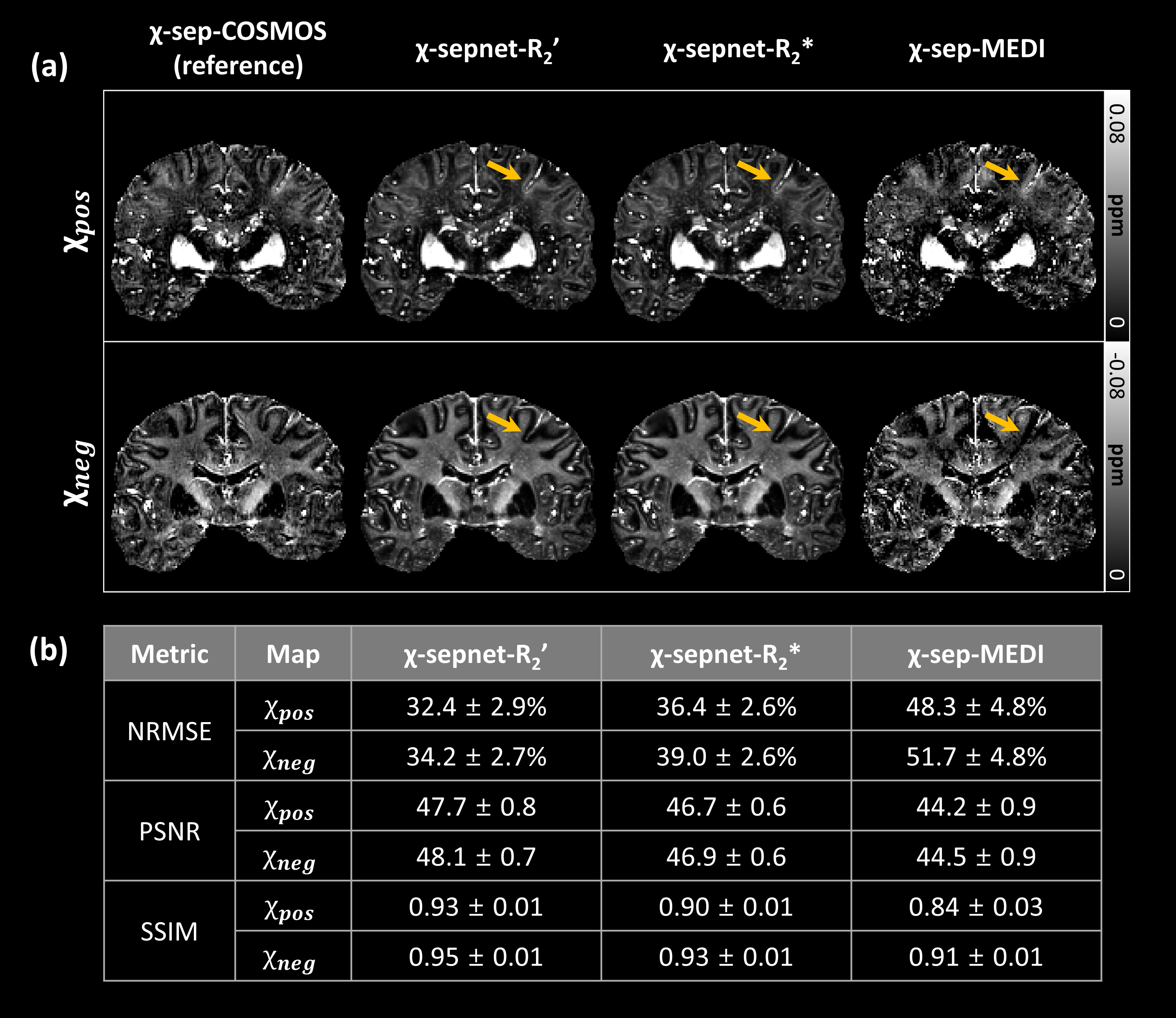
Fig. 2 (a) Results of the χ-separation methods. The χpos and χneg maps from two χ-sepnets are comparable or even cleaner than reference maps from χ-sep-COSMOS. The improvements may be from denoising nature of neural networks and registration issues in χ-sep-COSMOS data. Streaking artifacts are observed in χ-sep-MEDI maps (yellow arrows) but are less noticeable in other maps. (b) Quantitative metrics report χ-sepnet-R2’ generates the best results while χ-sepnet-R2* provides good outcomes. χ-sep-MEDI results reveal poor metrics against reference results.
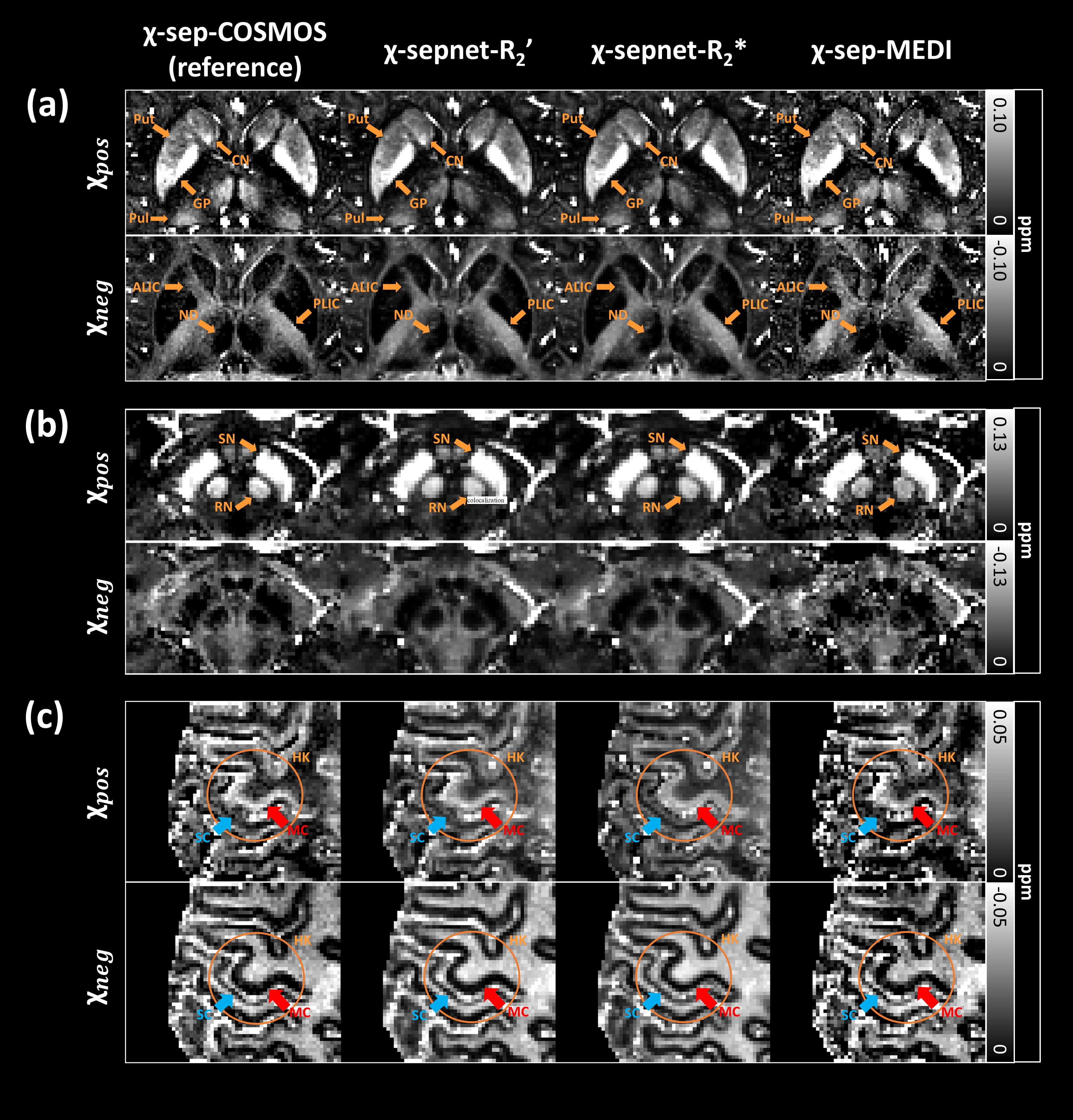
Fig. 3 Zoom-in maps of χ-separation methods at three locations. (a) Deep gray matter regions (CN, Put, GP, Pul, ND, ALIC, PLIC) clearly delineated in χpos and χneg maps. (b) In the brainstem region, SN and RN show high positive and low negative susceptibility concentrations. (c) In the region including HK (orange circles), χ-sepnet-R2’ and χ-sepnet-R2* results show that positive susceptibility maps report higher values in the motor cortex than in the sensory cortex. This finding can be confirmed in the χ-sepnet-COSMOS map.
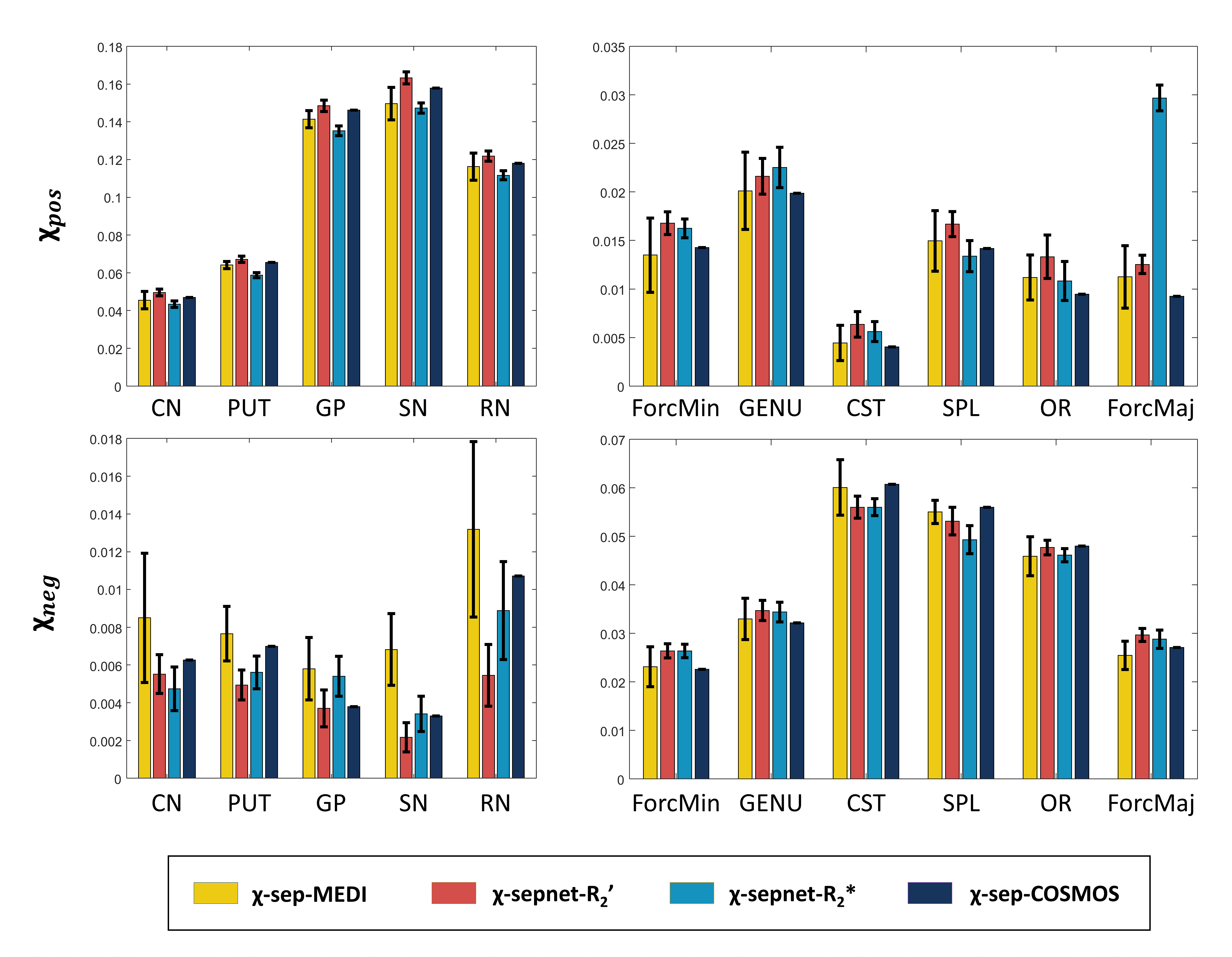
Fig. 4 Results of the ROI analysis, reporting the mean and standard deviation (std) in the five gray matter ROIs (CN: caudate nucleus, PUT: putamen, GP: globus pallidus, SN: substantia nigra, RN: red nuclues) and six white matter ROIs (ForcMin: forceps minor, ForcMaj: forceps major, GENU: genu, CST: corticospinal tract, SPL: superior parietal lobule, OR: optic radiation) over the six head orientations in one subject. This result demonstrates consistency in the χ-sepnet measurements, reporting much smaller std than that of χ-sepnet-MEDI.
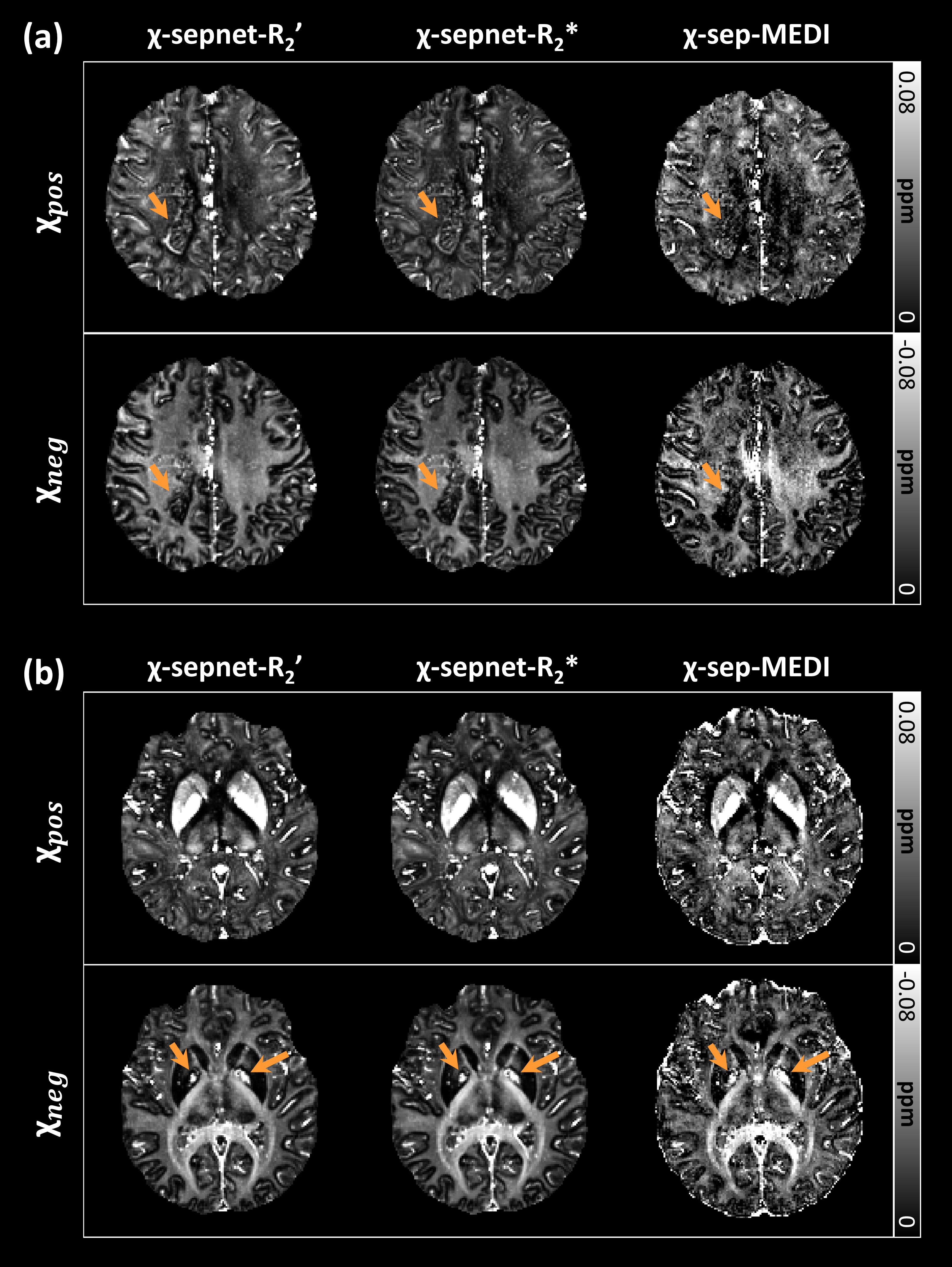
Fig. 5 Outcomes of χ-sepnets from two clinical data. (a) χ-sepnet results of MS patient clearly demonstrate lesions with increased paramagnetic and decreased diamagnetic changes, typical findings of MS lesions on χ-separation (arrows). (b) Focal diamagnetic hyperintensity in globus pallidus captured in negative susceptibility maps, which is a common location of calcification. The positive susceptibility maps of this patient reveal positive susceptibility in the same region, suggesting our methods can separate two sources, clearly distinguishing them from conventional QSM.