2449
Millimeter Spatial Resolution and Subsecond Temporal Resolution in Real-Time fMRI using Multi-Band Echo-Volumar Imaging1Neurology, U New Mexico, Albuquerque, NM, United States, 2Center for Magnetic Resonance Research, Minneapolis, MN, United States
Synopsis
In this study we develop highly undersampled multi-band slab-segmented echo-volumar imaging (MB_EVI), which combines the sampling efficiency of single-shot 3D encoding with the sensitivity advantage of multi-echo acquisition, and explore the feasibility of shortening the readout duration using segmented within-slab slice encoding. Kz-segmented Segmented MB-EVI enabled a nominal 1x1x1 mm isotropic voxel size with 64 slices and a temporal resolution of 618 ms with sub-second acquisition time.
INTRODUCTION
Achieving millimeter spatial and sub-second temporal resolution in fMRI holds promise for unraveling functional organization and connectivity at the laminar level[1], but requires compromises in spatial coverage that impair the performance of motion correction and the ability to characterize long-range connectivity across distant cortical areas. Recent advances in fMRI acquisition, using multi-band (simultaneous multi-slice) encoding[2], simultaneous image refocusing[3], and magnetic resonance encephalography (MREG)[4] have improved sensitivity for mapping functional connectomics[5, 6],[7] and for detecting task-based at frequencies above 0.2 Hz[8]. In this study we develop highly undersampled multi-band slab-segmented echo-volumar imaging (MB_EVI), which combines the sampling efficiency of single-shot 3D encoding with the sensitivity advantage of multi-echo acquisition [9, 10], and explore the feasibility of shortening the readout duration using segmented within-slab slice encoding.METHODS
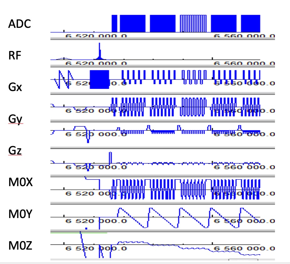
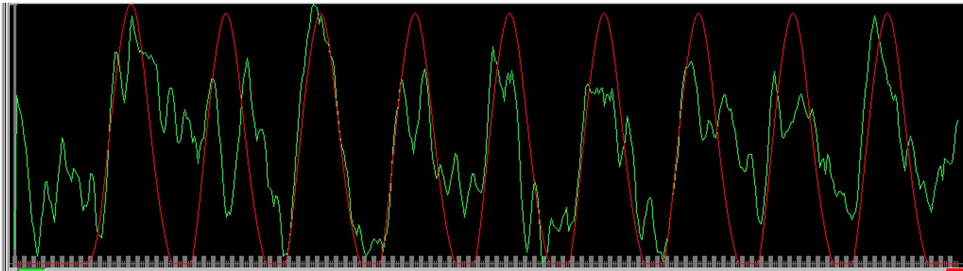
MB-EVI was developed based on a widely used multi-band EPI sequence that supports multi-echo acquisition[2, 11]. Simultaneous excitation of 2 or 3 slabs with blipped-controlled aliasing in parallel imaging, up to 4-fold in-plane undersampling, up to 5/8 in-plane partial Fourier acquisition and 6/8 slice partial Fourier encoding were supported (Figure 1). A slice-segmented interleaved acquisition was implemented by acquiring odd kz-lines in the first segment and even kz-lines in the second segment. TE-shifting between the 2 segments was supported. All slabs of first segment were acquired first, followed by all slabs of the second segment. Navigator-based phase correction could be performed either jointly for both segments jointly or separately. Offline reconstruction was performed in MATLAB. Online reconstruction was performed using regularized ‘leak-block’ slab-GRAPPA multiband-reconstruction. The online reordering of kz-lines from the 2 segments, the within-slab reconstruction of individual slices, slice reordering and slab concatenation was performed on an external workstation using custom software (TurboFIRE[10]). Task-based (motor/visual) and resting-state data were acquired in 5 healthy controls on a 3T Siemens Prisma scanner equipped with 32-channel head array coil. Informed consent was obtained. An isotropic resolution of 1.5 mm with 64 reconstructed slices was encoded without kz-segmentation, 6 kz-steps and an EVI readout duration of 86.64 ms, enabling a minimum TR of 334 ms. An isotropic resolution of 1 mm with 85 reconstructed slices was encoded using kz-segmentation, 3 kz-steps per segment and an EVI readout duration of 90 ms, enabling a minimum TR of 860 ms. Task- and resting-state fMRI analysis was performed using the TurboFIRE fMRI software tool[7, 12] with rigid-body motion correction and spatial normalization of MNI space into subject space. Real-time seed-based moving-average sliding window (15 s) correlation analysis was performed with regression of 8 signal time courses from 6 rigid body motion parameters and WM and CSF ROIs.RESULTS
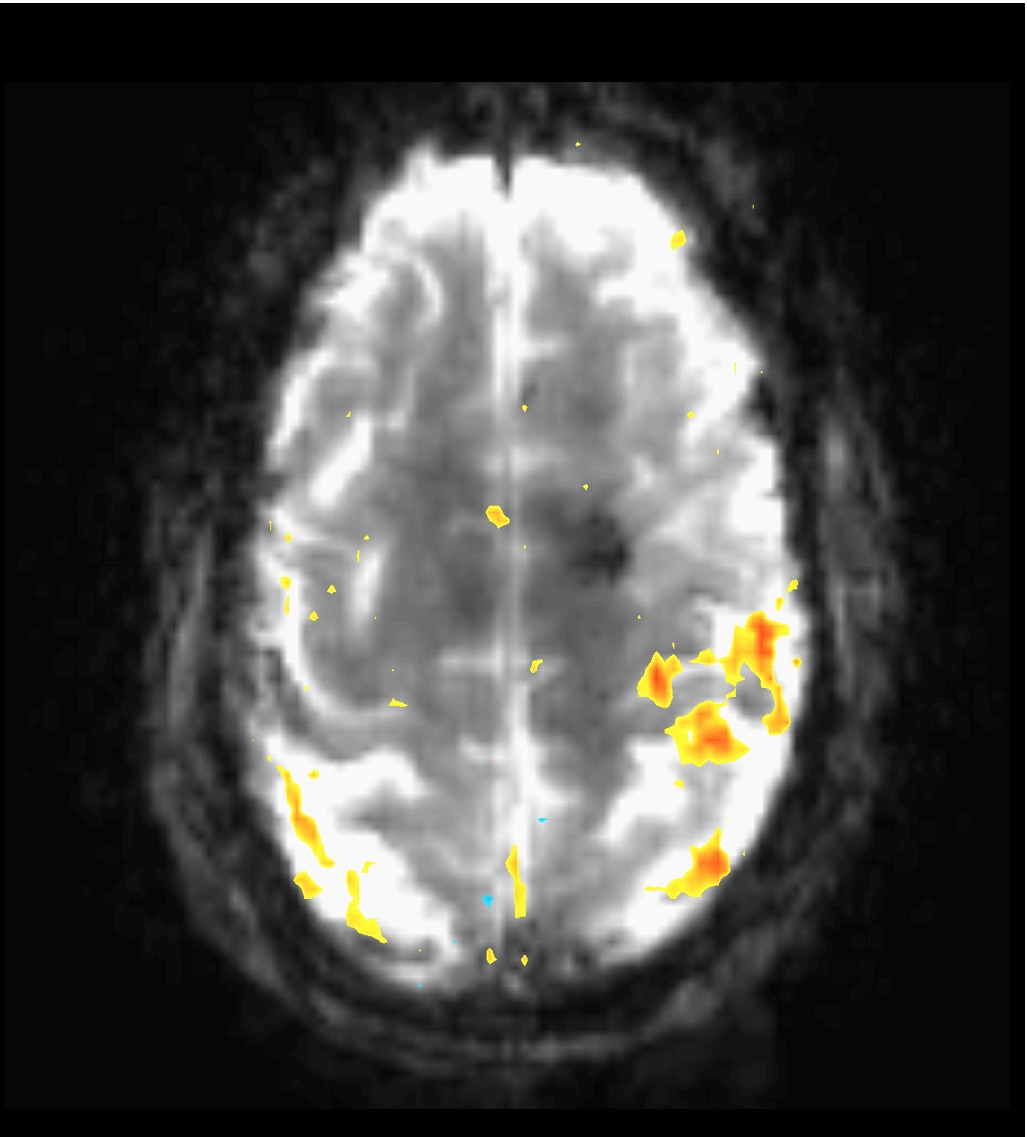
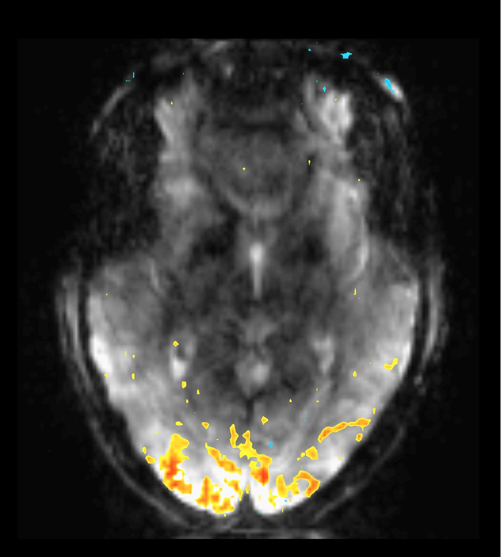
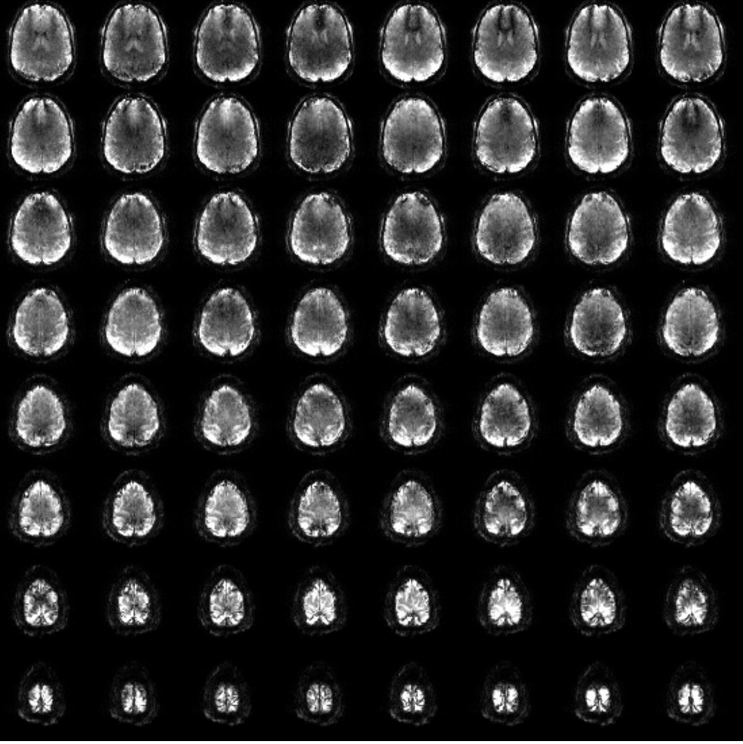
High spatial resolution (1.5 mm3 isotropic voxel size) MB-EVI improved high BOLD contrast (Figure 2) and delineation of neuroanatomy, enabling sensitive mapping of task-based activation (Figures 3 and 4). Kz-segmented Segmented MB-EVI enabled a nominal 1x1x1 mm isotropic voxel size with 64 slices and a temporal resolution of 618 ms (Figure 5). Real-time multiband and in-plane image reconstruction and real-time transfer to the external workstation with negligible time delay was verified. The performance of the reconstruction of the kz dimension and the real-time fMRI analysis in TurboFIRE are currently the rate-limiting steps.DISCUSSION
MB-EVI provides flexibility for maximizing spatial-temporal resolution, volume-coverage and BOLD-sensitivity for mapping task-activation and functional connectivity. Gains in spatial-temporal resolution were limited by increasing signal instability with MB accleration, sensitivity to respiratory signal pulsation, and increasing TE with descreasing voxel size. Further limitations of this approach include sensitivity loss at slab interfaces due to slab profile imperfections and T1 saturation, and Gibb’s ringing in the slice direction. Assessment of BOLD sensitivity of this methodology in comparison with MB_EPI is in progress.CONCLUSIONS
The MB_EVI approach provides an approximately 2-fold acceleration compared with state-of-the-art simultaneous multi-slice dual-echo echo-planar imaging.Acknowledgements
Supported by 1R21CA241714. We gratefully acknowledge Kunxiu Gao for TurboFIRE software development.References
1. Petridou, N. and J.C.W. Siero, Laminar fMRI: What can the time domain tell us? Neuroimage, 2019. 197: p. 761-771.
2. Moeller, S., et al., Multiband multislice GE-EPI at 7 tesla, with 16-fold acceleration using partial parallel imaging with application to high spatial and temporal whole-brain fMRI. Magn Reson Med, 2010. 63(5): p. 1144-53.
3. Chen, L., et al., Evaluation of highly accelerated simultaneous multi-slice EPI for fMRI. Neuroimage, 2015. 104: p. 452-9.
4. Hennig, J., et al., 15 Years MR-encephalography. MAGMA, 2020.
5. Feinberg, D.A., et al., Multiplexed echo planar imaging for sub-second whole brain FMRI and fast diffusion imaging. PLoS One, 2010. 5(12): p. e15710.
6. Smith, S.M., et al., Temporally-independent functional modes of spontaneous brain activity. Proc Natl Acad Sci U S A, 2012. 109(8): p. 3131-6.
7. Posse, S., et al., High-speed real-time resting-state FMRI using multi-slab echo-volumar imaging. Front Hum Neurosci, 2013. 7: p. 479.
8. Lewis, L.D., et al., Fast fMRI can detect oscillatory neural activity in humans. Proceedings of the National Academy of Sciences, 2016. 113(43): p. E6679-E6685.
9. Puckett, A.M., et al., Using multi-echo simultaneous multi-slice (SMS) EPI to improve functional MRI of the subcortical nuclei of the basal ganglia at ultra-high field (7T). Neuroimage, 2018. 172: p. 886-895.
10. Posse, S., et al., Enhancement of temporal resolution and BOLD sensitivity in real-time fMRI using multi-slab echo-volumar imaging. Neuroimage, 2012. 61(1): p. 115-30.
11. Xu, J., et al., Evaluation of slice accelerations using multiband echo planar imaging at 3 T. Neuroimage, 2013. 83: p. 991-1001.
12. Posse, S., et al., A new approach to measure single-event related brain activity using real-time fMRI: Feasibility of sensory, motor, and higher cognitive tasks. Human Brain Mapping, 2001. 12(1): p. 25-41.
Figures