2444
Abdominal 3D-T1-weighted imaging with the radial MP2RAGE sequence.1Centre de Résonance Magnétique des Systèmes Biologiques, UMR5536, CNRS, Bordeaux, France, Metropolitan
Synopsis
The Magnetization Prepared Two RApid Gradient Echo (MP2RAGE) sequence with a cartesian sampling is performed to obtain 3D-T1 weighted images of the brain. A radial encoding giving a better robustness to respiratory motion has been implemented in order to use the MP2RAGE sequence for abdominal imaging.
Introduction
The Magnetization Prepared Two RApid Gradient Echo (MP2RAGE) sequence is an advantageous way of 3D-T1-weighted imaging and for obtaining rapid 3D-T1 maps, by combining two images acquired at two different inversion times (TI). The MP2RAGE sequence with cartesian encoding is solely used for brain imaging. To extend its application to abdominal T1 mapping, it can be combined with encodings robust to respiration motion, like radial encoding. In these areas, the presence of fat tissues generates chemical shift artefact and high hyper intense signal. Consequently, the goal of our study is to implement a radial encoding into the MP2RAGE sequence in order to perform free-breathing abdominal T1 mapping at 3T.Methods
The sequence has been developed on a 3T Siemens scanner (Prisma) and reconstruction has been performed using Matlab. Firstly, the cartesian encoding has been replaced by a Kooshball radial one. Second, the excitation pulses within the echo trains were modified to perform a water selective excitation1. To do so, 3 types were tested in addition to the unique pulse : 1-1, 1-2-1, and 1-3-3-1, implying that TE was modified from 3.91 ms to 5.12 ms. In fact, the water selective excitation is based on a pulse decomposition. In the sequence, pulses are rectangular and at 3T, duration between two pulses is 1.15 ms.The MP2RAGE images were reconstructed as in Marques et al2. The T1-map was then built using the equations described by Faller et al3 in order to take into account the radial sampling in the MP2RAGE signal.Three healthy volunteers were scanned multiple times at variable days of interval with the radial MP2RAGE sequence. The sequence was acquired with a 1.3mm isotropic spatial resolution, an Echo Train Length (ETL) of 192 projections, MP2RAGETR of 6000ms and a total amount of projections of 23040, lasting 12min. Reconstruction was performed with a Density Control Function (DCF) and a fast non-uniform Fourier transform (nuFFT).
Results
Both TI images are mainly free from respiration artifacts, except at the interface between the liver and the lungs (Figure 1, 1a and 2a). The different tissues composing the kidneys are easily distinguishable. Blood vessels within the liver appear sharp (arrow, Figure 1, 3a).Then, the T1-maps were computed (Figure 2). The T1 values of kidney cortex and liver were 1215 ms ± 260 and 1092 ms ± 210 using the radial MP2RAGE without the water selective excitation. Using water-selective pulses (Figure 1b,c and 2b,c), the signal from adipose tissue from the skin and the abdomen is largely cancelled.
The 1-3-3-1 water selective pulse appears the most effective to suppress fat signal. The more pulses are used, the less fat is visible. However, fat can still be visible at the edge of the field of view. Images with the 1-3-3-1 pulse have a better contrast and organs are better defined. With the water selective excitation, T1 values for kidney cortex 1042 ms ± 93 and for liver 1075 ms ± 150 with smaller standard deviations than without the WS pulses (Figure 2). Compared with T1 values from literature4, these values are similar for the kidney cortex.
Discussion
The radial sampling enabled to obtain abdominal images and T1 maps without severe motion artefact. Inserting water-selective pulses enabled to obtain images without fat signal, enabling a better delineation of the internal organs. In addition, the T1 values might be improved by the water selective excitation to get closer to values reported with the standard inversion-recovery method.Nevertheless, some fat signal can still be present on the images. The Bo inhomogeneity on a large field of view (350 mm) could be responsable of that. Water excitation is estimated to be effective at a distance of 260 mm maximum. Thus, decreasing the field of view to get the T1-mapping of a unique organ is possible and can guarantee a better water selective excitation.
Conclusion
We developed a new sequence based on the MP2RAGE sequence with a radial encoding, giving the possibility to make free-breathing 3D-T1 mapping of the abdomen with a protocol lasting less than 15min and with a good spatial resolution.Future goals will be to reconstruct multiple images during the breathing cycle in order to maximize spatial resolution and to have better T1 values on organ boundaries. For that, reconstruction will be improved with parallel imaging and with other advanced reconstruction methods.
Acknowledgements
This study was achieved within the context of the Laboratory of Excellence TRAIL ANR-10-LABX-57. This work was also supported by the French National Research Agency (ANR-19-CE19-0014).References
1. Levitt M.H. ’Composite pulses’, Prog NMR Spectrosc, 1986.
2. Marques J. P et al. ‘MP2RAGE, a self bias-field corrected sequence for improved segmentation and T1-mapping at high field’, NeuroImage, 2010.
3. Faller T. et al. ‘Radial MP2RAGE sequence for rapid 3D T1 mapping of mouse abdomen: application to hepatic metastases’, European Radiology, 2019.
4. De Bazelaire C. et al. ‘MR imaging relaxation times of abdominal and pelvic tissues measured in vivo at 3.0 T: preliminary results’, 2004.
Figures
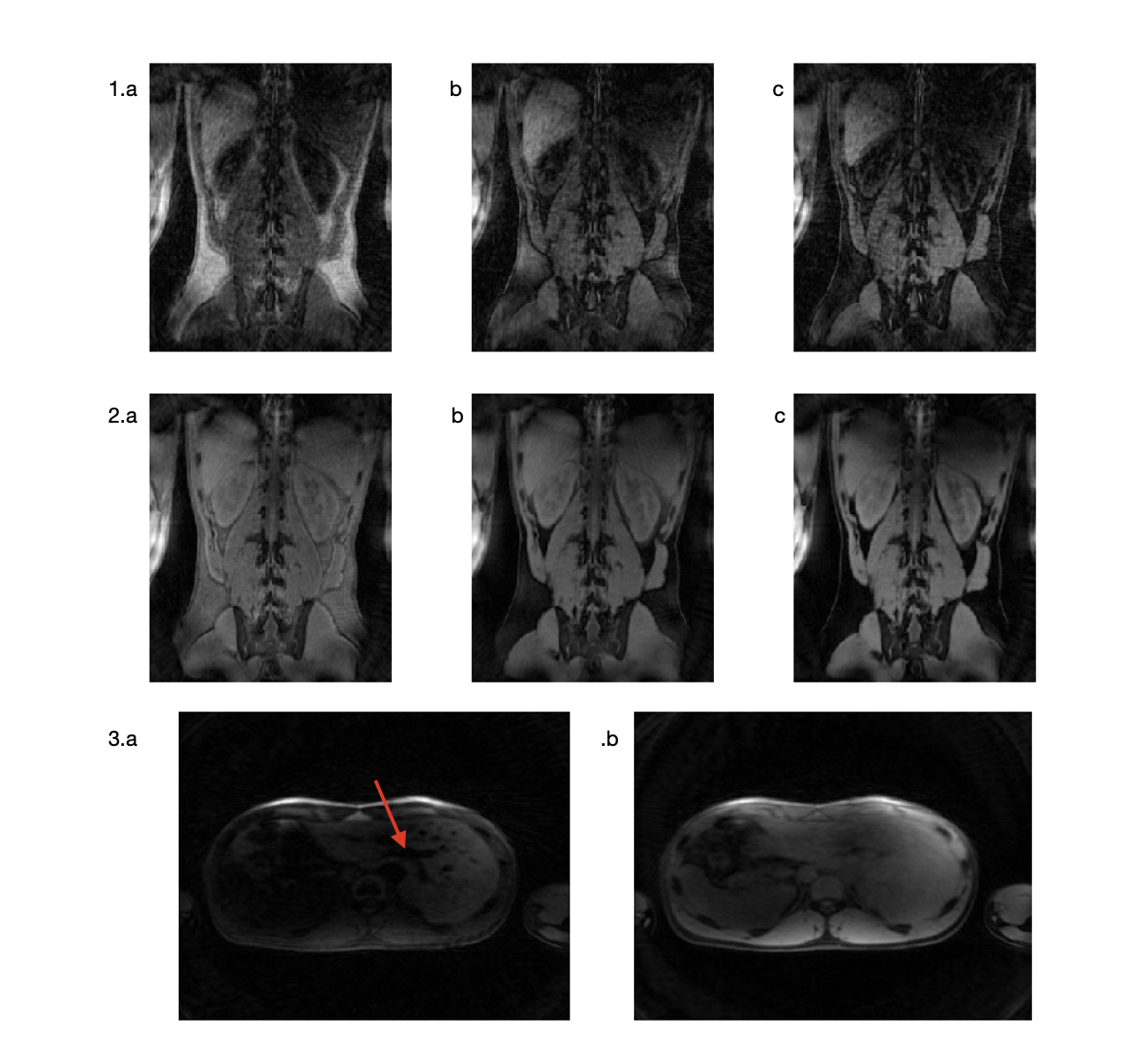
Figure 1 : Images GRE1 and GRE2 (1. and 2., respectively) obtained with 1 pulse, 1-2-1 pulses and 1-3-3-1 pulses (a. b. and c., respectively).
Acquisition time : 12 minutes, ETL : 192, MP2RAGETR = 6000 ms, TI1 = 1100 ms, TI2 = 3300 ms, resolution = 1.37mm x 1.37mm x 1.37mm.
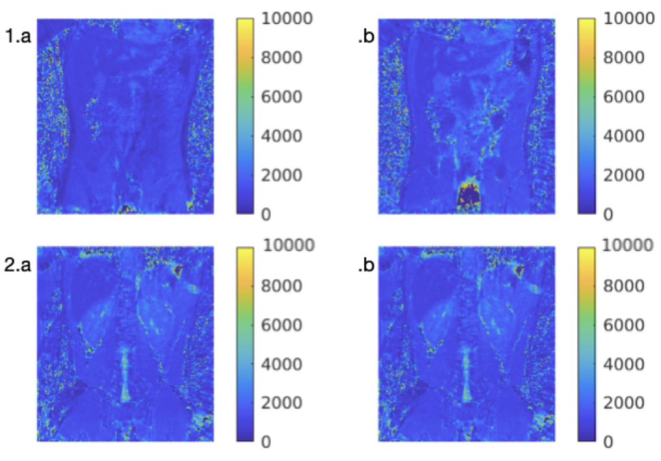
Figure 2 : T1-maps computed for the liver (1) and for the kidneys (2) with one pulse (1.a and 2.a) and with 1-3-3-1 pulses (1.b and 2.b).
Acquisition time : 12 minutes, ETL : 192, MP2RAGETR = 6000 ms, TI1 = 1100 ms, TI2 = 3300 ms, resolution = 1.37mm x 1.37mm x 1.37mm.