2442
Coil Selective Golden Angle DCE-MR Image Reconstruction using Mutual Dependence1Radiology, Boston Children's Hospital, Boston, MA, United States
Synopsis
MRI literature shows that it is possible to increase image reconstruction quality by removing coils that cause majority of the streaking artifacts. We introduce to measure the coil quality based on mutual information between a reference and each coil image. Specifically we apply this technique to DCE-MRI reconstruction from under-sampled radial stack of stars trajectory for kidney imaging and calculate mutual dependence between a dynamic image from each coil and a reference image to assess coil contribution to reconstruction. Experiments show mutual dependence based coil selection reduces artifacts and increases reconstructed image SNR by $$$16.84\%$$$ using $$$1/3$$$ of the coils.
Introduction
Radial MRI suffers from streaking artifacts caused by under-sampling of the k-space and gradient non-linearities. Coils that cover the periphery of the field of view cause majority of the artifacts, severely reducing the image quality. Specifically in stack of stars radial dynamic contrast enhanced (DCE) MRI which utilizes higher temporal resolution, enhanced under-sampling artifacts from certain coils deteriorate reconstruction quality. Therefore it is important to detect such coils and discard from reconstruction accordingly. However, removing a coil that does not contribute to the streaking artifact will reduce the SNR of the reconstructed image. Conventionally, coils with artifacts are removed based on their $$$\ell$$$ norm variations from a reference. In this work, we measure the coil quality with respect to mutual information (MI) that not only detects artifacts but also selects coils that contribute most information from important image regions.Method
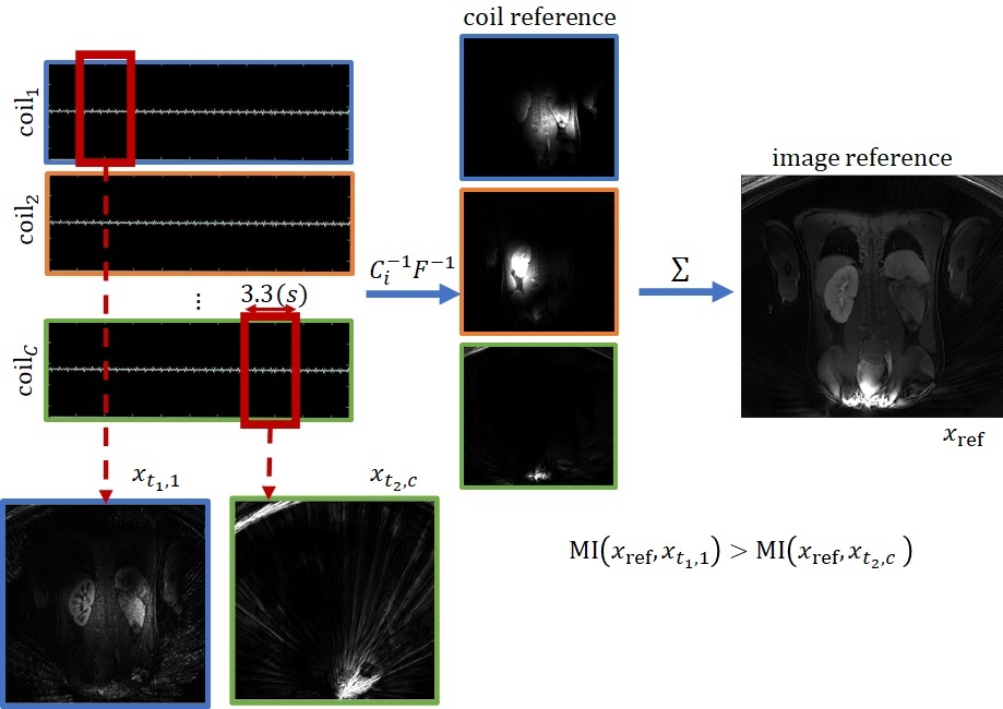
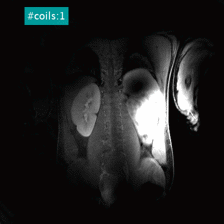
We acquired coronal DCE-MR images with a dynamic stack of stars sequence with golden angle radial sampling scheme from 12 patients (3 female, 9 male, age=12.2$$$.\pm$$$.6.9 years) with temporal resolution 3.3~(s/volume) [1] following an approved IRB protocol and after obtaining consent. DCE MRI data is highly under-sampled to match temporal resolution levels such as 3.3[s] to correctly measure the fast dynamics of contrast in aorta and to accurately estimate tracer kinetic model parameters. The under-sampled acquisition and gradient non-linearities cause large streaking artifacts in some coils. Respective artifacts appear as unexpected pixel intensity change patterns over time. We reconstruct a densely sampled, coil combined reference image using the entire k-space data acquired in 6 minutes. We observed that such a smoothed reference image illustrates target regions of interests without large under-sampling artifacts. We then, for each coil, compare each time image in the dynamic series to this reference image to assess the effect of the under-sampling artifacts in each time image [2]. We expect coil images that contribute most to this reference image will have higher MI value and should be prioritized and included in the image reconstruction. Accordingly, coil images with artifacts will have smaller MI value and will be discarded. Let $$$X=\left\lbrace x_{t,c} | x_{t,c}\in \mathbb{C}^{n\times n}, \forall t\in\lbrace1,2,\cdots, T \rbrace, \forall c\in\lbrace 1,2,\cdots, C \rbrace\right\rbrace$$$ represent the reconstructed complex $$$C$$$-coil image sequence for the time interval $$$[1,T]$$$. $$$x_{t,c} = \mathcal{C}_c F k_{t,c}$$$ where $$$k$$$ denotes frequency observations, $$$\mathcal{C}$$$ and $$$F$$$ denote the coil profile and Fourier operators respectively. The reference is computed by combining a densely reconstructed coil images $$$x_{\text{ref}}=\sum_c \mathcal{C}_c F [k_{t,c}]_t$$$. We compute a MI score for each coil, that is the average of individual MI values between each temporal image $$$x_{t,c}$$$ and $$$x_{\text{ref}}$$$. The coil score for coil $$$c$$$ is $$$\text{sc}_c = \frac{1}{T}\sum_t MI(x_{t,c}, x_{\text{ref}})$$$ [3]. Calculating MI between dynamic time reconstructions with the reference is illustrated in Figure-1. We sort the coils based on their MI scores and select a subset of coils with highest MI scores as the reconstruction coil set $$$\tilde{C}\subseteq C$$$. We use the corresponding frequency domain observations $$$\lbrace k_{t,c} | \forall t\in\lbrace1,2,\cdots,T\rbrace , \forall c \in \tilde{C} \subseteq C \rbrace$$$ from the reconstruction coil set to reconstruct a dynamic series of volumes. In our experiments we compare the proposed approach with $$$\ell_2$$$ norm coil selection influenced by [2]. SNR was measured by dividing the mean of a region of interest to the standard deviation of a background region.Results
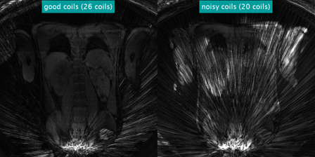
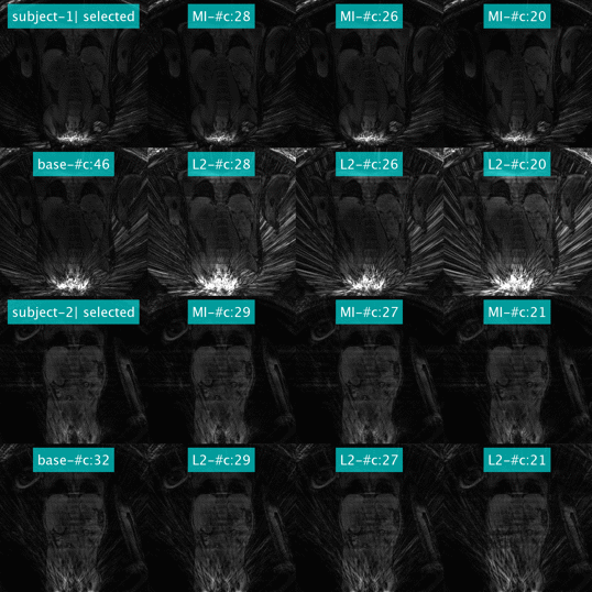
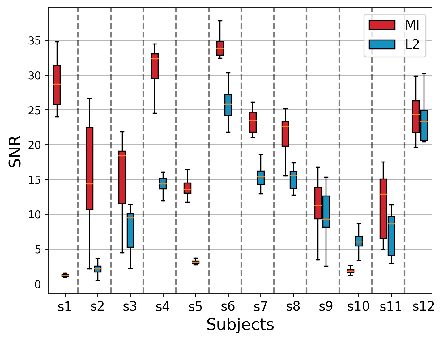
Figure-2 illustrates the effects of adding different coils on a reconstruction. Each coil covers a portion of the scanned area, once combined serves as a complete reconstruction. The coils are added based on the order of increasing MI score in the illustration and observe that the latter coils, with smaller MI scores inhibit artifacts, once they are added to the reconstruction. Figure-3 illustrates the dynamic reconstructions performed with hand picked "good" coils and "noisy" coils on left and right panels respectively. Observe that at this temporal resolution, some of the coils have low SNR, mostly capturing noise. Figure-4 illustrates 2 examples with each reconstruction method. "Selected" refers to reconstructions with hand-picked coils and and "base" refers to reconstructions using all coils without removal. Number of coils for "base" reconstructions are 46 and 32 for subject 1 and subject 2 respectively. As observed MI based selection outperforms the competitor and results similar visual quality for same number of coils. Figure-5 compares SNRs of images reconstructed using two alternative coil selection methods for all 12 subjects. Red bars and blue bars represent MI and $$$\ell_2$$$ selections respectively. For this experiments, we only kept top scoring $$$1/3$$$th of all the coils in reconstruction. Results show that the proposed method outperforms the competitor method. However, in a very rare scenario, where the majority of coils are corrupted, such as subject-10, MI based selection matches the coil artifacts on the reference image reducing the overall SNR. Through our experiments we observed that proposed approach increased overall SNR by $$$16.84\%$$$ by reducing the artifacts.Conclusion and Discussion
Proposed score for coil selection outperforms the competitor method that uses an $$$\ell_2$$$ driven score. Results show that the proposed approach can sort coils based on their quality reflecting a coil contribution that results in an improved quality image reconstruction.Acknowledgements
This work was supported partially by the Society of Pediatric Radiology Multi-center Research Grant 2019, Crohn’s and Colitis Foundation of America’s (CCFA) Career Development Award and by the NIDDK and NIBIB of the National Institutes of Health under award numbers R01DK125561, R21DK123569, R21EB029627, and by the grant number 2019056 from the United States-Israel Binational Science Foundation (BSF), and a pilot grant from National Multiple Sclerosis Society under Award Number PP-1905-34002, and Society of Pediatric Radiology, Multicenter Research Award.References
[1] Coll‐Font, Jaume, et al. "Bulk motion‐compensated DCE‐MRI for functional imaging of kidneys in newborns." Journal of Magnetic Resonance Imaging 52.1 (2020): 207-216.
[2] Feng, Li, et al. "RACER‐GRASP: respiratory‐weighted, aortic contrast enhancement‐guided and coil‐unstreaking golden‐angle radial sparse MRI." Magnetic resonance in medicine 80.1 (2018): 77-89.
[3] Maes, Frederik, Dirk Vandermeulen, and Paul Suetens. "Medical image registration using mutual information." Proceedings of the IEEE 91.10 (2003): 1699-1722.
Figures