2357
Comparison of distortion correction with Echo Planar Imaging Correction and the 2D navigator based multi-shot SENSE EPI schemes1BU-MR Clinical Science, Philips Healthcare, Suzhou, China, 2Philips Healthcare (China), Shenzhen, China, 3Philips Healthcare (China), Beijing, China, 4BU-MR Clinical Application, Philips Healthcare, Suzhou, China, 5BU-MR R&D, Philips Healthcare, Suzhou, China, 6BU-MR Clinical Science, Philips Healthcare, Tokyo, Japan
Synopsis
Brain diffusion imaging by single shot EPI has remained challenging for the local B0 field variation, which leads to geometry distortion and signal inhomogeneity in the resulting images. In this study, we show that the brain single shot EPI image quality could be improved by correction of the geometric distortion and increased resolution, when compared to the traditional acquisition scheme. In-plane resolution of 1.2x1.2 mm2 was achieved and the distortion correction was effectively reduced.
Introduction
While diffusion weighted imaging (DWI) has been widely used in brain imaging for both microstructure studies and oncological applications, the conventional single-shot EPI (ssEPI) remains suffering from geometric distortion and blurring due to B0 inhomogeneity and tissue susceptibility. Recent development in distortion reduction techniques, such as Zonally magnified oblique multislice (ZOOM)1, multiplexed sensitivity-encoding (MUSE)2 multi-shot DWI3 and Echo Planar Imaging Correction (EPIC)4, has shed light on research studies. EPIC combines the top-up strategy with parallel imaging to reduce the geometric distortion with high efficiency. The Image Reconstruction with Image space Sampling (IRIS) scheme by 2D navigated multi-shot SENSE EPI3 applies segmented k-space acquisition with reduced phase-encoding FOV. In this study, we examined the distortion correction performance of the above two strategies and compared against the conventional ssEPI with SENSE on healthy volunteers.Methods
The MR imaging was performed on a 3.0 T Ingenia Elition system (Philips Healthcare, the Netherlands) with volunteers under informed consent. The 16-channel head and spine array coil was used to acquire the anatomical T2-weighted images with in-plane resolution of 0.6 x 0.6 mm2. The diffusion-weighted axial images were acquired using three strategies -- the conventional single-shot EPI sequence, the EPIC method, and the IRIS method with increased spatial resolution, respectively. For ssEPI and EPIC acquisitions, the scanning parameters are as following: FOV = 220 x 220 mm2, voxel size = 1.6 x 2.0 mm2, 24 slices with thickness = 5 mm, TE/TR = 82/2958 ms, and b-value = 0, 1000 s/mm2, with SENSE factor set to 2 (default for the clinical scan). For IRIS scanning, we used the same parameters except for TE/TR = 60/2902 ms, voxel size = 1.2 x 1.2 mm2. 2D navigator was also acquired following each image acquisition train for motion correction for each k-space segment, and data was reconstructed as described in Jeong H et al [3]. The ROIs for the brain tissue boundary on each subject were delineated manually by experienced operators, and mapped to all DW images from the three imaging strategies for comparison.Results
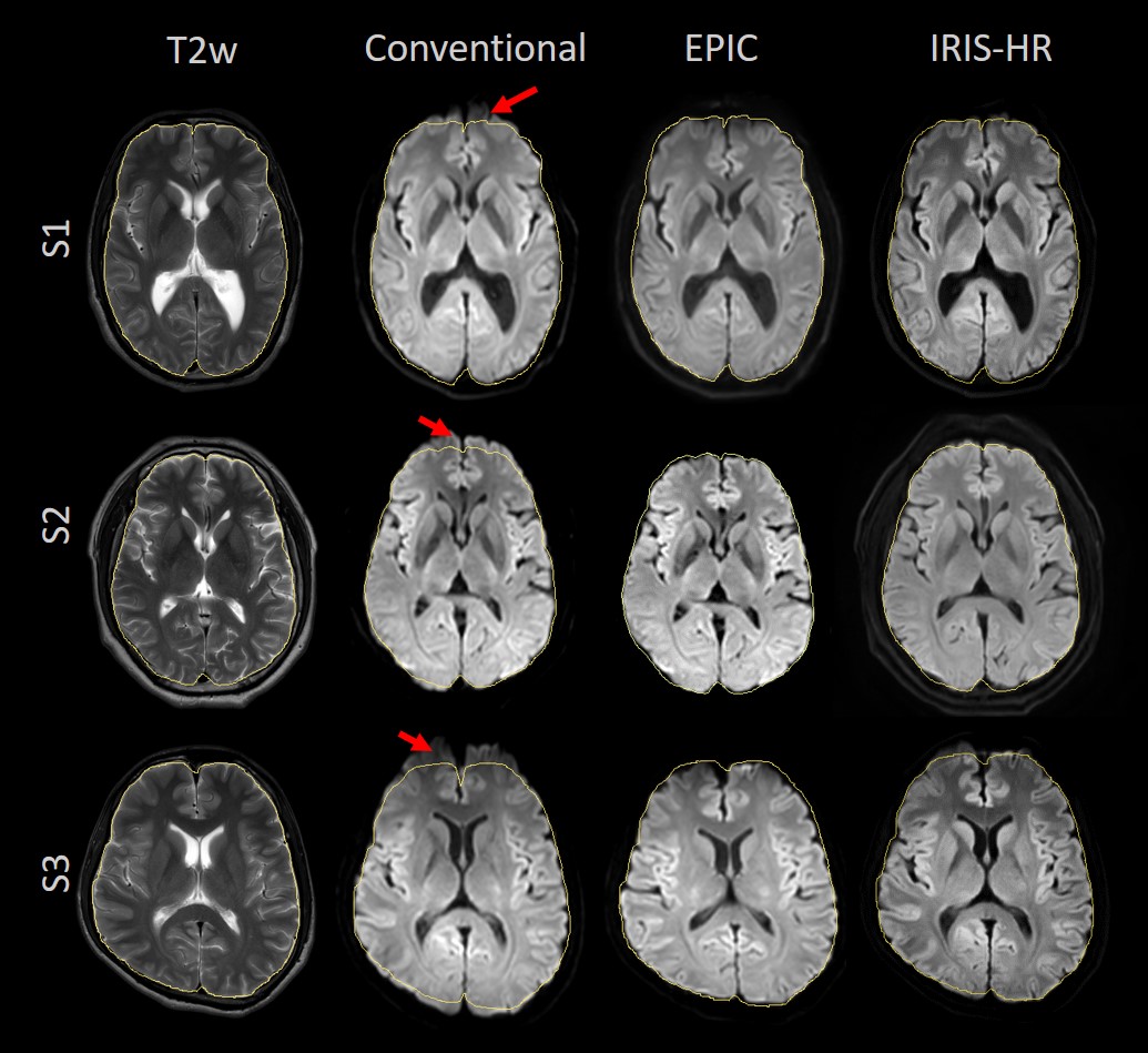
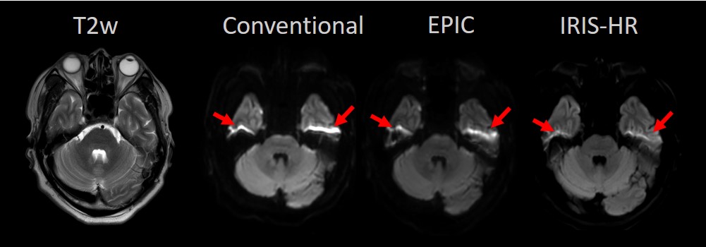
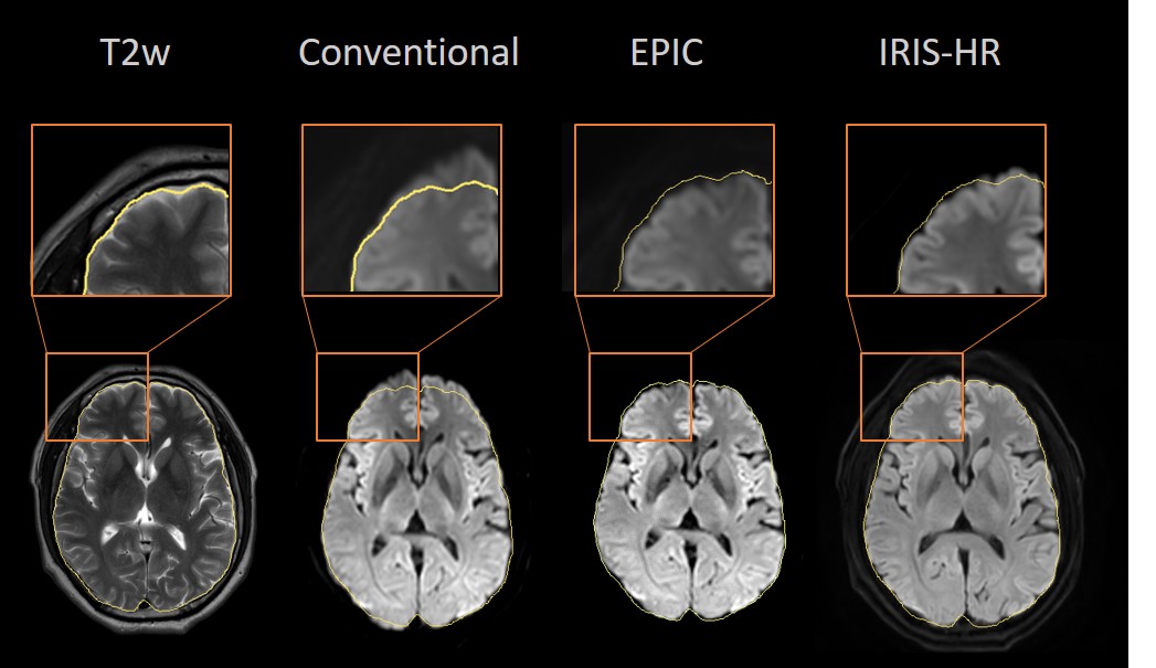
Figure 1 showed the images acquired using the conventional, EPIC and IRIS method with increased spatial resolution (i.e. IRIS-HR), with b = 0, 1000 s/mm2 respectively. Imaging time for the EPIC scan was 1min8s with in-plane resolution 1.6 x 2 mm2, and IRIS-HR 3min with in-plane resolution 1.2 x 1.2 mm2. The severe distortion from conventional method was observed in all cases (column 2, red arrow). EPIC reduced the geometric distortion in a large degree, and the IRIS method simultaneously reduce the distortion and increase the spatial resolution with comparable image quality. Figure 2 further showed the images acquired using the three strategies in the lower part of the brain. Moreover, the detailed observation was performed as shown in Figure 3, where the morphological information of the brain gyri were better presented with high resolution through the IRIS-HR result.Discussion
Both strategies can provide effective distortion reduced images. EPIC provides effective distortion corrected result within relatively short time, which has advantage on a few clinical applications where the patients cannot afford long lasting scans, and it is also useful for fMRI. On the other hand, IRIS is potential to achieve better morphological appearance with high spatial resolution diffusion with relatively longer but acceptable scanning time. EPIC and IRIS are both good solutions for high resolution diffusion with significant distortion reduction. The combination of EPIC and IRIS could be useful as a future direction.Acknowledgements
No.References
1. Wheeler-Kingshott, C.A.M., et al. Investigating Cervical Spinal Cord Structure Using Axial Diffusion Tensor Imaging. NeuroImage 2002, Vol16, Issue1: 93-102.
2. Chu ML, et al. POCS-Based reconstruction of multiplexed sensitivity encoded MRI (POCSMUSE): a general algorithm for reducing motion-related artifacts. Magn Reson Med 2014; DOI: 10.1002/mrm.25527
3. Jeong, H., et al. High resolution human diffusion tensor imaging using 2D navigated multi-shot SENSE EPI at 7 Tesla. Magn Reson Med. 2013, 69 (3): 793-802.
4. Andersson JLR, Skare S, Ashburner J, How to correct susceptibility distortions in spin-echo echo-planar images: application to diffusion tensor imaging, NeuroImage, 2003; 20(2):870-888.
Figures