2352
Estimating B0 changes in Oscillating Steady State Imaging (OSSI) using an Artificial Neural Network1Biomedical Engineering, University of Michigan, Ann Arbor, MI, United States, 2Electrical Engineering and Computer Science, University of Michigan, Ann Arbor, MI, United States
Synopsis
Oscillating steady state imaging (OSSI) is a novel fMRI acquisition method which produces a high SNR T2*-weighted signal. Physiological and drift induced B0 changes cause undesired signal distortions which can significantly diminish functional contrast in OSSI fMRI. Here, we investigated a method of rapidly estimating quantitative B0 field changes from OSSI images using an artificial neural network (ANN) model. Our results demonstrated that this technique can be used to rapidly measure field changes, which has the potential to be used for prospective and retrospective B0 field correction in OSSI.
Introduction
OSSI uses a quadratic phase progression in combination with balanced gradients to produce a high SNR oscillatory signal that oscillates with a periodicity of $$$n_c\times TR$$$1, where $$$n_c$$$ is the number of RF phase cycles used ($$$\sim$$$6-10).The oscillations can be combined across $$$n_c$$$ to produce a more uniform signal that is BOLD sensitive. The phase progression in OSSI induces a frequency-dependent phase dispersal that makes it sensitive to $$$T2^*$$$ changes and consequently ideal for high SNR fMRI 1. This frequency sensitivity also makes OSSI susceptible to respiration and drift which causes unwanted perturbations in $$$B_0$$$ resulting in a reduction in fMRI temporal resolution2. Given this sensitivity, quantitative information about the magnetic field distribution is essential for the reconstruction of high-fidelity OSSI fMRI images. A conventional approach for quantifying $$$B_0$$$ field is to measure the phase differences between gradient echo (GRE) images acquired at two different echo times. This requires an additional scan which may be too time consuming especially in prospective $$$B_0$$$ field compensation applications. Here, we used a deep fully-connected feed-forward neural network called OSSI-ANN to estimate dynamic field changes due to respiration and drift in OSSI images.Methods
[Simulations]We trained our neural network with Bloch simulated OSSI signals using the steady state magnetization OSSI model described in prior work1. We simulated 626 isochromats with frequencies ranging between $$$\frac{-1}{2TR}:\frac{1}{2TR}$$$Hz, and randomly sampled the parameters in Figure 2A to produce 4200 transverse magnetization timecourses.
[Phantom & In Vivo Experiments]
OSSI signals from phantom and human subjects were acquired using a single-slice, single-shot variable density spiral-out trajectory ($$$n_c$$$ =10, $$$TR$$$=16ms, flip angle =10$$$^\circ$$$, FOV =24x24cm$$$^2$$$, matrix size =92x92). For our phantom experiment, we modulated the $$$B_0$$$ field by physically rolling another uniform spherical phantom in and out of the scanner at 30,50,80 and 100s during a 2 minutes scan. For our fMRI human experiment, the functional task was a finger-tapping experiment where the human subject was asked to tap their fingers for 20s and rest for 20s. The task was repeated 5 times (200s).
[Neural Network Architecture]
Our neural network consists of one convolutional layer followed by five dense layers as shown in Figure 1. The input to our network is the OSSI signal and the output is the corresponding respiration and drift induced $$$B_0$$$ field changes. Each full simulation timecourse/voxel was segmented into multiple overlapping temporal patches of size 950x1. Each patch was further reshaped into $$$n_c$$$x95 ($$$n_c$$$=10) which served as the input of the network. The output size was 95x1. Ultimately, the output signal was recombined to form the original time course by using the Hann window function as shown in Figure 2B and 2C.
Results
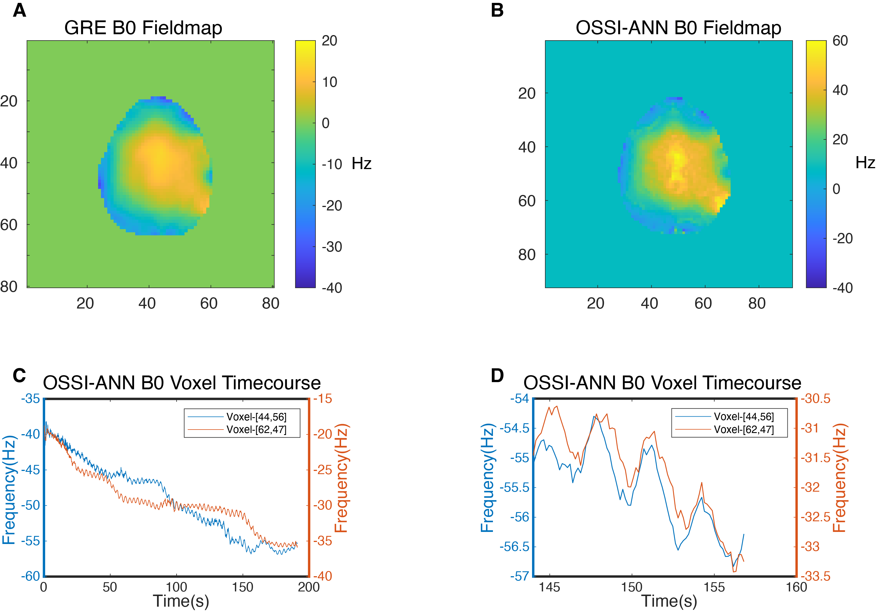
Figure 3 show examples of OSSI-ANN timecourses from simulated test data compared to the ground truth. Our OSSI-ANN results were in good agreement with the ground truth in all unseen test examples. Figure 4A and 4B compare $$$B_0$$$ fieldmap from the double-echo gradient echo (GRE) field estimation technique to OSSI-ANN fieldmap. The spatial profile of the OSSI-ANN $$$B_0$$$ field is similar to the GRE fieldmap with a constant shift in frequency. Figure 4C shows off-resonance timecourses from the OSSI-ANN fieldmap indicating field modulations (at approximately 30,50,80 and 100s) as a result of physically rolling another phantom near the phantom we were imaging. Figure 5A and 5B show the double-echo GRE $$$B_0$$$ map versus OSSI-ANN $$$B_0$$$ map in a human subject, as in the phantom, the spatial pattern is similar for both methods except that they differ by a constant frequency offset($$$\sim$$$25Hz). Figure 5C shows off-resonance timecourses for two voxels generated from the OSSI-ANN. OSSI-ANN captures the respiratory cycle and drift, as can be seen more clearly in Figure 5D (zoomed in version of 5C).Discussion & Conclusion
In this study, we demonstrated the feasibility of using OSSI-ANN to directly measure dynamic $$$B_0$$$ field changes from voxels of OSSI images without the need for an additional scan. Our trained network provided quality fieldmaps that are comparable to the GRE fieldmaps with high sensitivity to field changes as depicted in the results from our in-vivo experiments. Future work will focus on incorporating our field measurements in a retrospective/prospective $$$B_0$$$ field compensation scheme3 to correct for respiration induced field distortions in OSSI functional images.Acknowledgements
We wish to acknowledge the support of NIH Grant U01EB026977.References
[1] Shouchang Guo and Douglas C Noll. Oscillating steady-state imaging (ossi):A novel method for functional mri. Magnetic Resonance in Medicine,84(2):698–712, 2020.
[2] Amos A Cao and Douglas C Noll. A retrospective physiological noise correction method for oscillating steady-state imaging. Magnetic Resonance in Medicine, 85(2):936–944, 2020.
[3] Wallace TE, Afacan O, Kober T, Warfield SK. 2020 Rapid measurement and correction of spatiotemporal B0 field changes using fid navigators and a multi-channel reference image.Magnetic resonance in medicine 83, 2, 575–589
Figures
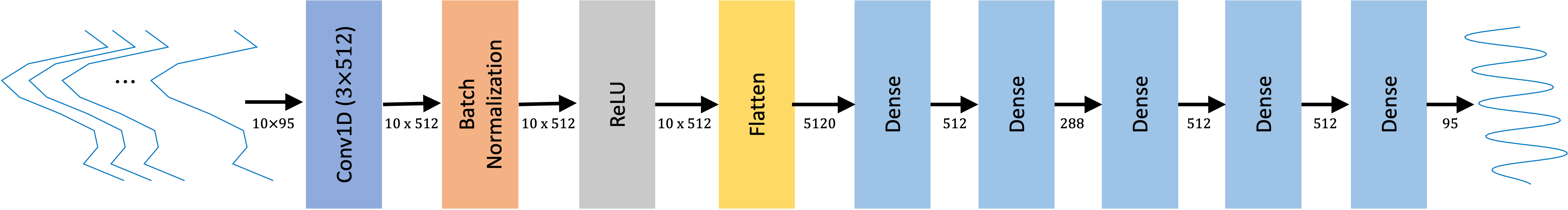
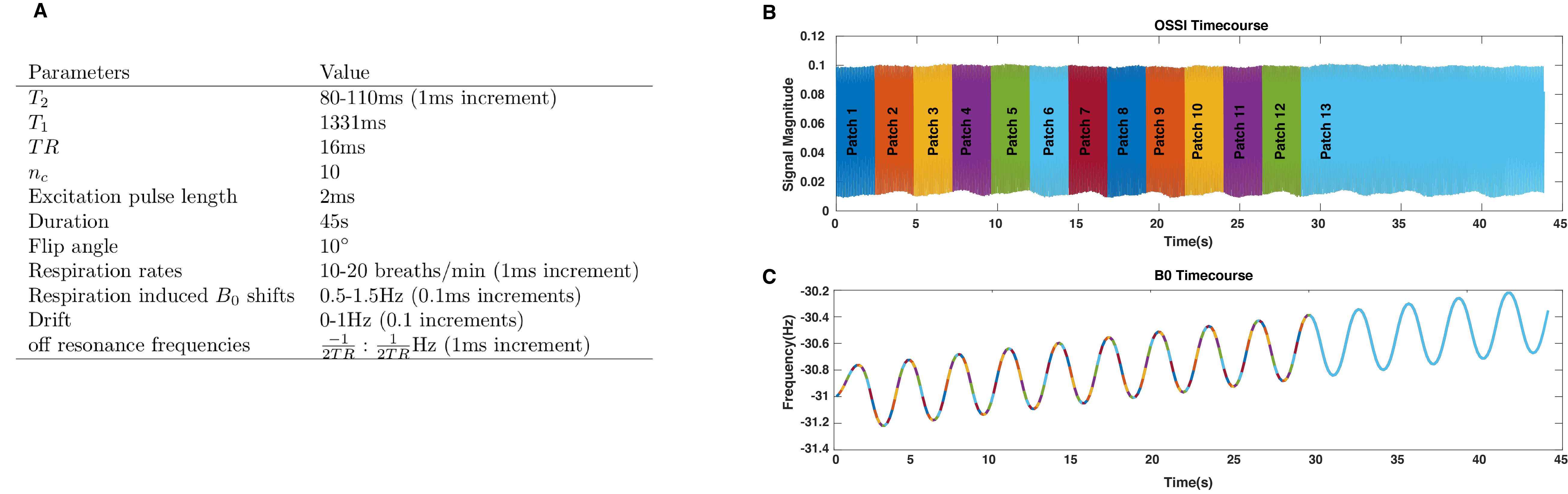
Figure 2: A, Sequence parameters that we used for simulating the training data. B shows an example of the OSSI temporal patches(network input) and the recombined B0 temporal patches (network output) in C.
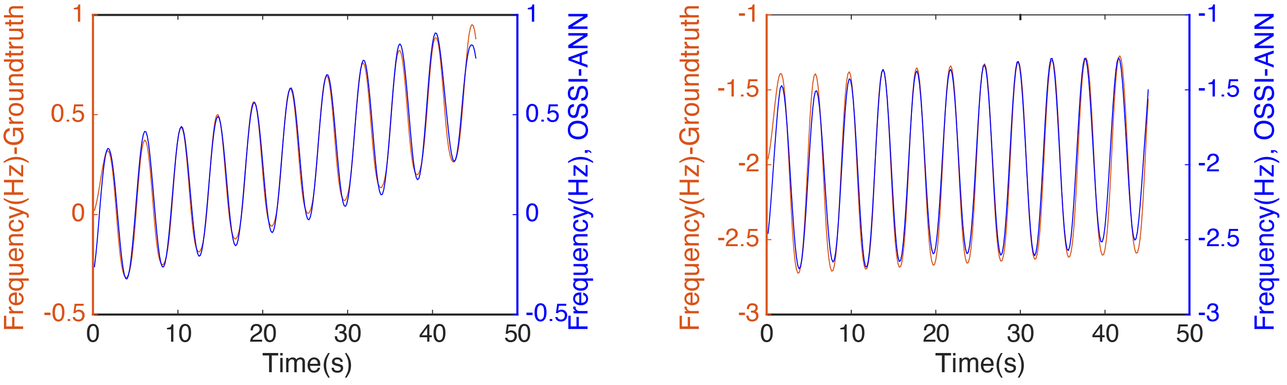
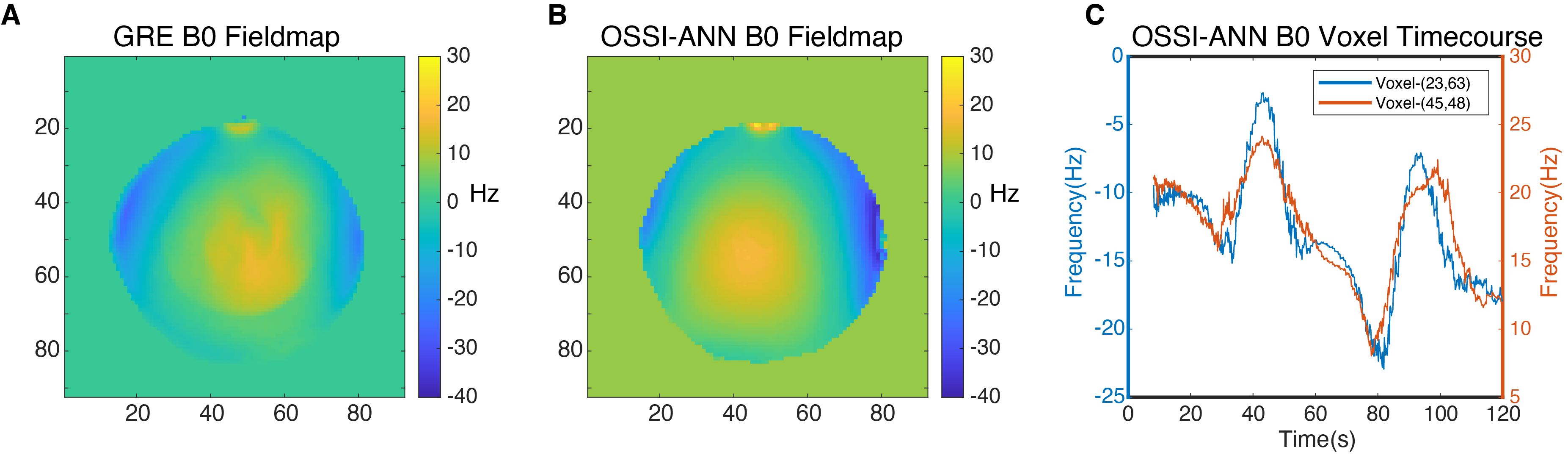
Figure 4: Results from phantom experiments. A, Field map from double-echo GRE field estimation method where TE1=0ms and TE2 =2.3ms. B, Field map from OSSI-ANN. C, B0 time course from selected voxels illustrating signal changes at ~30,50,80 and 100s.