2343
SENSE-based multipeak water/fat separation for diffusion-weighted imaging using self-navigated interleaved EPI1C.J. Gorter Center for High Field MRI, Department of Radiology, LUMC, Leiden, Netherlands, 2Division of Image Processing, Department of Radiology, LUMC, Leiden, Netherlands, 3University and ETH Zurich, Zurich, Switzerland, 4Philips Research, Hamburg, Germany
Synopsis
The presence of fat signals is one challenge for diffusion-weighted EPI, especially when considering the multi-peak spectrum nature of fat. In this work, we propose an improved SENSE-based water/fat separation algorithm to suppress multi-peak fat signals and apply this specifically to diffusion-weighted multi-shot EPI. The motion-induced shot-to-shot phase variations, an inevitable challenge in multi-shot DWI, are incorporated into the signal model using either a self-navigation or an extra-navigated method. The results show that the proposed SENSE-based algorithm yields good water/fat separation for non-diffusion and diffusion data with a multi-peak fat spectrum model.
Introduction
Multi-shot interleaved EPI (msh-EPI) provides significant improvement for diffusion-weighted imaging (DWI) to allow images with higher spatial resolution and less geometric distortions1. However, multi-shot acquisitions for DWI applications are susceptible to physiological motion-induced phase variations between the shots. This difficulty is usually addressed by 1) acquiring additional navigator images1,2, or 2) employing self-navigation approaches3,4. Moreover, residual fat artefacts represent another difficulty in DWI due to the large water-fat shift in phase-encoding direction. Fat suppression5 techniques, widely used for eliminating fat, often fail for large B0 inhomogeneities. Moreover, they do not fully consider the multipeak nature of the fat spectrum (e.g., Olefinic peak, 0.61 ppm to water). Recently, chemical-shift encoding has been proposed to jointly separate water and fat images while estimating the B0 inhomogeneities6-8. Nevertheless, the need to acquire images at multiple TEs may limit its efficiency in clinical applications. Alternatively, Uecker et al. has proposed an approach exploiting the chemical-shift-induced spatial shift of fat using sensitivity encoding (SENSE) to disentangle water and fat signals. The method is validated with a single-peak fat model and single-shot EPI images.In this work, we propose to include the multipeak nature of the fat spectrum into the approach by Uecker et al.9 to separate water and fat in EPI based on SENSE. This approach is extended to multi-shot, in-vivo DWI using a novel water-fat phase self-navigation method to compensate for motion-induced phase errors, making the need for measuring an extra-navigator echo obsolete.
Methods
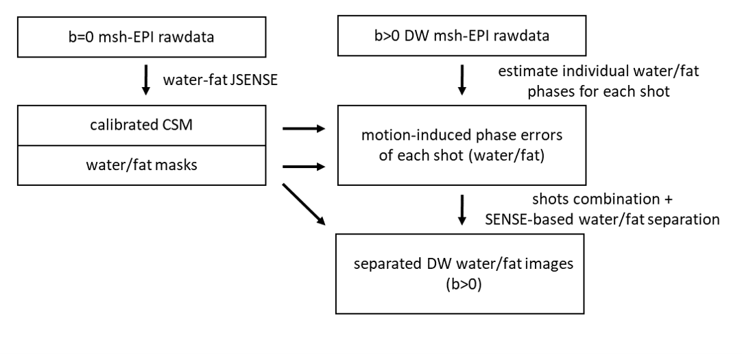
The forward model of the DW msh-EPI data at a certain b-value can be written as:$$s=\sum_{n}^{N}Q_{n}\left(\hat{F}\hat{C}\hat{\Phi}_{w,n}\rho_{w}+\alpha_{m}\sum_{m=1}^{M}\hat{\Psi}_{f,m}\hat{F}\hat{C}\hat{\Phi}_{f,n}\rho_{f}\right)\tag{1}$$where $$$\hat{F}$$$ is the Fourier transform operator, $$$\hat{C}$$$ the SENSE operator, $$$\hat{\Phi}_{w,n}$$$ and $$$\hat{\Phi}_{f,n}$$$ the operators of motion-induced phase errors for water and fat, respectively. $$$\alpha_{m}$$$ is the relative amplitude weight, $$$\hat{\Psi}_{f,m}$$$ denotes the fat off-resonance operator for peak $$$m$$$, and $$$Q_{n}$$$ represents the k-space trajectory for each shot $$$n$$$. $$$s$$$ and $$$\rho_{w}/\rho_{f}$$$ are the vectorized representations of the measured multi-shot k-space signal and the target water/fat images. The water/fat images can be calculated by minimizing:$$\left\{\rho_{w},\rho_{f}\right\}=\underset{\rho_{w},\rho_{f}\in\mathbb{C}}{\operatorname{argmin}}\|\hat{A}X-s\|_{2}^{2}\tag{2}$$where $$$X=\left[\rho_{w},\rho_{f}\right]^{T}$$$ and $$$\hat{A}$$$ is the coefficient matrix, containing all the operators described above. For multi-shot acquisition, the relatively small spatial shift of fat compared to single-shot EPI makes the accuracy of coil-sensitivity maps (CSM) particularly important. Therefore, the advanced JSENSE described in Shin et al.10 is used to self-calibrate the CSM for b=0 s/mm2. Two threshold water/fat masks can be calculated using the separated $$$\rho_{w}$$$ and $$$\rho_{f}$$$ images to mask the CSM and stabilize the phase estimation step.For the diffusion-weighted cases, the SENSE-based water/fat separation is performed for each shot data to calculate motion-induced phase errors for water/fat individually. Inspired by MUSE3, the water/fat-adapted SENSE formalism is used to recover ghost-free images for each shot. First, this can be done by solving the equation:$$s_{n}=Q_{n}\left(\hat{F}\hat{C}\rho_{w,n}+\alpha_{m}\sum_{m=1}^{M}\hat{\Psi}_{f,m}\hat{F}\hat{C}\rho_{f,n}\right)\tag{3}$$using conjugate gradient (CG), to estimate $$$\rho_{w,n}$$$ and $$$\rho_{f,n}$$$ for each individual shot $$$n$$$ from the measured data. Then, the motion-induced phase $$${\Phi}_{w,n}$$$ and $$${\Phi}_{f,n}$$$ can be calculated separately for water and fat by:$$e^{i\phi_{w/f,n}}=\frac{\rho_{w/f,n}}{\left|\rho_{w/f,n}\right|}.\tag{4}$$In addition, k-space triangular window11 is applied individually on $$$e^{i\phi_{w,n}}$$$ and $$$e^{i\phi_{f,n}}$$$ to enforce smoothness and construct the operators $$$\hat{\Phi}_{w,n}$$$ and $$$\hat{\Phi}_{f,n}$$$ in Eq.1. The window width is chosen to be half of the matrix size. Finally, the DW water/fat images can be calculated by solving Eq. 2 using CG. The whole pipeline is illustrated in figure 1.
DW msh-EPI spin-echo data of the lower leg of a normal volunteer was acquired using a 3T-MRI (Philips Healthcare, Best, Netherlands) with the following parameters: TR/TE=2000/60ms, resolution 1.5×1.5×4 mm3, three b-values (0, 300, 600 s/mm2), 3 shots, an 8-channel knee coil. For comparison, an extra-navigator2 was acquired for each shot. In addition, another fat-suppressed DWI scan was acquired using SPIR5 and reconstructed through IRIS algorithm2.
Results and Discussion
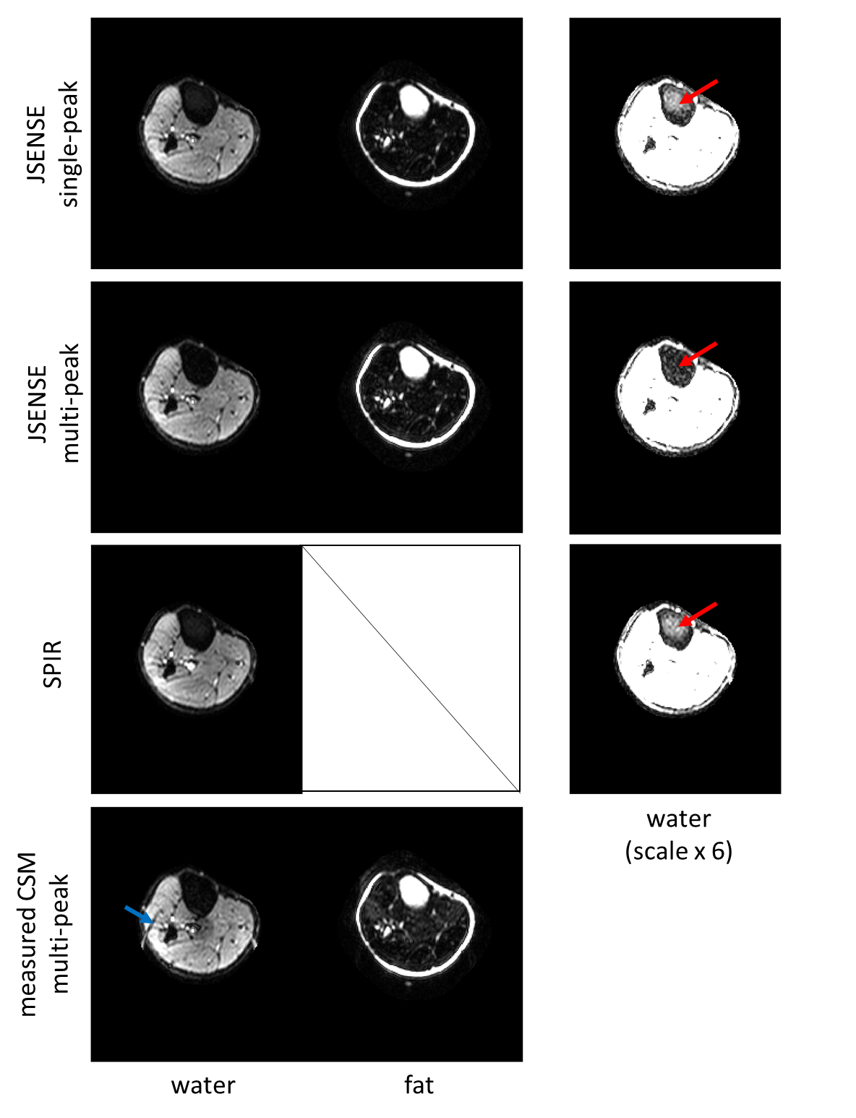
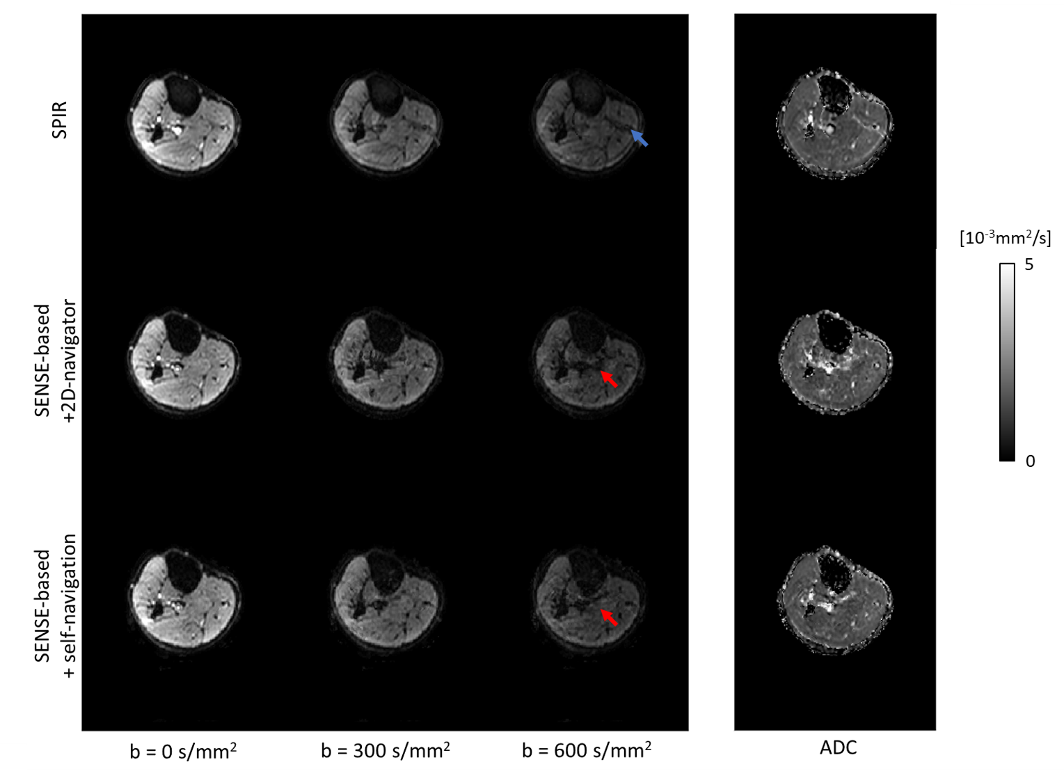
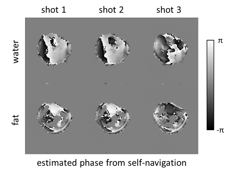
Figure 2 shows the non-diffusion dataset comparing the JSENSE-calibrated CSM with single-peak/multi-peak8 fat model and the SPIR scan. The result for a conventional pre-scan CSM with a multi-peak fat model is shown for comparison. The JSENSE results show improved water images compared to the pre-scan for both spectral fat models. This is mainly due to the geometric distortion mismatch between the EPI images and the reference scan. Besides, the water images with adjusted level/window show the capability of the algorithm to deal with the multi-peak fat spectrum, which is superior to fat suppression by SPIR. Figure 3 shows water images with different b-values and the associated ADC maps for (1) SPIR, (2) the SENSE-based water/fat separation with extra-navigators and (3) with self-navigation. SPIR shows an unsuppressed fat rim, probably arising from strong B1+ inhomogeneities. Some artefacts can be seen in the SENSE-based water image with extra-navigators, which are not present for the self-navigated results. Figure 4 shows the estimated water/fat phases derived for self-navigation.Conclusion
The proposed method allows for an efficient SENSE-based water/fat separation of non-DW and DW msh-EPI data. The SENSE-based solution requires only one acquisition for each b-value to separate water/fat with a multi-peak fat model. This directly increases the flexibility and is important when different b-values/diffusion directions are desired. Future studies will include undersampling for acceleration and use of phased array coils with more channels to support higher segmentation factors.Acknowledgements
The authors would like to acknowledge NWO-TTW (HTSM-17104).References
1. Butts, K., Pauly, J., De Crespigny, A. and Moseley, M. (1997), Isotropic diffusion-weighted and spiral-navigated interleaved EPI for routine imaging of acute stroke. Magn. Reson. Med., 38: 741-749.
2. Jeong H-K, Gore JC, Anderson AW. High-resolution human diffusion tensor imaging using 2-D navigated multishot SENSE EPI at 7 T. MRM. 2013;69(3):793-802.
3. Chen NK, Guidon A, Chang HC, Song AW. A robust multi-shot scan strategy for high-resolution diffusion weighted MRI enabled by multiplexed sensitivity-encoding (MUSE). Neuroimage. 2013 May 15;72:41-7.
4. Guo H, Ma X, Zhang Z, Zhang B, Yuan C, Huang F. POCS-enhanced inherent correction of motion-induced phase errors (POCS-ICE) for high-resolution multishot diffusion MRI. Magn Reson Med. 2016 Jan;75(1):169-80.
5. Zee CS, Segall HD, Terk MR, et al. SPIR MRI in spinal diseases. J Comput Assist Tomogr 1992;16:356-360.
6. Hernando D, Karampinos DC, King KF, Haldar JP, Majumdar S, Georgiadis JG, Liang ZP. Removal of olefinic fat chemical shift artifact in diffusion MRI. Magn Reson Med. 2011 Mar;65(3):692-701.
7. Burakiewicz J, Charles-Edwards DG, Goh V, Schaeffter T. Water-fat separation in diffusion-weighted EPI using an IDEAL approach with image navigator. Magn Reson Med. 2015 Mar;73(3):964-72.
8. Dong Y, Koolstra K, Riedel M, van Osch MJP, Börnert P. Regularized joint water–fat separation with B0 map estimation in image space for 2D-navigated interleaved EPI based diffusion MRI. Magn Reson Med. 2021; 00: 1– 18. https://doi.org/10.1002/mrm.28919
9. Uecker, M. & Lustig, M. Making SENSE of Chemical Shif: Separating Species in Single-Shot EPI using Multiple Coils. In Proc. Intl. Soc. Mag. Reson. Med., 20, 2490 (Melbourne, 2012).
10. Shin PJ, Larson PE, Uecker M, Reed GD, Kerr AB, Tropp J, Ohliger MA, Nelson SJ, Pauly JM, Lustig M, Vigneron DB. Chemical shift separation with controlled aliasing for hyperpolarized (13) C metabolic imaging. Magn Reson Med. 2015 Oct;74(4):978-89.
Figures