2329
High resolution Multi-voxel spectroscopy using CSI-semi-LASER for mouse brain preclinical studies1Department of Biochemistry and Molecular Biology, Universitat Autonoma de Barcelona, Bellaterra, Spain, 2Servei de Ressonància Magnètica Nuclear, Universitat Autonoma de Barcelona, Bellaterra, Spain, 3Department of Imaging Physics, University of Texas, Houston, TX, United States, 4Centro de Investigación Biomédica en Red en Bioingeniería, Biomateriales y Nanomedicina (CIBER-BBN),, Universitat Autonoma de Barcelona, Bellaterra, Spain, 5Institut de Biotecnologia i de Biomedicina (IBB), Universitat Autonoma de Barcelona, Bellaterra, Spain
Synopsis
This work describes an improved multi-voxel Chemical shift imaging (CSI) pulse sequence that uses the semi-LASER localization approach. Bruker CSI sequence containing PRESS localization block (CSI-PRESS) was modified by replacing the aforementioned PRESS block with a semi-LASER one resulting in a CSI-semi-LASER sequence. This work was developed on a 7T Bruker BioSpec 70/30 USR spectrometer running ParaVision 5.1 which does not contain neither built-in blocks for semi-LASER sequence nor adiabatic pulses. The sequence was tested in wt C57/BL6 mouse brain and compared with the stock CSI-PRESS sequence. The results show improved homogeneity and reduction in chemical shift displacement error (CSDE).
INTRODUCTION
Proton (1H) Chemical Shift Imaging (CSI) is a versatile diagnostic technique that provides non-invasive metabolic information about living tissues. Our group has a long track of using CSI-based nosological images to provide metabolomic information for treatment monitoring in murine brain tumors [1]. However, tumor inhomogeneities and the small mouse brain volume may compromise spectrum quality when using the Bruker commercially available PRESS-CSI sequences. The semi-LASER sequence is a robust localization technique for 1H-MRS and can be used as an alternative to PRESS sequence for single voxel localization [2]. In the semi-LASER sequence, the single voxel localization is achieved by using two pairs of adiabatic full passage (AFP) hyperbolic secant refocusing pulses instead of the two 180o refocusing pulses for PRESS. For the same amount of volume excitation, the adiabatic refocusing pulses perform uniform refocusing of the spins producing a larger net magnetization vector [3]. The semi-LASER sequence was implemented in-house in a 7T Bruker BioSpec 70/30 USR using ParaVision 5.1 for single voxel and multivoxel spectroscopy. Our goal was to increase signal to noise ratio (SNR) for the acquired spectra, which may allow us to increase the spatial resolution, as well as to have better spatial distribution homogeneity in order to improve pattern spectral classification in our murine brain tumor models.METHODS
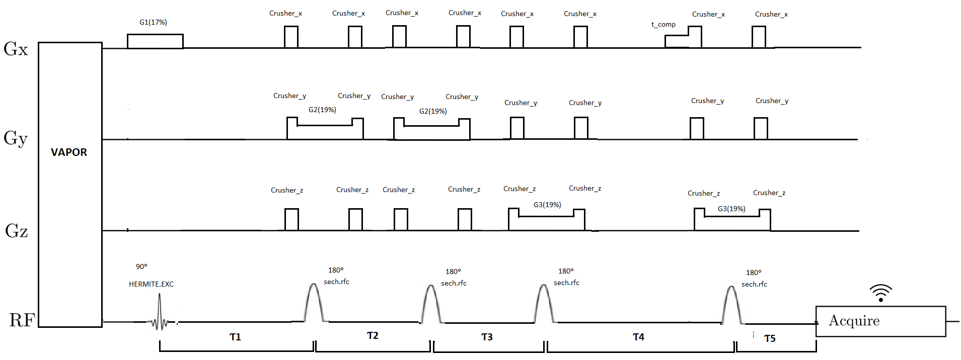
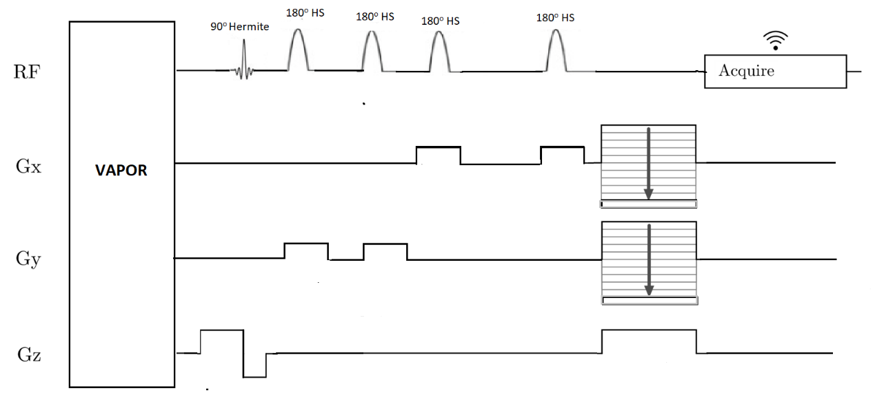
All experiments were performed on a 7T Bruker Biospec 70/30 USR preclinical scanner using ParaVision 5.1 (Bruker BioSpin GmbH, Ettlingen, Germany) equipped with a mini-imaging gradient set (400 mT/m). For mouse brain in vivo studies, a 72-mm inner diameter linear volume coil was used as transmitter and a dedicated mouse brain surface coil as receiver. For the implementation of the semi-LASER sequence, the PRESS localization block was replaced by a semi-LASER localization block (Figure 1). For excitation, 90o Hermite pulses were used in both PRESS and semi-LASER sequences. For refocusing, 180o adiabatic full passage Hyperbolic secant pulses were used in the semi-LASER block instead of the conventional Hermite pulses. The spin echo condition was maintained if Ƭ1+Ƭ3+Ƭ5 = Ƭ2+Ƭ4, with time delays defined in Figure 1. In our implementation, we kept the intervals Ƭ2= Ƭ3 for all values of TE, hence the spin echo condition was satisfied when Ƭ5=Ƭ4-Ƭ1. A pair of crusher gradients were also positioned symmetrically around AFP pulses in order to suppress unwanted signals. The 180o refocusing adiabatic pulses were generated using the TopSpin stdisp tool. In PRESS, the excitation pulse bandwidth (BW) = 9 kHz, pulse length = 600 µs, flip angle = 90o and refocusing pulse bandwidth (BW) = 5.7 kHz, pulse length = 600µs and flip angle = 180o. In the semi-LASER sequence, the excitation pulse is the same as PRESS. The two pairs of refocusing pulses have bandwidth (BW) = 9 kHz, pulse length = 2000 µs and flip angle = 180o. The CSI-semi-LASER pulse sequence was implemented likewise but incorporating stepped phase-encoding gradients along 2 axes, as shown in figure 2.RESULTS
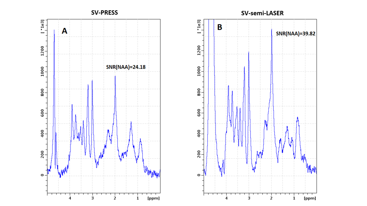
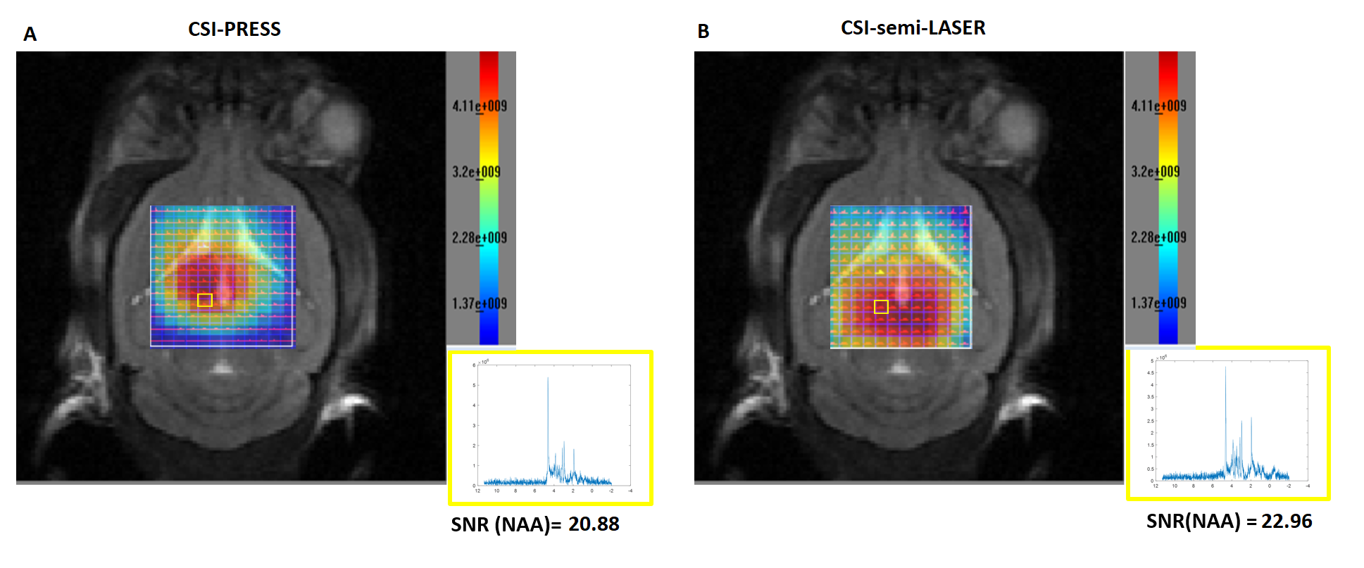
Figure 3 shows the comparison of single voxel PRESS versus semi-LASER acquisitions in a wt C57/BL6 mouse brain. Water was sufficiently suppressed in both sequences using a VAPOR water suppression block, and both sequences provided accurately localized spectra. Semi-LASER spectra showed a 1.6-fold SNR increase compared to PRESS. CSI-semi-LASER was also successfully implemented and a mouse brain study (figure 4). CSI grids overlaid on T2-weighted mouse brain MRI for PRESS and semi-LASER are shown in panels A and B respectively and insets show representative spectra from the same location. A SNR increase of 9.9% with respect to PRESS CSI was observed. A reduced chemical shift displacement error (CSDE) is obtained in CSI semi-LASER. For the NAA peak at 2.0 ppm, the CSDE was calculated in the x, y and z directions and values found were 9%, 8.9% and 8.9% for the CSI-semi-LASER and 9%, 14.2% and 14.2% for CSI-PRESS. Moreover, the CSI-semi-LASER showed a more spatially homogenous profile when compared to the CSI-PRESS sequence as it can be seen in the NAA metabolic maps (Figure 4).DISCUSSION
Single voxel and CSI-semi-LASER sequences were successfully implemented at 7T for ParaVision 5.1. The implemented CSI-semi-LASER sequence provided superior quality spectra when compared to the conventional CSI-PRESS sequence, as a result of the better excitation profile of the adiabatic refocusing pulses and the reduction in chemical shift displacement errors. With sharp selection profiles and a small CSDE, voxels closer to the edge of the VOI will be suitable evaluation, which is not the current situation. Furthermore, the relative insensitivity to B1 inhomogeneities offered by the adiabatic refocusing pulses is an additional advantage when using the semi-LASER technique as a substitute to the conventional CSI-PRESS sequence. The higher signal-to-noise ratio (SNR) of the CSI-semi-LASER sequence, when compared to the conventional CSI-PRESS, could be used to achieve a higher resolution or to reduce the acquisition time.CONCLUSION
The implemented CSI-semi-LASER sequence for Bruker Biospec preclinical scanner with Paravision 5.1 is suitable for high resolution multi-voxel spectroscopic acquisitions in mouse brain studies, providing an alternative to the conventional CSI-PRESS sequence, and producing spectra with improved resolution and higher SNR. Further work will be devoted to make these sequences available to be fully tested elsewhere.Acknowledgements
This work is funded by the INSPiRE-MED project 2019-2022 (H2020/MCSA COFUND, Grant agreement ID: 813120
All studies involving animals were approved by the local ethics committee, according to regional and state legislations (protocol references CEA-OH-9685/CEEAH-3665).
References
1. Arias-Ramos N, Ferrer-Font L, Lope-Piedrafita S, Mocioiu V, Julià-Sapé M, Pumarola M, Arús C, Candiota AP. Metabolomics of Therapy Response in Preclinical Glioblastoma: A Multi-Slice MRSI-Based Volumetric Analysis for Noninvasive Assessment of Temozolomide Treatment. Metabolites. 2017 May 18;7(2):20
2. Weiss, K., Melkus, G., Jakob, P.M. et al. Quantitative in vivo 1H spectroscopic imaging of metabolites in the early postnatal mouse brain at 17.6 T. Magn Reson Mater Phy 22, 53 (2009). https://doi.org/10.1007/s10334-008-0142-2
3. Scheenen, T.W.J., Klomp, D.W.J., Wijnen, J.P. and Heerschap, A. (2008), Short echo time 1H-MRSI of the human brain at 3T with minimal chemical shift displacement errors using adiabatic refocusing pulses. Magn. Reson. Med., 59: 1-6. https://doi.org/10.1002/mrm.21302
Figures