2319
Development of in vivo dynamic nuclear polarization (DNP)-MRI at 16 mT for large animals and redox metabolic imaging of acute hepatitis rat model1Gifu University, Gifu, Japan, 2Kyushu University, Fukuoka, Japan, 3Japan REDOX Limited, Fukuoka, Japan
Synopsis
To use the current methods in clinical practice, the development of a prototype in vivo DNP-MRI system for preclinical examinations of large animals is indispensable for clarifying the problems peculiar to the increase in size of the DNP-MRI device. Therefore, we developed a in vivo DNP-MRI (Overhauser MRI) system with a sample bore size of 20 cm and a 16-mT magnetic field using a U-shaped permanent magnet. The in vivo DNP-MRI system developed was used to non-invasively image the redox reaction of a carbamoyl-PROXYL probe in the livers of large rats weighing 800 g and hepatitis-model rats.
Introduction
In in vivo DNP-MRI, also known as Overhauser enhanced MRI (OMRI) or proton-electron double resonance imaging (PEDRI), EPR irradiation at the resonant frequency of the in vivo free radical molecule induces DNP causing increased MRI signal. It is necessary to apply a resonant frequency for electron spin that is 658 times higher than that for nuclear spin because of the higher magnetic moment of the unpaired electrons. For eventual clinical application, it is necessary to carry out preclinical studies using larger animals to demonstrate the proof of concept (POC). Therefore, it is essential to develop DNP-MRI systems for larger animals to clarify the problems unique to large devices. In this study, we developed a 20-cm sample-bore DNP-MRI system using a permanent magnet with a low magnetic field (16 mT) that employs the excitation of electron and proton nuclear spins under the same magnetic field. Using this system, we succeeded in obtaining DNP contrast and visualizing the redox reaction in the liver of large rats of around 800 g in mass. Furthermore, we successfully visualized redox alterations in the livers of hepatitis-model rats.Methods
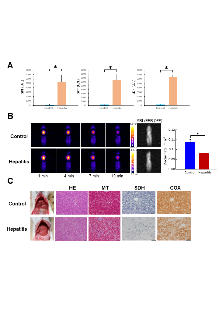
We developed a in vivo DNP-MRI system with a sample bore size of 20 cm and a 16-mT magnetic field using a U-shaped permanent magnet. Because the NMR frequency is very low, we adopted a digital radiofrequency transmission/reception system with excellent filter and dynamic range characteristics and equipped with a digital eddy current compensation system to suppress large eddy currents. The pulse sequence was based on the fast spin-echo sequence, which was improved for low frequency and large-eddy current equipment.Two EPR irradiation coils of the modified Alderman Grant (MAG) type were designed for body imaging of rats on the DNP-MRI system, as shown in Figure 2. The elliptical-form coils had a major axis diameter of 90 mm, a minor axis diameter of 60 mm, and a central parallel part length of 46 mm, having enough space to insert a rat weighing 800 g. In vivo imaging of large-size rats was performed using the L-size MAG coil for EPR irradiation. DNP-MRI of the upper abdomen was started immediately after intravenous administration of carbamoyl-PROXYL solution (300 mM, 9 mL/g body mass). Pharmacokinetic DNP-MRI images were obtained at 2.0, 8.5, 15.0, and 21.5 minutes after injection. Finally, a normal MRI image was obtained without EPR irradiation. Acute hepatitis rat models were created according to previous literature. Rats under isoflurane anesthesia (2%) were intraperitoneally injected with 400 mg/kg D-GaIN and 50 µg/kg LPS in saline. In vivo DNP-MRI was performed 19–24 hours later. Untreated normal rats were used as controls. The rats were fasted overnight before the imaging study.Results
We first evaluated the homogeneity of MRI images obtained by 16-mT DNP-MRI using a large uniform phantom. The area where the image intensity was consistently 90% of the maximum value in the x, y, and z direction measured 59.4 × 93.8 × 115.6 mm, respectively. The overall homogeneity of the ROI calculated using the NEMA method was 5.9% for the y-axis direction, 7.4% for the z-axis direction, and 4.9% for the x-axis direction. To evaluate the DNP enhancement efficacy and the enhanced region on the two sizes of MAG coils, phantom imaging was performed using a 200-g phantom containing 2 mM carbamoyl-PROXYL solution. The DNP-enhanced regions were shown to be 71.9 mm in the x-axis direction and 50.0 mm in the z-axis direction for the L-size MAG coil, and 56.3 mm in the x-axis direction and 40.6 mm in the z-axis direction for the R-size MAG coil. This indicated that the DNP enhancement coverage of either of the coils was sufficient to monitor rat liver. The enhancement efficacy (EPR ON/OFF image) of the L-size MAG coil was 16.65 (±0.35) times, and that of the R-size MAG coil was 36.18 (±0.34) times, without heating. The DNP images showed negative enhancement of image intensity in the liver region of large rats. Difference images were created by subtracting the EPR ON image from the EPR OFF image, and these clearly showed that the enhancing region represented the liver. The enhancement decreased over time because of radical reduction of carbamoyl-PROXTYL. Redox maps were made from the four DNP kinetic images. In the DNP-MRI, the DNP images of rats showed a positive enhancement of image intensity region in the upper abdomen with a surrounding region showing negative enhancement of image intensity. In the DNP-MRI, the DNP images of rats showed a positive enhancement of image intensity region in the upper abdomen with a surrounding region showing negative enhancement of image intensity.Disucussion
The advantages of using a permanent magnet include its low maintenance cost, the possibility of using a highly-sensitive solenoid receiver coil, and the ability to install the equipment in any laboratory because it does not require a high-precision magnetic shield. Furthermore, because the same external magnetic field is used for excitation of electrons and nuclear spin, there is no loss of the DNP effect due to switching of the external magnetic field, and the DNP effect can be maximized.Acknowledgements
This work was supported by Medical field research results development business (Advanced measurement analysis technology/Device development program) from the Japan Agency for Medical Research and Development, AMED Grant Number 19hm0102038h0004.
References
Hinako Eto, Tatuya Naganuma , Motonao Nakao, Masaharu Murata, Abdelazim Elsayed Elhelaly, Yoshifumi Noda, Hiroki Kato , Masayuki Matsuo, Tomohiko Akahoshi, Makoto Hashizume, Fuminori Hyodo* Free Radic Biol Med. 2021 Jun;169:149-157. doi: 10.1016/j.freeradbiomed.2021.04.017. Epub 2021 Apr 15.Figures