2310
Clinical assessment of the degenerative lumbar osseous morphology using zero echo time magnetic resonance imaging (ZTE-MRI)1Radiology, Tongji Hospital, Tongji Medical College, Huazhong University of Science and Technology, Wuhan, Hubei Province, China, China, 2GE Healthcare, Beijing, China
Synopsis
Lumbar degenerative disease increases with age and is common in the elderly population. The symptoms caused by the osseous degeneration affect the quality of life. Zero echo time (ZTE) sequence could capture and reveal short-T2 materials, allowing to obtain osseous information without ionizing radiation. The ZTE performed similarly to multi-slice computed tomography (MSCT) on the ordinal variables and most measurements of osseous spine. ZTE-MRI could offer more cortical bone details than conventional MRI images and might be a valid alternative to CT for assessment of lumbar osseous morphology to some extent.
Introduction
Lumbar degenerative disease increases with age and is common in the elderly population. The symptoms caused by the osseous degeneration affect the quality of life1. Radiographic manifestations serve as a link to symptoms and clinical findings for degeneration diagnosis. Multi-slice computed tomography (MSCT) is used for surgical planning and more detailed assessment of osseous morphology although radiation exposure poses a threat to health. Cortical bone shows no signal on conventional MRI images due to highly-organized structures with low-proton contents2. An advanced MRI technique, zero echo time (ZTE) sequence, could capture and reveal short-T2 materials, allowing to obtain osseous information without ionizing radiation. The purpose of this study was to determine and compare the performance of ZTE and conventional MRI sequences on lumbar osseous morphology in patients who were suspected with lumbar degeneration with MSCT as standard reference.Materials and Methods
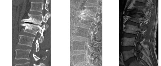
This study was approved by the institutional review board in our hospital and written informed consent was obtained. 22 subjects with concerned lumbar degeneration were recruited and scanned with ZTE sequence after routine conventional MR sequences on a 3.0T MR (Signa Pioneer, GE Healthcare) and also received MSCT examination. The image quality as well as quantitative and qualitative assessment of lumbar osseous morphology on MSCT, ZTE and MRI images were evaluated by three musculoskeletal radiologists independently.Results
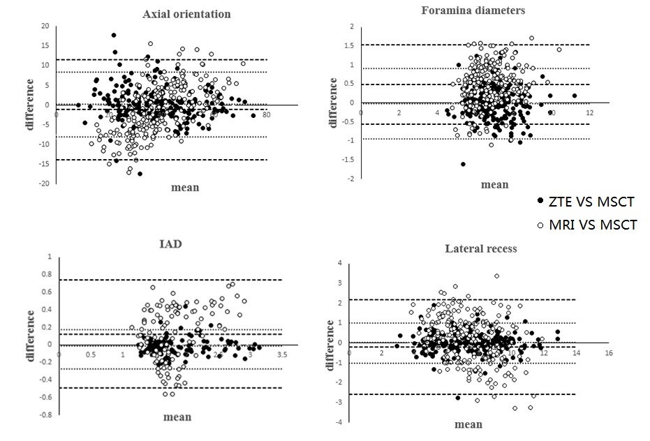
There was no difference for the osseous parameters between modalities, including axial orientation (p=0.444), IAD (p=0.381), lateral recess (p=0.370), and osteophyte grade (p=0.052). The measurements of the foramina diameter were statistically different between conventional MRI and MSCT (p<0.05) but not between the MSCT and ZTE (p=0.660). Cortical bone abnormalities were more likely to be misdiagnosed on conventional MRI. Cortical bone appeared blurrier on ZTE than MSCT especially in cases with severe lumbar degeneration. The inter-reader agreement between MSCT and ZTE-MRI was higher than between MSCT and conventional MRI.Discussion
With the concern about less medical resource burden and safety demand for no-radiation lumbar examination, 3D ZTE imaging sequence arouses interests in the depiction of the osseous structures. The performance of the ZTE-MRI and conventional MRI on the lumbar osseous at different degeneration levels were evaluated using MSCT as reference standard. Higher measurement consistency and no statistical difference of the lumbar osseous morphometry were found between ZTE-MRI and MSCT but the foramina measurements between conventional MRI and the MSCT was statistically different due to lack signal of cortical bone on the conventional MRI images. There was no difference of qualitative assessment between the MSCT and ZTE images but significant difference of the facet joint OA grade between the MSCT and conventional MRI. Compared with CT images, blurred appearance of cortical bone on ZTE images especially in severe cases may exaggerate the severity assessment of higher-degeneration facet joints OA. Irregularly blurred cortical bone on ZTE images might be caused by the changes of fat content and surrounding soft tissues and thus possibly made the degeneration degree of facet joint OA overestimated especially at higher degeneration level.Conclusions
ZTE-MRI could be a promising tool to depict the bone morphology and offer more cortical bone details compared to conventional MRIs and might be a valid alternative to CT for assessment of lumbar osseous morphology to some extent without additional ionizing radiation exposure.Acknowledgements
Funding: This study was supported by the National Natural Science Foundation of China (NSFC) (No. 31630025 and 81930045).References
1. Airaksinen O, Brox JI, Cedraschi C, et al. European guidelines for the management of chronic nonspecific low back pain. Eur Spine J. 2006; 15 (Suppl 2):S192-300.
2. Du J, Carl M, Bydder M, et al Qualitative and quantitative ultrashort echo time (UTE) imaging of cortical bone. J Magn Reson. 2010; 207:304-311
Figures