2278
Automatic DTI measures in the superficial white matter associated with cognitive decline and CSF amyloid-β42 in neurodegenerative dementia1Fondazione IRCCS Ca' Granda Ospedale Maggiore Policlinico Milano, Milano, Italy, 2Department of Pathophysiology and Transplantation, Università degli Studi di Milano, Milano, Italy, 3Image Sciences Institute, University Medical Center Utrecht, Utrecht, Netherlands, 4Department of Electronics, Information and Bioengineering, Politecnico di Milano, Milano, Italy
Synopsis
Diffusion tensor imaging (DTI) studies showed superficial white matter (SWM) alterations in Alzheimer’s disease (AD). The study aims to investigate the relationship between DTI in the SWM and CSF biomarkers in neurodegenerative dementia (ND). 93 ND patients were retrospectively recruited and images were processed for automatic segmentation of lobar white matter and Diffusion tensor imaging (DTI). Spearman’s correlation tests were performed between lobar DTI measures in the SWM, CSF biomarkers and clinical data. DTI measures in the SWM strongly correlated with Mini-Mental State Examination (MMSE) and amyloid-β42 suggesting SWM DTI as a candidate in-vivo non-invasive clinical and preclinical biomarker.
INTRODUCTION
According to the retrogenesis model, the WM tracts that are the last to myelinate are the first to degenerate in AD and other dementia [1]. Diffusion tensor imaging (DTI) studies showed superficial white matter (SWM) alterations correlated with cognitive decline in Alzheimer’s disease (AD) [2,3,4]. Correlations with CSF biomarkers were not demonstrated. The study aims to investigate the relationships between DTI measures in the SWM and CSF biomarkers in neurodegenerative dementia (ND).METHODS
From a database of 323 suspected dementia cases, 93 ND patients were retrospectively recruited and clinical and neuropsychological data, CSF biomarkers, and magnetic resonance images were collected. Automatic segmentation of the white matter in the frontal, parietal, occipital and temporal lobes was performed in Freesurfer and the SWM was obtained deleting from each lobe the voxels belonging to the coregistered ICBM-DTI-81 WM atlas [5]. Diffusion tensor imaging (DTI) was performed in ExploreDTI [6] and DTI maps were coregistered to T1-weighted images in each subject. DTI parameters were measured as mean value in each SWM lobar region. Spearman’s correlation tests were performed between lobar DTI measures in the SWM, CSF biomarkers and clinical data.RESULTS
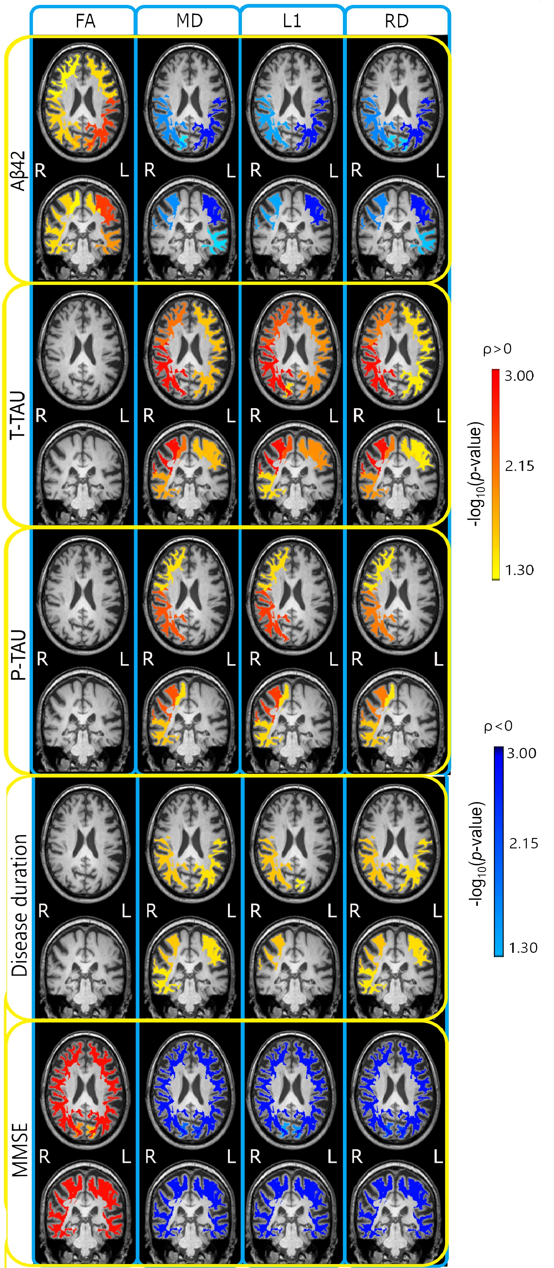
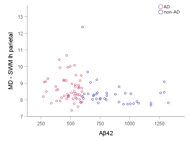
In all the four lobes, DTI measures in the SWM strongly correlated with Mini-Mental State Examination (MMSE) and amyloid-β42, less extensively with total-tau and phosphorylated tau, and with disease duration in the parietal lobe bilaterally, as shown in Fig.1. The mean diffusivity in the parietal SWM is plotted against amyloid-β42 in Fig.2.DISCUSSION
The automatic DTI measures in the SWM are strongly linked not only to the cognitive decline but also to the CSF amyloid-β42. We may argue that in vivo measures of the SWM alterations based on user-independent DTI contribute to filling the gap of knowledge between the abnormal protein deposition, that occurs very early in the preclinical phase, and the cognitive decline that may come decades later. Indeed, the decline in CSF amyloid-β42 level begins before the clinical phase in AD and approaches a plateau when symptoms are observable [7]. The association with CSF amyloid-β42 opens up opportunities for automatic DTI measures in the SWM to be further evaluated as an in-vivo non-invasive clinical biomarker as well as a candidate biomarker in the preclinical phase.Acknowledgements
No acknowledgement found.References
[1] Reisberg B, Franssen EH, Souren LEM, Auer SR, Akram I, Kenowsky S. Evidence and mechanisms of retrogenesis in Alzheimer’s and other dementias: Management and treatment import. Am J Alzheimer’s Dis Other Dementiasr. 2002;17(4):202-212. doi:10.1177/153331750201700411
[2] Reginold W, Luedke AC, Itorralba J, Fernandez-Ruiz J, Islam O, Garcia A. Altered Superficial White Matter on Tractography MRI in Alzheimer’s Disease. Dement Geriatr Cogn Dis Extra. 2016;6(2):233-241. doi:10.1159/000446770
[3] Phillips OR, Joshi SH, Piras F, et al. The superficial white matter in Alzheimer’s disease. Hum Brain Mapp. 2016;37(4):1321-1334. doi:10.1002/hbm.23105
[4] Bigham B, Zamanpour SA, Zemorshidi F, Boroumand F, Zare H. Identification of Superficial White Matter Abnormalities in Alzheimer’s Disease and Mild Cognitive Impairment Using Diffusion Tensor Imaging. J Alzheimer’s Dis Reports. 2020;4(1):49-59. doi:10.3233/ADR-190149
[5] Mori S, Wakana S, Van Zijl P, Nagae-Poetscher L. MRI Atlas of Human White Matter.; 2005. doi:10.1002/cmr.a
[6] Leemans A, Jeurissen B, Sijbers J, Jones D. ExploreDTI : a graphical toolbox for processing , analyzing , and visualizing diffusion MR data. In: 17th Annual Meeting of Intl Soc Mag Reson Med. ; 2009:3537.
[7] Selkoe DJ, Hardy J. The amyloid hypothesis of Alzheimer’s disease at 25 years. EMBO Mol Med. 2016;8(6):595-608. doi:10.15252/emmm.201606210
Figures