2259
A 4-channel flexible array with integrated B0 shimming for 3 T1High Field MR Center, Center for Medical Physics and Biomedical Engineering, Medical University of Vienna, Vienna, Austria, 2A.A. Martinos Center for Biomedical Imaging, Massachusetts General Hospital/Harvard Medical School, Charlestown, MA, United States, 3A.A. Martinos Center for Biomedical Imaging, Massachusetts General Hospital, Harvard Medical School, Charlestown, MA, United States
Synopsis
Integrated B0 shimming capability with RF coil arrays has already shown advantages in dense configurations. For highly flexible arrays this approach is a challenge due to the bulky RF chokes required to bypass the segmenting capacitors of classical loop coils. In this work we present a flexible 4-channel coaxial coil array module with integrated B0 shimming requiring only two RF chokes per shim channel and thereby demonstrate the feasibility of integrating B0 shim capability into flexible arrays.
Introduction
Image quality can be improved by increased RF coil sensitivity and better B0 homogeneity. Flexible RF coils offer the potential to improve signal-to-noise ratio (SNR) by form-fitting to the subject. Local B0 shimming integrated with the RF coil is capable of improving strongly localized B0 inhomogeneities that cannot be compensated for by the large shim coils integrated in the scanner bore [1,2]. Coaxial coil elements as highly flexible receive elements [3-6] have already proven to be advantageous to fit individual patient anatomies, whereas shim coil inserts are rigid [7]. Adding shimming capability to a highly flexible array offers the possibility to further improve image quality, in particular in applications where high B0 homogeneity is required, such as EPI-based or strongly frequency-dependent methods like fat-suppression techniques.Following our work on a single flexible coaxial coil channel with on-coil shimming [8], we present a 4-channel module of flexible coaxial receive coils [9] combined with on-coil shimming capability.Methods
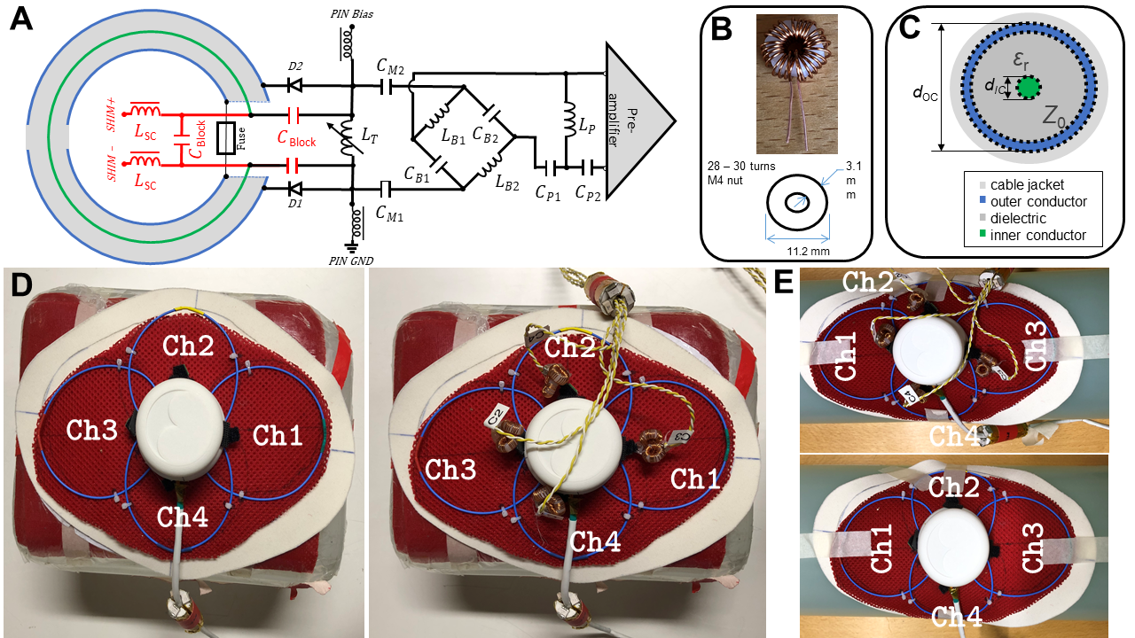
The 4-ch module is made of 80 mm diameter coaxial loop elements (Molex 047SC-2901, Lisle, USA), with a 5 mm gap in the outer conductor (Fig.1D-E) opposite the coil port with a gap in the inner conductor (Fig.1C). All four coil elements are connected to a custom designed PCB [10] with onboard low-noise preamplifiers (MwT MSM-123281, Microwave Technology, Inc., Fremont, USA). The loops have a self-resonance close to the Larmor frequency; fine-tuning is achieved with a hand-wound toroidal inductor LT (Fig. 1A). All loops were tuned to 123.2 MHz and matched to 50 Ω using a vector network analyzer (Keysight Technologies E5071C, Santa Rosa, USA). A controlled 1 A DC shim current was fed to the center conductor of each coaxial loop via two toroidal chokes made of 28-30 windings of 0.5 mm diameter insulated wire wound around a plastic M6 nut (Fig. 1B). The toroidal chokes block RF and limit the interaction with the self-resonant coaxial element. The shim current is supplied via long cables to the 4-ch module using a linear power supply, located outside the scanner room.To block the supplied DC shim current from biasing the receive loop’s PIN diodes D, blocking capacitors CBlock were introduced to the coil port entrance (Fig.1).
Qloaded, Qunloaded, active detuning and preamplifier decoupling were measured using a double pick-up probe (S21<-75dB) and a custom built rectangular 5 liter phantom (280 x 280 x 130 mm3, σ = 0.2 S/m DC) on the bench, before and after adding the shim chokes to each loop.
MR data were acquired on a 3T scanner (MAGNETOM Prisma Fit, Siemens Healthineers, Erlangen, Germany) using the rectangular phantom (for flat configuration of the array) and a 5.3 liter cylindrical leg phantom with 3.75 g NiSO4 x 6H2O + 5 g NaCl / 1000 g H2O (10606530 K2305, Siemens Healthineers, Erlangen, Germany) for a bent configuration of the array to demonstrate the functioning of the shim despite flexibility.
On the rectangular phantom, B0 field maps from each single element were acquired using ASPIRE [11] (TR = 9ms, TE1 / TE2 / TE3=2.2 / 4.6 / 6.9ms, FA = 6°, in-plane resolution=1.3 x 1.3 mm2, slice thickness = 2.3 mm, matrix = 208 x 156). The B0 fields created by each shim loop were computed from the difference in B0 between an acquisition with and without 1 A DC of shim current applied. Phase unwrapping was performed in MATLAB (The MathWorks Inc., USA) using ROMEO [12] [https://github.com/korbinian90/ROMEO].
Results
Bench measurements showed preamplifier decoupling >10 dB, ΔS21 for active detuning >35 dB, and a Q-ratio = Qunloaded / Qloaded of 157 / 54 = 2.9 without shim chokes and 137 / 51 = 2.7 with added chokes, corresponding to a loss of 12.7% (Qunloaded), 6.6% (Qloaded) and 7.6% (Q-ratio) due to the added shimming components.In flat configuration on the rectangular phantom, each shim element produced a ωΔB0 of about ±150 Hz at approximately 30 mm depth below the conductor inside the phantom. In bent configuration, showing the flexibility of the loop, the ωΔB0 offset was about ±190 Hz at approximately 25 mm depth, which is in good agreement to analytical calculations using Biot-Savart’s law.
Discussion and Conclusion
In this work we have demonstrated that creating B0 shim fields with a highly flexible 4-ch module made of coaxial cable is feasible with minimal hardware addition and Q-factor loss. The moderate degradation of Q is acceptable and could be optimized in future implementations.Due to the usage of the inner conductor of the coaxial cable as a shim loop, no lumped components, like shim chokes, are necessary along the coil conductor to integrate the known “AC/DC” approach [2], thus, keeping the flexibility of the array. The small footprint and weight of the required hardware addition opens the possibility to add B0 shim capability even to large flexible receive arrays.
Global and slice-wise shimming using current optimization software (http://rflab.martinos.org/index.php?title=Multi-coil_B0_shimming) and spatial tracking of several modular 4-channel arrays or the integration of B0 shimming capability into existing flexible arrays, will be tested upon the completion of the shim amplifier with four of the 8-channel revision C current driver boards (http://rflab.martinos.org/index.php?title=Current_driver:Current_driver).
Acknowledgements
This work was funded by the Anniversary Fund of the Oesterreichische Nationalbank Nr. 17980 “FlexShim“ and a joint Austrian/French grant (Austrian Science Fund FWF Nr. I-3618/Agence Nationale de Recherche ANR-17-CE19-0022) “BRACOIL“.References
[1] C. Juchem, T. W. Nixon, S. McIntyre, V. O. Boer, D. L. Rothman and R. A. de Graaf, "Dynamic multi-coil shimming of the human brain at 7 T," Journal of Magnetic Resonance, vol. 212, no. 2, pp. 280-288, 2011.
[2] J. P. Stockmann, T. Witzel, B. Keil, J. R. Polimeni, A. Mareyam, C. LaPierre, K. Setsompop and L. L. Wald, "A 32-channel combined RF and B0 shim array for 3T brain imaging," Magnetic Resonance in Medicine, no. 75 (1), pp. 441 - 451, 2016.
[3] L. Nohava, R. Czerny, S. Roat, M. Obermann, A. Kuehne, R. Frass-Kriegl, J. Felblinger, J.-C. Ginefri and E. Laistler, "Flexible Multi-Turn Multi-Gap Coaxial RF Coils: Design Concept and Implementation for Magnetic Resonance Imaging at 3 and 7 Tesla," IEEE Transactions on Medical Imaging, no. 40 (4), pp. 1267-1278, April 2021.
[4] B. Zhang, D. K. Sodickson and M. A. Cloos, "A high-impedance detector-array glove for magnetic resonance imaging of the hand," Nature Biomed. Eng., 2018.
[5] T. Ruytenberg, A. Webb and I. Zivkovic, "Shielded-coaxial-cable coils as receive and transceive array elements for 7T human MRI," Magn. Reson. Med., pp. 1-12, 2019.
[6] M. S. Mollaei, C. C. van Leeuwen, A. J. E. Raaijmakers and C. R. Simovski, "Analysis of high impedance coils both in Transmission and Reception regimes," IEEE Transactions on Medical Imaging, vol. 8, pp. 129754-129762, 2020.
[7] J. W. Pan, K. M. Lo and H. Hetherington, "Role of very high order and degree B0 shimming for spectroscopic imaging of the human brain at 7 Tesla," Magnetic Resonance in Medicine, vol. 68, no. 4, pp. 1007-1017, 2012.
[8] B. Gruber, M. Zaitsev, E. Laistler, “On-coil B0 shimming with a flexible coaxial coil element at 3 T”, Proceedings of the ISMRM 29th Annual Meeting and Exhibition, Virtual Meeting, p. 4535, 2021
[9] M. Obermann, S. Roat, and E. Laistler, "Coaxial coil modules as building blocks of individually arranged receive-only coil arrays", Proceedings of the ISMRM 29th Annual Meeting and Exhibition, Virtual Meeting, p. 3567, 2021
[10] M. Obermann, L. Nohava, R. Czerny, R. Frass-Kriegl, S. Roat, M. Pichler, J. Felblinger, J.-C. Ginefri and E. Laistler, "Optimization and miniaturization of Rx-only coaxial coil interfacing," Proceedings of the ISMRM 28th Annual Meeting and Exhibition, Virtual Meeting, p. 4042, 2020.
[11] K. Eckstein, B. Dymerska, B. Bachrata, W. Bogner, K. Poljanc, S. Trattnig and S. D. Robinson, "Computationally Efficient Combination of Multi-channel Phase Data From Multi-echo Acquisitions (ASPIRE)," Magnetic Resonance in Medicine, no. 2018 (79), pp. 2996-3006, 2018.
[12] B. Dymerska, K. Eckstein, B. Bachrata, B. Siow, S. Trattnig, K. Shmueli and S. D. Robinson, "Phase unwrapping with a rapid opensource minimum spanning tree algorithm (ROMEO)," Magnetic Resonance in Medicine, no. 85 (4), pp. 2294-2308, April 2021.
Figures