2253
BraCoil – preliminary performance evaluation of a wearable breast coil array for 3 T MR mammography1High Field MR Center, Center for Medical Physics and Biomedical Engineering, Medical University of Vienna, Vienna, Austria, 2IADI, Inserm, Université de Lorraine, Nancy, France, 3CHRU-Nancy, Inserm, Université de Lorraine, CIC, Innovation Technologique, Nancy, France
Synopsis
A dedicated radio frequency coil design for MR mammography needs to optimize sensitivity, reduce scan time, and improve patient comfort to ultimately facilitate diagnosis. We investigate signal-to-noise ratio and parallel imaging performance differences between widely used commercial 16-/18-channel receive coils for breast MRI at 3 T in prone and supine position and an in-house developed 28-channel wearable coil array (“BraCoil”). An SNR gain of up to a factor of 6 with BraCoil was found in this phantom study.
Introduction
The goal in breast MRI is high sensitivity for a large range of different breast sizes, and the minimization of the scan time employing parallel imaging techniques. Therefore, the use of dedicated RF receive arrays with a high number of individual elements appears to be a viable strategy. We hypothesize that the combination of close form-fitting coil design enabled by high mechanical flexibility1, a coil element size optimized for the desired penetration depth, and the choice of a measurement position where the breast tissue is flattened, i.e. the supine position or the prone position, when the breast is compressed by the subject’s own weight, leads to favorable conditions for strongly increasing the achievable signal-to-noise ratio (SNR) in the breast.In this work we perform a phantom study at 3 T to investigate the SNR and parallel imaging performance differences between widely used commercial breast MRI coils for prone and supine patient positioning and an in-house developed wearable breast coil array.
Methods
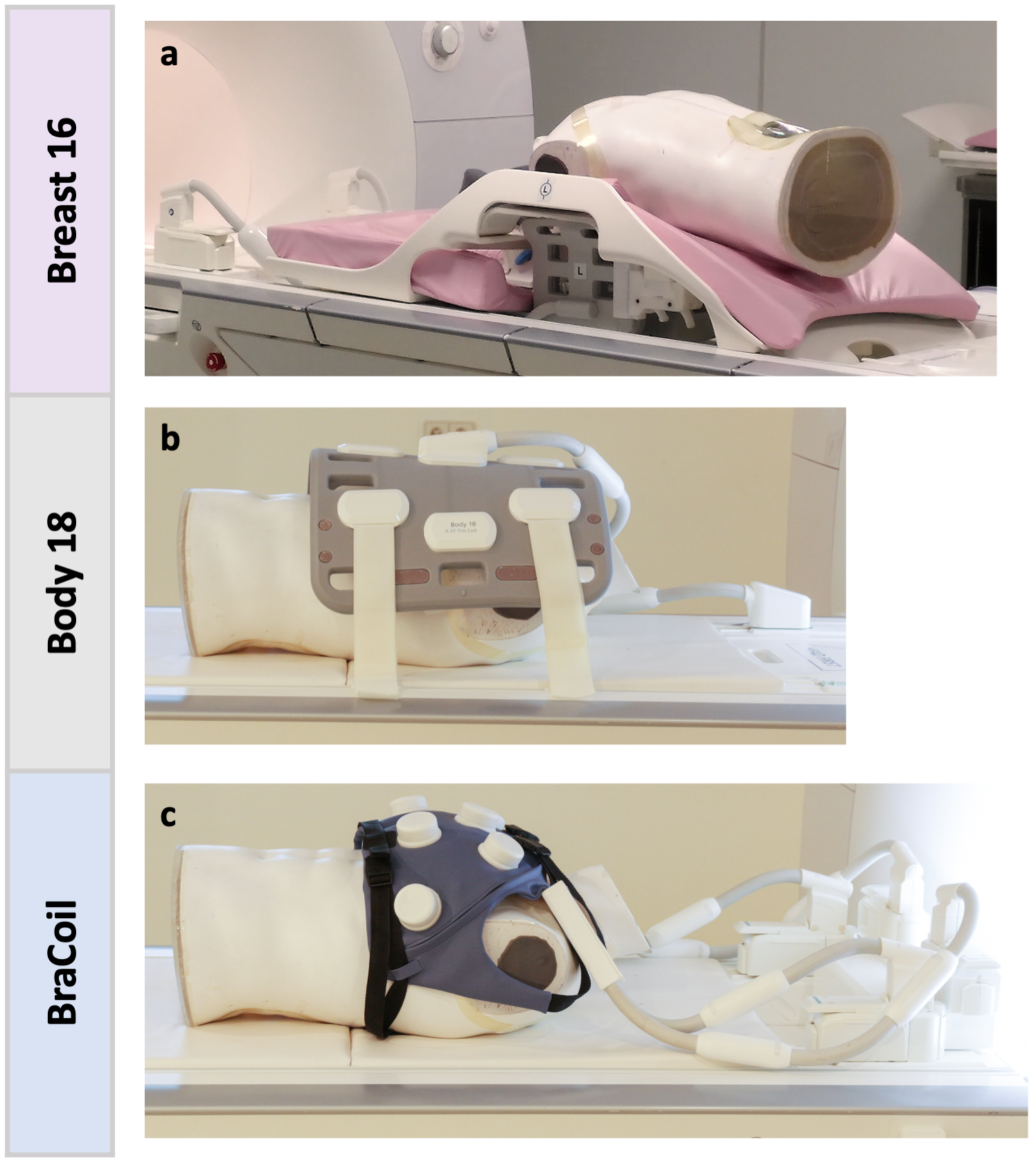
RF HardwareIn this study we use an ultra-flexible wearable 28-channel coil array that can be worn like a vest (“BraCoil”, Center for Medical Physics and Biomedical Engineering, Medical University of Vienna, Austria). The coil design is presented in a companion abstract by Obermann et al. Semi-flexible coils such as the 18-channel - “Body 18” - coil (Siemens Healthineers, Erlangen, Germany) allow for proof-of-concept supine breast MRI. Nonetheless, neither the size or number of receive elements nor the layout is optimized for the breast. The commercial reference coils used for comparison purposes are the 16-channel - “Breast 16” - Sentinelle coil (Siemens Healthineers, Erlangen, Germany) for prone and the before-mentioned Body 18 coil for supine measurements. All coils and their positioning under/on the phantom are shown in Fig. 1.
MRI experiments
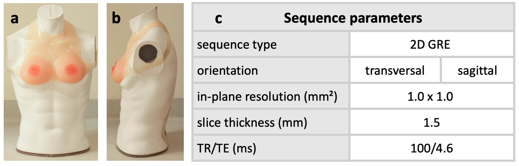
A torso phantom (filled with acrylic gel, σ = 0.60 S/m, ε = 62) with strap-on silicone breasts (“C-cup realistic strap-on breast”, Vollence) (see Fig. 2a,b) was used for both prone and supine measurements. All acquisitions were performed on a 3 T MR scanner (Magnetom Prisma Fit, Siemens Healthineers, Erlangen, Germany). With each coil, non-accelerated 2D gradient echo (GRE) images in the transversal and sagittal plane, and noise only scans were acquired. Sequence parameters are summarized in Fig. 2c.
Post processing
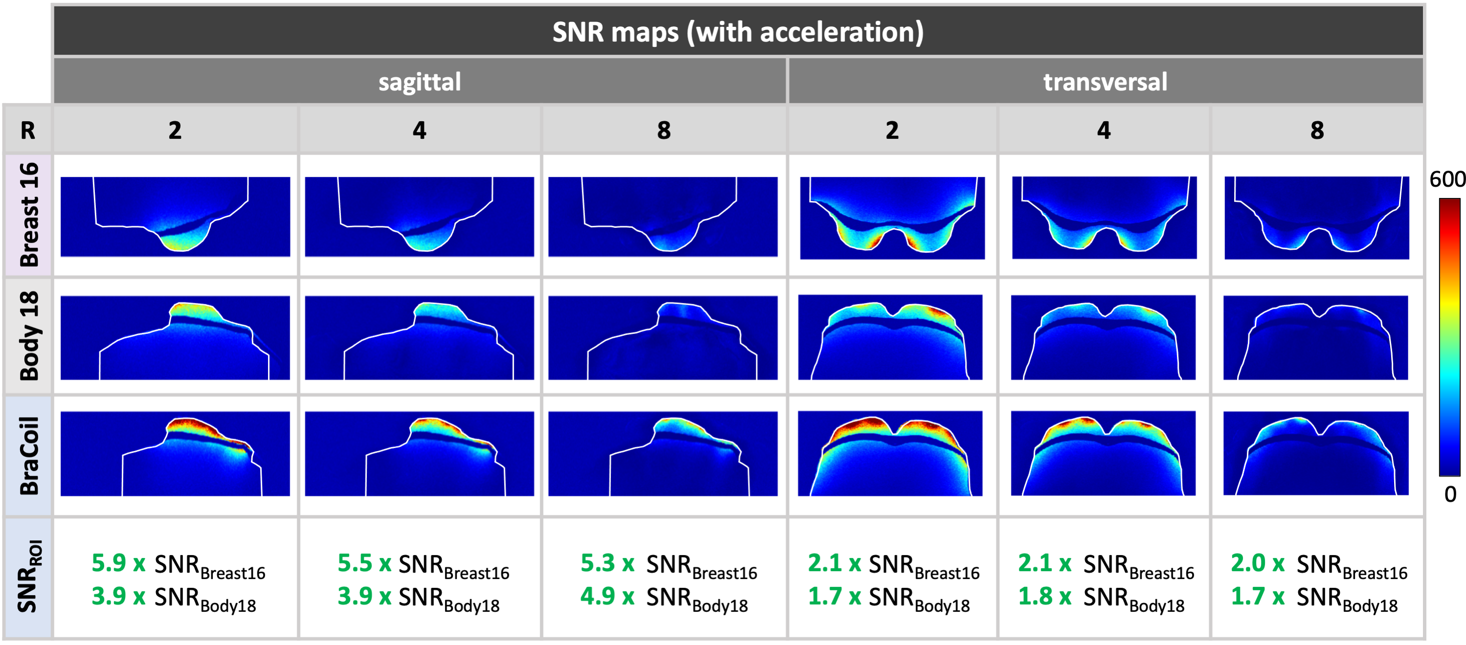
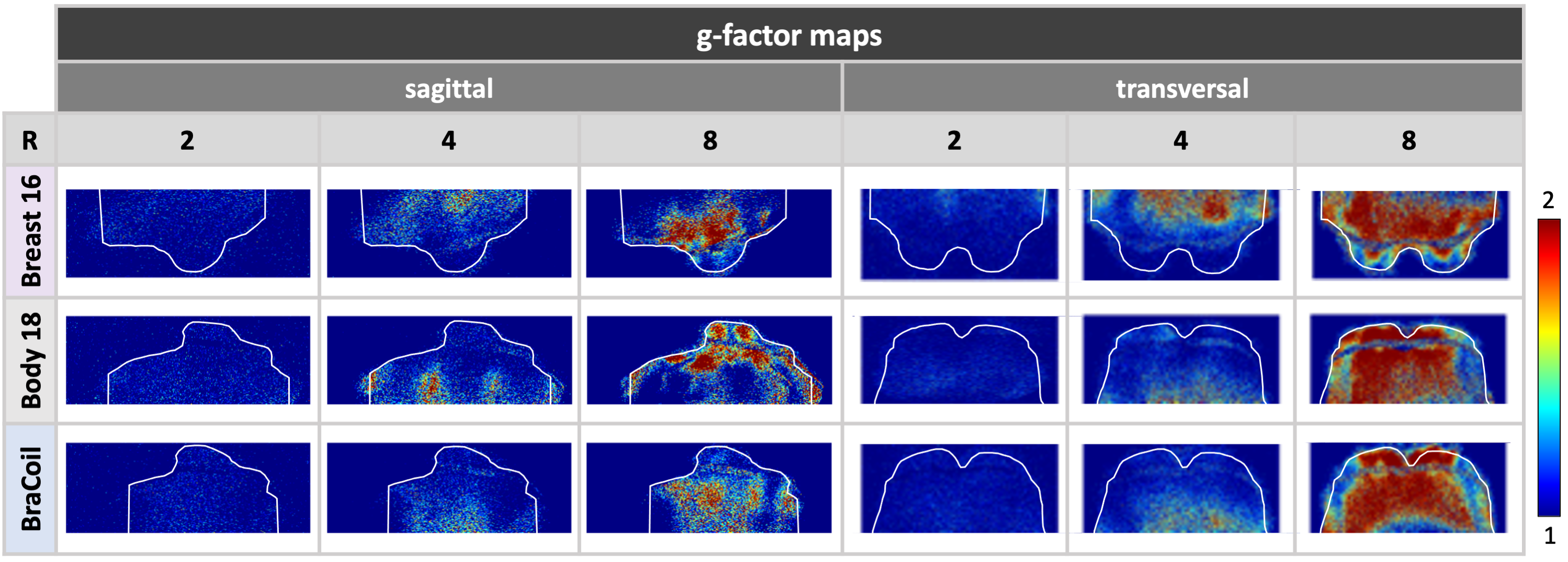
SNR maps for the non-accelerated, i.e., fully-encoded case as well as g-factor and SNR maps for an acceleration factor R = 2, 4 and 8 were calculated offline2. The phase-encoding direction was L-R in the transversal and H-F in the sagittal plane. Further, the SNR was evaluated in a ROI covering the strap-on breast only, i.e. excluding the torso (Fig. 3+4).
Results
The non-accelerated SNR maps in Fig. 3 demonstrate a significant SNR gain, up to a factor of 6, with the BraCoil compared to the investigated commercial coils. The non-conformal design of the rigid Breast 16 coil leads to suboptimal loading of the RF coil and, therefore, sensitivity loss, which is also partly present with the not ideally form-fitted Body 18 coil. The SNR map comparison calculated with acceleration factors to characterize the parallel imaging performance in Fig. 4 demonstrates the superiority in SNR of the BraCoil, up to high acceleration in L-R or H-F direction (R≤8). Corresponding g-factor maps for all examined accelerations and coils are shown in Fig. 5.Discussion
This work confirms the hypothesis that SNR in the breast can be strongly improved using a wearable coil array in comparison to the standard acquisition setup with hanging breasts in prone position. Also, in comparison to the semi-flexible Body 18 coil, a strong SNR gain could be demonstrated due to the smaller elements of the present coil and the even better form-fitting.A certain flattening of the breast with the Body 18 coil was observed (see Fig.3). No foam wedges to create a coil-sample distance and, therefore preserve the breast shape, were employed for a fair SNR comparison to the BraCoil. Supine imaging with the BraCoil is a new approach in MR mammography compared to standard prone imaging. The advantage with the BraCoil is not only the possible combination of superior image quality and acquisition speed but also the fact that the breast is only slightly deformed or compressed which will facilitate correlation to other imaging modalities in vivo.
We will also pursue strategies for motion management in the supine position based on a combination of miniature on-coil sensors and a reconstruction-based motion correction method (GRICS), which has already delivered very promising results for free-breathing abdominal and cardiac imaging3,4.
A patent is filed5, and the regulatory steps for in vivo usage are currently underway to enable future studies focused on in vivo high-speed imaging in prone and supine position to demonstrate the usability of the BraCoil with a large variety of breast sizes in comparison to commercial reference coils.
Conclusion
As intended, the combination of form-fitting, dense coil array layout, and flattening of the breast tissue in the BraCoil concept was shown to strongly increase sensitivity in comparison to commercial breast MRI coils. The measured factor 2 or more in SNR will enable drastically shorter measurement times and/or higher resolution in MR mammography.Acknowledgements
This work was funded by a joint Austrian/French grant (Austrian Science Fund FWF Nr. I-3618/Agence Nationale de Recherche ANR-17-CE19-0022) “BRACOIL“.References
1. Obermann, M., Roat, S. & Laistler, E. Coaxial coil modules as building blocks of individually arranged receive-only coil arrays. in Proc. Intl. Soc. Mag. Reson. Med. 29 (2021) 1610.2.
2. Robson, P. M. et al. Comprehensive quantification of signal-to-noise ratio and g-factor for image-based and k-space-based parallel imaging reconstructions. Magn. Reson. Med. 60, 895–907 (2008).
3. Chen, B. et al. Design and Validation of a Novel MR-Compatible Sensor for Respiratory Motion Modeling and Correction. IEEE Trans. Biomed. Eng. 64, 123–133 (2017).
4. Odille, F., Vuissoz, P.-A., Marie, P.-Y. & Felblinger, J. Generalized reconstruction by inversion of coupled systems (GRICS) applied to free-breathing MRI. Magn. Reson. Med. 60, 146–157 (2008).
5. Laistler, E., Obermann, M., Nohava, L., Roat, S. Coil module for magnetic resonance imaging applications. Patent. Application number EP21020242.0 - 1126 (2021).
Figures