2252
Feasibility evaluation of virtual elastography based on diffusion-weighted imaging (vMRE) at 1.5 Tesla
Alicia Palomar Garcia1, Valentin H. Prevost2, Alba Iruela Sanchez1, Wolter de Graaf3, and Bruno Triaire2
1Canon Medical Systems Spain and Portugal, Barcelona, Spain, 2Canon Medical Systems Corporation, Tochigi, Japan, 3Canon Medical Systems Europe, Zoetermeer, Netherlands
1Canon Medical Systems Spain and Portugal, Barcelona, Spain, 2Canon Medical Systems Corporation, Tochigi, Japan, 3Canon Medical Systems Europe, Zoetermeer, Netherlands
Synopsis
This study evaluated the feasibility of performing virtual elastography based on diffusion-weighted MRI (vMRE) on a 1.5T system with two different acceleration methods, SPEEDER and EXSPER. Proton density fat fraction (PDFF) imaging was also acquired. The vMRE and PDFF measurements were consistent with healthy values that can be found in the literature. This study demonstrated the feasibility of performing vMRE at 1.5T for the estimation of overall liver elasticity. SPEEDER and EXSPER vMRE implementations provided consistent stiffness estimations and both would be adequate for conducting future studies at 1.5T systems on patients.
Introduction
Chronic liver diseases are one of the leading causes of mortality globally. For these patients, an early diagnosis and correct staging is crucial for improving prognosis. Although liver biopsy is the reference technique for disease staging, many non-invasive alternatives have been proposed to provide stiffness estimation while avoiding the possible risk of complications such as hemorrhage or infection. Some of these methods, such as MR elastography (MRE) and ultrasound techniques like 2D shear wave elastography (SWE), provide good diagnostic accuracy, but present some limitations in clinical context because they require dedicated hardware and provide local estimates of liver stiffness, respectively. More recently, virtual elastography based on diffusion-weighted MRI (vMRE) has been introduced as an alternative to quantify tissue stiffness in the whole liver in a single acquisition without the need of additional equipment1. In all previous studies using vMRE, the MRI was performed on 3T systems1,2,3. This study evaluated the feasibility of performing vMRE on a 1.5T system, comparing two different parallel imaging implementations (SPEEDER and EXSPER). In addition, the relation between vMRE measurements and proton density fat fraction (PDFF) was evaluated.Methods
Five healthy volunteers underwent whole-liver MRI sessions on a Vantage 1.5T Orian XGO system (Canon Medical Systems Corporation, Tochigi, Japan) with a 16-channel body coil combined with a spine coil. Scanning sessions included diffusion-weighted imaging (DWI) with the following parameters: 2D SE-EPI; PASTA fat suppression; TR=4614ms; TE=67ms; in-plane resolution=2x2mm; slice thickness=6mm, parallel imaging=SPEEDER or EXSPER, acceleration factor=3; two b-values with different number of averages: 200 s/mm2 (NAQ=2) and 1500 s/mm2 (NAQ=4); respiratory triggering and total scanning time of 5:15 min or 6 min for SPEEDER and EXSPER, respectively. A multi-echo breath-hold 3D FE sequence for fat fraction quantification was also acquired with the same resolution and slice thickness.A dedicated Olea Sphere plugin was used to compute the virtual elasticity maps from the DWI data, based on the linear relationship between shifted ADC and stiffness modulus stated in previous studies1,2,3. For quantification analysis, five measurements were done in each subject, placing ROIs in the same position by coregistering PDFF and elasticity maps. Spearman’s rank correlation was used to evaluate the relation between vMRE-based stiffness and PDFF. Measurements for a volunteer with high PDFF, but no reported liver disease, were discarded, so a total of 20 measures were considered for the correlation.
Results
Resulting virtual elasticity maps (with SPEEDER and EXSPER) and PDFF map are shown in Figure 1. Mean values of PDFF and vMRE with both acceleration techniques (SPEEDER and EXSPER) for the five healthy volunteers are reported in Figure 2. Stiffness measurements based on vMRE with SPEEDER were in the range between 1.11 ± 0.35 kPa and 3.31 ± 0.51 kPa, and measurements using EXSPER were within 1.46 ± 0.53 kPa and 2.75 ± 0.54 kPa. No significant correlation was found between vMRE measurements and PDFF.Discussion
The range of values obtained with vMRE is aligned with previous studies reporting normative stiffness in healthy subjects1,2. In addition, there is no significant difference between the two different acceleration methods that were tested. Both SPEEDER and EXSPER implements would be could be then used for vMRE imaging. However, in specific scenarios such as protocols with small field of view, EXSPER would be more robust to unfolding artifacts4.The PDFF percentage obtained for the sample studied is consistent with the values reported for healthy subjects5,6. In this study, no significant correlation was found between fat fraction and virtual elasticity, possibly due to the limited sample size and the short variability between measurements. Further studies with larger samples and including patients with liver diseases would be necessary to proper study the relation between these metrics.
Conclusion
This study demonstrated the feasibility of performing vMRE at 1.5T for the estimation of overall liver elasticity. Data acquired with SPEEDER and EXSPER provided consistent stiffness measurements on healthy livers and both would be adequate for vMRE studies. Future works with larger samples and the inclusion of patients with liver disease would be needed to better estimate the relation between liver elasticity and fat fraction.Acknowledgements
No acknowledgement found.References
- Le Bihan D, Ichikawa S, & Motosugi U. Diffusion and intravoxel incoherent motion MR imaging–based virtual elastography: a hypothesis-generating study in the liver. Radiology. 2017; 285(2), 609-619.
- Kromrey M L, Le Bihan D, Ichikawa S, et al. Diffusion-weighted MRI-based virtual elastography for the assessment of liver fibrosis. Radiology. 2020; 295(1), 127-135.
- Ota T, Hori M, Le Bihan D, et al. Diffusion-Based Virtual MR Elastography of the Liver: Can It Be Extended beyond Liver Fibrosis? J Clin Med. 2021; Sep 30; 10(19):4553.
- Deshmane A, Gulani V, Griswold M A, et al. Parallel MR imaging. Journal of Magnetic Resonance Imaging. 2012; 36(1), 55-72.
- Idilman I S, Aniktar H, Idilman R, et al. Hepatic steatosis: quantification by proton density fat fraction with MR imaging versus liver biopsy. Radiology. 2013; 267(3), 767-775.
- Caussy C, Reeder S B, Sirlin C B, et al. Noninvasive, quantitative assessment of liver fat by MRI‐PDFF as an endpoint in NASH trials. Hepatology. 2018; 68(2), 763-772.
Figures
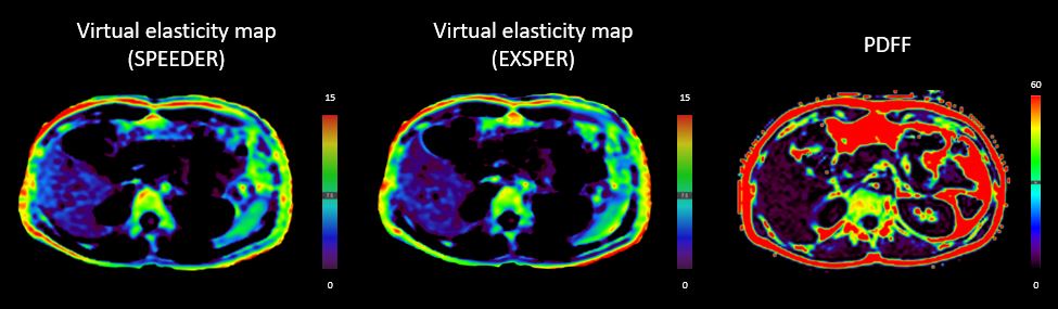
Illustrative
images of computed virtual elasticity maps with both acceleration techniques
(SPEEDER and EXSPER) and proton density fat fraction map.
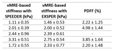
Mean
stiffness vMRE-based measurements with both acceleration techniques (SPEEDER
and EXSPER) and mean PDFF of the sample studied. These values are expressed as
mean ± standard deviation.
DOI: https://doi.org/10.58530/2022/2252