2246
Impact of bowel preparation methods on pancreatic Magnetic Resonance Elastography1Radiology and Nuclear Medicine, Amsterdam UMC location AMC, Amsterdam, Netherlands, 2Department of Radiology, Charité – Universitätsmedizin Berlin, Berlin, Germany
Synopsis
Pancreas magnetic resonance elastography (MRE) allows non-invasice estimation of tissue stiffness for multiple pathophysiological diseases. MRE reproducibility and the effect of bowel-preparation (butylscopolaminebromide and drinking water) for increased MRE data quality were assessed for the pancreas. Shear wave speed and MRE quality were determined for the pancreas, liver and kidneys. Intrasession and intersession reproducibility showed a coefficient-of-variation of 6.59% and 13.10%. Repeated measures ANOVA showed no significant difference in SWS of all organs (f=3/11,p>0.05). Bowel-preparation methods for pancreatic MRE do not increase MRE quality. Drinking water increases the MRE quality for kidneys and liver whilst not altering the measured SWS.
Introduction
Magnetic resonance elastography (MRE) in the pancreas has the potential to elucidate multiple pancreatic pathologies[1], such as pancreatic adenocarcinoma. To characterize tumor microenvironment and estimate stromal disposition in the pancreas a high quality and reproducible MRE is imperative. However, its central abdominal location presents two potential issues as bowel movement may interfere with the shear wave resulting in low quality MRE data while the presence of gas in the gastrointestinal tract surrounding the pancreas inhibits shear wave propagation.To address these challenges, we hypothesize the effects of two MRE patient preparation methods: 1) drinking 0.5L of water decreases the amount of air in the stomach and increases wave penetration, and 2) using butylscopolaminebromide (Buscopan:20mg/ml,Eureco-PharmaB.V) increases MRE quality through inhibition of smooth-muscle contraction. The aim of this study was to assess test-retest of reproducibility of non-prepared pancreatic MRE and to see if patient preparation improves MRE quality.
Methods
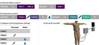
MRE reproducibility of the pancreas was studied in a group of 10 healthy volunteers (♀=6,mean age=26±3years). Both intrasession and intersession reproducibility were investigated (figure.1a). Different subject preparations were evaluated in 15 healthy volunteers (♀=7,mean age=41±16years), following the preparation steps as indicated in Figure 1b; No preparation (TOMO1), Buscopan (TOMO2), water (TOMO3), water and Buscopan (TOMO4). Buscopan was injected via IV to temporarily (~4min) decrease bowel movement.All scanning was performed at 3.0T (Ingenia,Philips,Best,Netherlands) in combination with body transmit, anterior, and posterior receive coils. Volunteers were positioned prone, head first. Four compressed-air driven MRE-transducers were placed on the lower thoracic cage, two anterior and two posterior (figure.1d)[2]. Elastography images were acquired with a multi-frequency free-breathing SE-EPI sequence at four frequencies (MREfreq=30,40,50,60Hz)[2]. Sequence parameters were: nslices=29, voxelsize=2.7x2.7x5mm3, SENSE=2.5, TE/TR=55/2400ms, number of MRE-offsets=8, MEGdir=3, and acquisition-time≈4 min. All subjects fasted four hours prior to scanning.
Postprocessing was accomplished using the kMDEV inversion algorithm resulting in shear-wave-speed (SWS) maps of the abdomen[3]. Volumes-of-interest were manually drawn on the mean magnitude images to assess the mean pancreatic SWS, along with the kidneys and liver to assess influence on other organs. Strain-SNR and attenuation of the shear wave were evaluated as quality parameters[4].
The reproducibility and repeatability of TOMO1 was assessed by Bland-Altman plots. Repeated measures ANOVA and pairwise comparison (p<.05) were used to determine intra-subject variability for all parameters for the preparation methods (SPSS,Statistics,Version26,IBM,USA). The differences in pancreatic SWS throughout preparation methods were compared to the within- and between-session reproducibility.
Results
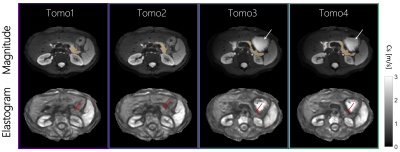
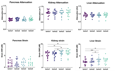
Intrasession and intersession reproducibility are shown using Bland-Altman plots in Figure 2, with an mean SWS of 1.10±0.10m/s overall and coefficient-of-variation: CVintrasession=6.59% and CVintersession=13.10%. Figure 3 shows magnitude images and corresponding elastograms for a single volunteer over all preparation scans. Repeated measures ANOVA showed no significant difference for SWS of all organs (f=3/11,p>.05). However, exploratory pairwise comparison for the pancreas showed significant difference between TOMO1 and TOMO2 (p=.005). Mean SWS of the pancreas were 1.13±0.26, 1.19±0.28, 1.15±0.27 and 1.18±0.27m/s for TOMO1,2,3 and 4, respectively (figure.4). Strain-SNR and attenuation showed no significant differences between different scans for the pancreas (figure.5). However, there is a difference between before and after drinking water in the kidneys and liver, with the exception between TOMO2 and TOMO4 in the liver. There was no significant difference in attenuation of the shear wave for all scans.Discussion
Mean SWS in the pancreas were in line with previously published literature[2]. Pairwise comparison showed that there is a significant difference between the SWS in the pancreas of TOMO1 and TOMO2. Buscopan injection increases the mean SWS of the pancreas compared to nonprepared-MRE. However, this difference was not observed between other preparation scans or in the kidneys and liver. The pancreas is surrounded by the hepatopancreatic ampulla, which is a smooth-muscle sphincter controlling inflow and inhibiting reflux of duodenal substances into the ampulla. Buscopan could have an influence on the ductal pressure and thereby apparent SWS, because it inhibits spontaneous smooth-muscle activity and thereby the hepatopancreatic ampulla. Nevertheless, the difference observed between the nonprepared-MRE and Buscopan SWS is within the limits of the repeatability, indicating that this effect is negligible.Quality parameters showed no significant difference in the pancreas, suggesting that there is no increased wave penetration when drinking 0.5L of water or decrease of shear-wave interference using Buscopan. However, there is an observed difference in the strain-SNR in the kidney and liver. For the kidneys there seems a difference between before and after drinking water, with an increase in strain-SNR after drinking water. In the liver this is also observed, except between TOMO2 and TOMO4. Previous work showed similar results for the mean SWS in the pancreas (1.20±0.12m/s) and no change in SWS of the pancreas, liver and kidney at different hydration states (an hour after drinking water)[2].
Conclusion
Bowel-preparation methods for pancreatic MRE does not increase MRE quality. Repeated measures ANOVA showed no significant difference in SWS for all organs(f=3/11,p>0.05). Exploratory pairwise comparison for the pancreas showed significant difference between TOMO1 and TOMO2(p=.005). This could be due to Buscopan having an effect on the hepatopancreatic ampulla. However, this effect is within the limits of the repeatability of the nonprepared-MRE and therefore clinically irrelevant. Drinking water increases the MRE quality for kidneys and liver whilst not altering the measured SWS.Acknowledgements
This research was funded by KWF Kankerbestrijding.References
[1] MacCurtain BM, Quirke NP, Thorpe SD, Gallagher TK. Pancreatic Ductal Adenocarcinoma: Relating Biomechanics and Prognosis. Journal of Clinical Medicine. 2021; 10(12):2711. https://doi.org/10.3390/jcm10122711
[2] Dittmann F, Tzschätzsch H, Hirsch S, et al. Tomoelastography of the abdomen: Tissue mechanical properties of the liver, spleen, kidney, and pancreas from single MR elastography scans at different hydration states. Magn Reson Med. 2017;78(3):976-983. doi:10.1002/mrm.26484
[3] H. Tzschatzsch, J. Guo, F. Dittmann, S. Hirsch, E. Barnhill, K. Johrens, J. Braun, I. Sack, Tomoelastography by multifrequency wave number recovery from time-harmonic propagating shear waves, Medical Image Analysis 30 (2016) 1-10.
[4] Bertalan, G, Guo, J, Tzschätzsch, H, et al. Fast tomoelastography of the mouse brain by multifrequency single-shot MR elastography. Magn Reson Med. 2019; 81: 2676– 2687. https://doi.org/10.1002/mrm.27586
Figures


