2203
Relationship between internal carotid artery hemodynamics and cerebrovascular events in moyamoya angiopathy based on 4D Flow MRI1The Affiliated Drum Tower Hospital of Nanjing University Medical School, nanjing, China, 2Philips Healthcare, Shanghai, China, 3The Affiliated Drum Tower Hospital of Nanjing University Medical School, Nanjing, China
Synopsis
Little is known of distal internal carotid artery (ICA) hemodynamics in patients with moyamoya angiopathy (MMA) and its relationship with cerebrovascular events. In this study, distal ICA hemodynamics were analyzed using 4D Flow MRI in 70 MMA patients (61 infarct hemispheres and 63 non-infarct hemipheres). Vmax was found to be associated with cerebrovascular event in MMA patients.
Introduction
Moyamoya angiopathy (MMA) is a progressive stenosis or occlusion of the terminal internal carotid artery (ICA), proximal middle cerebral artery (MCA) and anterior cerebral artery (ACA), which occurs around artery collaterals at the base of the skull1. Cerebrovascular events in patients with MMA commonly present as ischemic stroke or cerebral hemorrhage. Intracranial vessel wall enhancement of the distal ICA is a risk factor for stroke in patients with MMA2. However, little information is available about distal ICA hemodynamics and its relationships with stroke. The aim of this study is to characterize distal ICA hemodynamics by 4D Flow MRI and its relationship with cerebrovascular events.Materials and Methods
This prospective study enrolled 70 MMA patients from July 2020 to August 2021 in a single center on an Ingenia 3.0T CX MR scanner (Philips Healthcare, the Netherlands). 4D Flow MRI and conventional MRI was performed in all subjects. Distal ICA hemodynamics, including Vmax, Vavg, flowavg, wall shear stess (WSS), pulsatility index (PI) were determined using GT flow (version 2.2.15). The cerebrovascular events of each hemisphere were identified according to conventional brain MR images, which were diagnosed as cerebral infarction or hemorrhage. Hemispheres with ischemic or hemorrhagic lesions were defined as positive hemispheres. The independent sample T test were used to evaluate differences between groups (positive hemispheres, hemispheres with cerebrovascular events; negative hemispheres, hemispheres without cerebrovascular events). The univariate and multivariate logistic regression analyses were used to evaluate the relationship of internal carotid artery hemodynamics and cerebrovascular events.Results
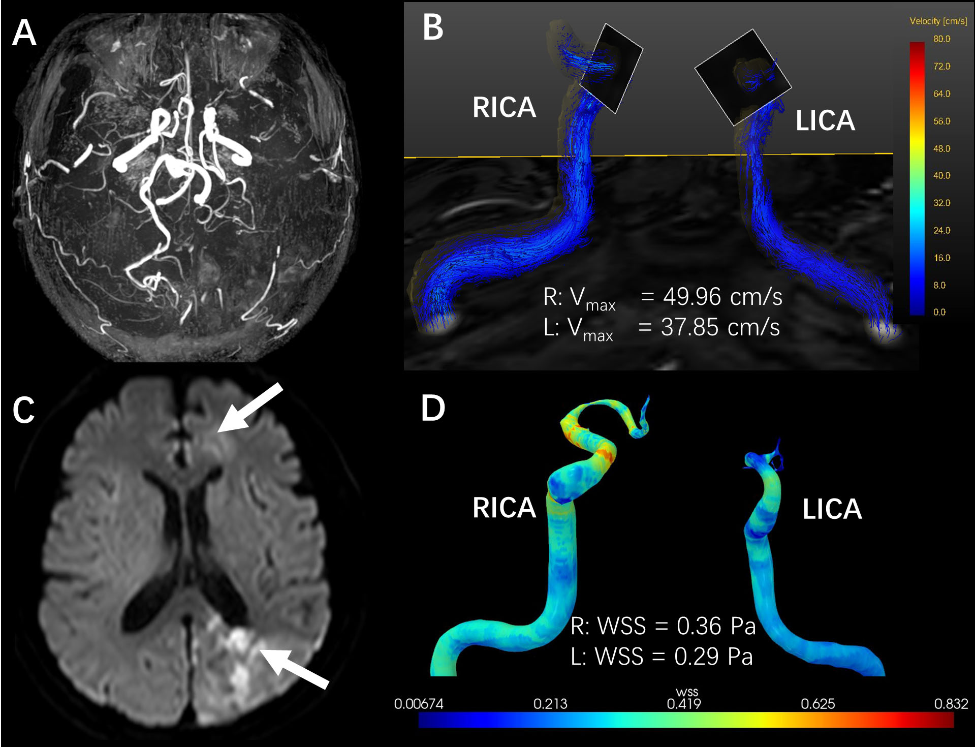
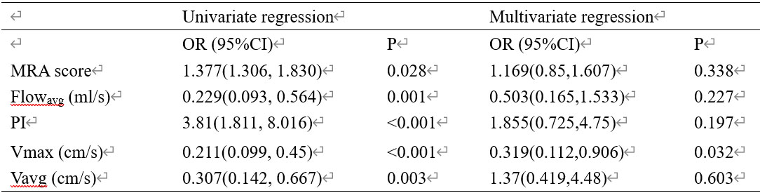
Totally 70 MMA patients with 124 hemispheres were evaluated, including 61 positive hemispheres (50 years old ± 9; 30 males) with cerebral events and 63 negative hemispheres (49 years old ± 9, 24 males) without events. Positive hemispheres had lower flow dynamics (Vmax: 38.27cm/s±15.13 vs 48.24 cm/s±11.31; Vavg: 17.23cm/s±9.32 vs 21.94cm/s±7.95; flowavg: 17.23ml/s±9.32 vs 21.94ml/s±7.95) and higher PI (1.06±0.59 vs 0.76±0.38) compared with negative hemispheres (all p<0.05). Vmax (OR: 0.319; 95% CI: 0.112, 0.906; P=0.032) was significantly associated with cerebrovascular events (Table 1). Figure 1 shows a patient with bilateral MMA whose left hemisphere had acute cerebrovascular events in the left frontal and parietal lobes with lower flow velocity.Conclusion
To our certain knowledge, this is the first study to evaluate the relationship between internal carotid artery hemodynamics and cerebrovascular events in MMA by using 4D Flow MRI. Our results suggest that the low blood flow in distal ICA may lead to stroke in MMA patients. This association may help identify high-risk patients by monitoring ICA hemodynamics.Acknowledgements
No acknowledgement found.References
1. Herve D, Kossorotoff M, Bresson D, et al. French clinical practice guidelines for Moyamoya angiopathy. Rev Neurol (Paris) 2018;174(5): 292-303.
2. Kathuveetil A, Sylaja PN, Senthilvelan S, et al. Vessel Wall Thickening and Enhancement in High-Resolution Intracranial Vessel Wall Imaging: A Predictor of Future Ischemic Events in Moyamoya Disease. AJNR Am J Neuroradiol 2020;41(1): 100-105.
Figures