2181
Evaluation of an Ultra High Dielectric Constant (uHDC) Package for Enhancement of a Knee Phased Array1Radiology, Pennsylvania State University College of Medicine, Hershey, PA, United States, 2Neurosurgery, Pennsylvania State University College of Medicine, Hershey, PA, United States, 33HyQ Research Solutions, LLC, College Station, TX, United States
Synopsis
Prior work has explored the use of dielectric materials to enhance imaging of the brain and spine with rectangular blocks. One area that has not been examined to date is knee imaging, where space constraints limit the use of blocks. In this work we evaluate the performance of an ultra-high dielectric constant (uHDC) curved plate (εr ~ 4500) for knee imaging in a clinical array at 3T. SNR gains of 9 to 71% percent were observed across the patella and patellar cartilage. Transmit efficiency increased up to 100% close to the material, with an increase in transmit inhomogeneity.
Introduction
Prior work has examined the use of uHDC materials for enhancing SNR and reducing transmit power in several scenarios at 3T, with rectangular blocks around the brain1 & spine2, or a conformal helmet3. There are imaging configurations that could benefit from the application of uHDC material but require a different geometrical package. One configuration that has not been examined to date is that of a knee phased array. Typically knee arrays integrate a single local transmit channel alongside a receive phased array with 15 – 30 coil elements within a cylindrical former. Space constraints within the coil limit the usage of rectangular uHDC blocks, where a curved geometry would be more ideal. This work evaluates the use of a curved uHDC plate to enhance the performance a clinical knee array at 3T.Methods
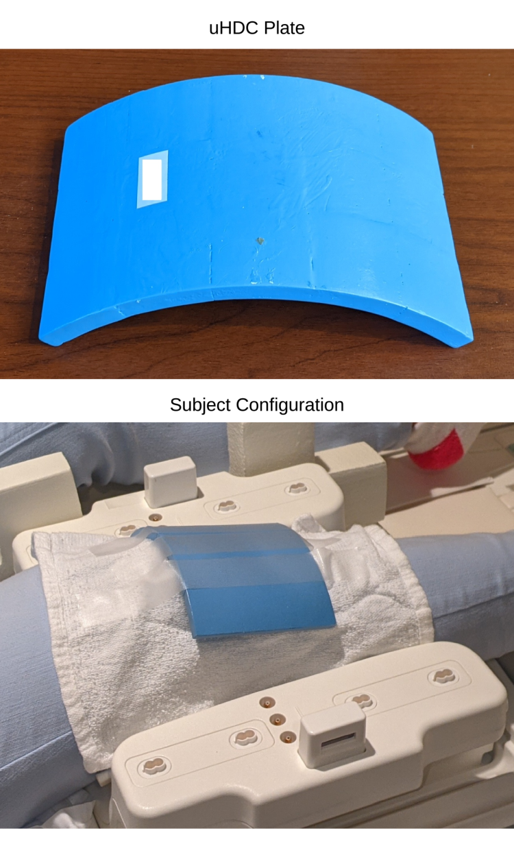
The uHDC configuration is depicted in Fig. 1. A single curved uHDC plate was placed on top of a subject’s knee. The uHDC plate is an experimental sample provided by HyQ Research Solutions, LLC, with dimensions of 150 x 98 x 8 mm and a stated permittivity of 4500. The plate was nearly flush with the top half of the knee array once the knee array is assembled (not pictured). Experiment: A single healthy subject was scanned, with and without the uHDC plate. B1+ maps (1.83 x 1.83 mm) were collected with a 2D multi-slice pre-saturated TurboFlash sequence4 to quantify transmit efficiency. A 2D multi-slice small tip-angle GRE sequence (1 x 1 mm) with an integrated noise-prescan was acquired to calculate signal-to-noise ratio with the SNR units method5. Both sequences acquired 50 slices at 3 mm thickness. The SNR maps were subsequently divided by the transmit sensitivity to remove the influence of the flip angle on the measurement. Data was collected on a Siemens 3T PrismaFit (Siemens Healthineers, Erlangen, Germany) with a 15-Ch QED transmit/receive knee array.Results
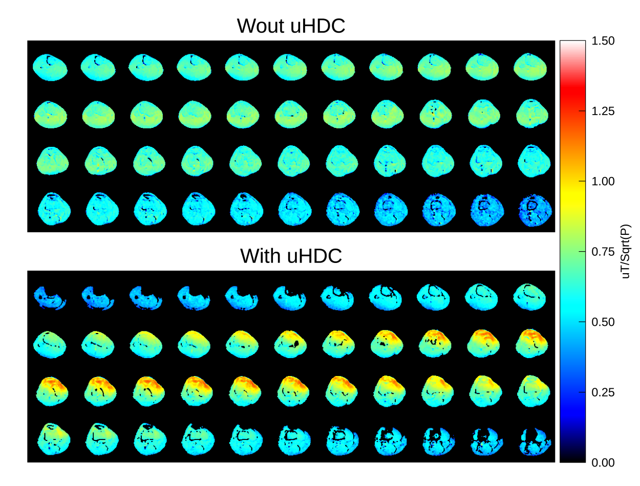
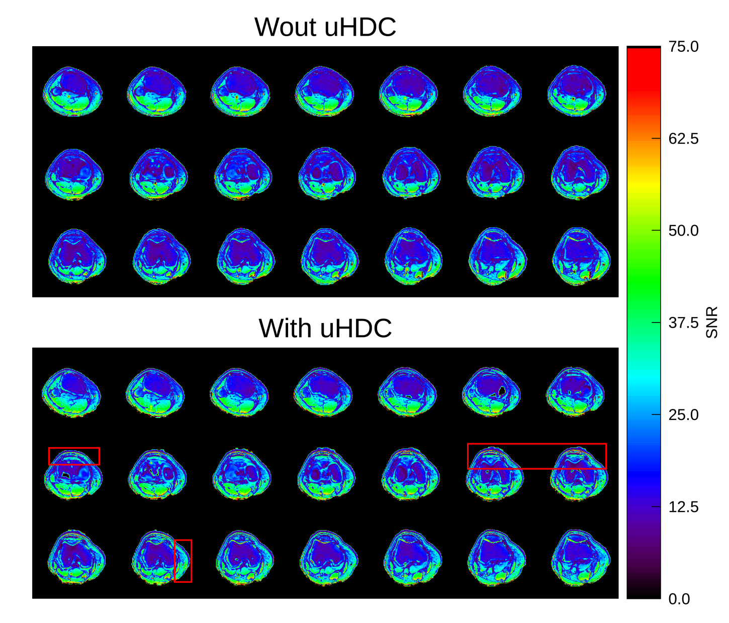
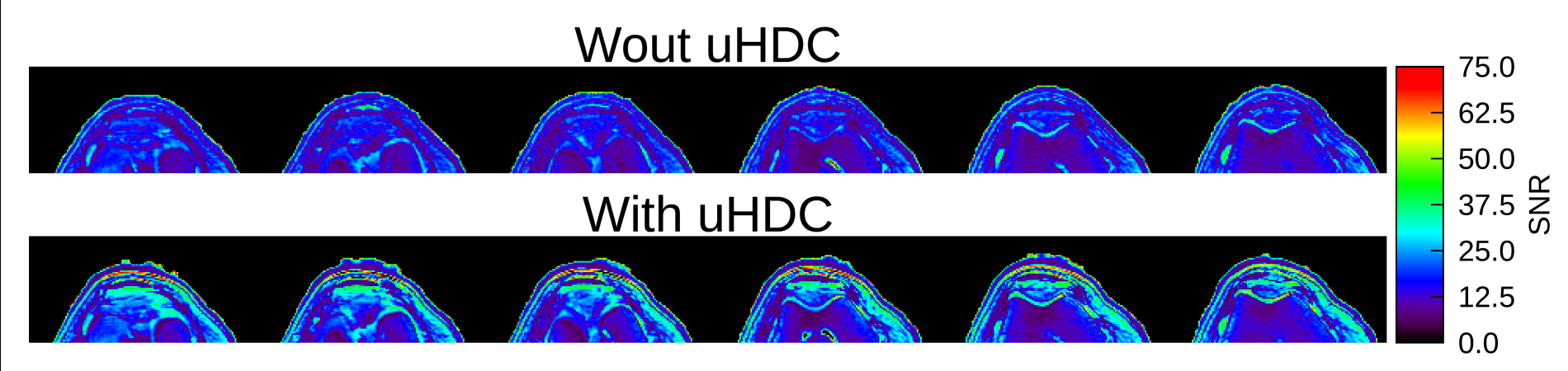
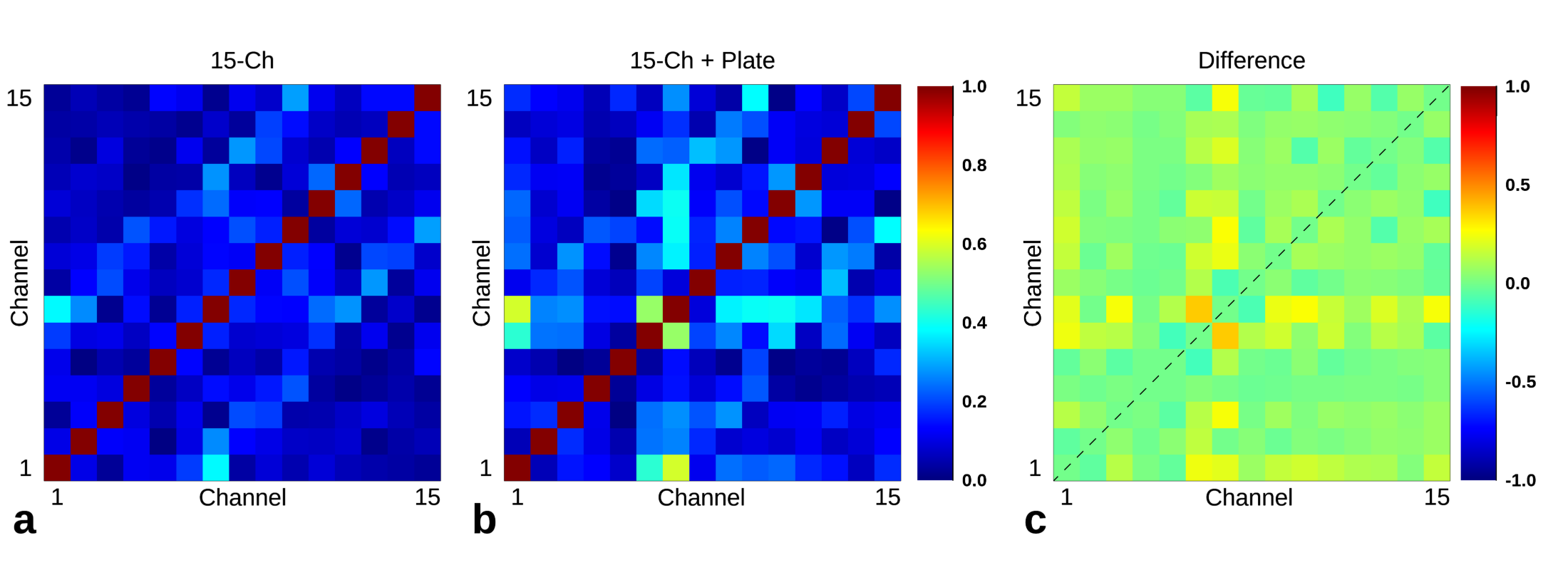
Transmit efficiency maps are displayed in Fig. 2. There is a strong enhancement of the transmit efficiency in the region close to the plate, with some local values reaching nearly double that of baseline. Regions that are distant to the plate suffer from a loss of transmit efficiency relative to baseline. Figure 3 displays a selection of SNR maps in a mosaic format. General regions of enhancement are marked with red rectangles, and the enhancement extends to adjacent slices. Figure 4 displays a cropped view of several slices. Within the cropped view, significant enhancement is observed in the patellar region up the top of the knee. Within the patella, the SNR enhancement ranged between 45 to 71%, measured across several slices. In the patellar cartilage, the enhancement ranged between 9.5 to 39.9%. Figure 4 displays the noise correlation matrix. A slight increase in noise correlation was observed between most neighboring pairs, save for the correlation between elements 6 and 7 which has an increase of about 0.5. Dielectric material overlapping neighboring coil elements can potentially cause strong increases in noise correlation.Discussion and Conclusion
The application of a curved uHDC plate within a clinical knee array offers SNR increases between 9 – 71% within a region of interest close to the material. The uHDC plate was constructed with a curvature suitable for this application but was not designed specifically for the knee array used in this study. Further SNR gains may be achievable with optimization of the uHDC permittivity and geometry. Gains in SNR are potentially useful for higher-resolution imaging of the surfaces within the knee. The B1+ mapping scans acquired in this study used approximately the same transmit power (both with and without the uHDC plate), which led to higher flip angles close to the dielectric (80 – 90 degrees) compared to baseline (50 degrees). When the flip angle is correctly calibrated in a region close to the material, a power reduction of about 70% would be expected. Additionally, there was an increase of B1+ inhomogeneity with the uHDC plate. The gradient echo scans acquired were resilient to the B1+ inhomogeneity, though this will pose an issue for spin-echo or RARE techniques. Careful calibration of the flip angle in a region of interest, or consideration of the uHDC permittivity and geometry, will be necessary to overcome this issue.Acknowledgements
We would like to acknowledge HyQ Research Solutions for providing the dielectric material used in this study. Sebastian Rupprecht and Qing X.Yang are affiliated with HyQ Research Solutions.References
[1] Rupprecht et al, “Improvements of transmit efficiency and receive sensitivity with ultrahigh dielectric constant (uHDC) ceramics at 1.5T and 3T”, MRM 2018; 79(5): 2842-51
[2] Koolstra et al, “Improved Image Quality and Reduced Power Deposition in the Spine at 3 T Using Extremely High Permittivity Materials”, MRM 2018; 79(5): 1192-1199
[3] Sica et al, “Toward whole‐cortex enhancement with an ultrahigh dielectric constant helmet at 3T”, MRM 2020; 83:1123-1134
[4] Chung et al, Rapid B1+ Mapping Using a Preconditioning RF Pulse with TurboFLASH Readout MRM“ MRM 2010; 64:439-446
[5] Kellman et al, “Image reconstruction in SNR units: a general method for SNR measurement”, MRM 2005; 54(6): 1439-47
Figures