2121
Comparison of different multiband acceleration factors on rhyming-task fMRI on a compact 3T scanner1Department of Radiology, Mayo Clinic, Rochester, MN, United States
Synopsis
Multiband acquisition can improve the temporal resolution of task-based fMRI at the cost of increased noise and artifacts. The optimal multiband acceleration depends on the task being studied, specific imaging protocol, and hardware involved. In the current work, multiband acceleration factors of 4, 5, and 6 were compared for a rhyming-task fMRI study using a 32-channel head coil on a compact 3T scanner with high-performance gradients. Multiband factors of 4 and 6 provided significant improvement over no acceleration, whereas a multiband factor of 5 was inconsistent and did not provide a significant improvement in half of the volunteer cohort.
Introduction
Multiple slices can be simultaneously acquired in a single RF excitation using multiband technique to increase the temporal resolution in fMRI. Multiband (i.e., simultaneous-multislice1) acquisition is becoming increasingly popular in fMRI studies as the increased temporal resolution typically results in greater statistical power.2–6 The multiband factor determines the number of slices that are acquired simultaneously. Larger multiband factors achieve better temporal resolution at the cost of reduced signal to noise ratio (SNR) and slice leakage artifacts. The impact of different multiband factors has been characterized for resting-state fMRI where optimal multiband factors tend to be between 4 to 8.7 However, these findings cannot be directly applied to task-based fMRI where the optimal multiband factor may depend on the brain region and specific task being studied.3,4The aim of the current study was to evaluate different multiband factors on rhyming-task fMRI performed on a compact 3T (C3T) scanner with high-performance gradients (80 mT/m and 700 T/m/s).8 Echo-planar-imaging scans on the C3T benefit from shortened echo spacing, leading to less signal dropout, and reduced distortion compared to conventional whole-body scanners.9 Rhyming-task fMRI is a well-established technique for visualizing language regions for neurosurgery planning.10 Task-based fMRI studies have indicated that a multiband factor of 4 is a suitable conservative value3,4 whereas rhyming-task fMRI studies have shown promising results with multiband factors of 52 and 6.6 In the current work, multiband factors of 1 (no acceleration), 4, 5, and 6 are compared in a clinical setting using FDA-approved analysis software.
Methods
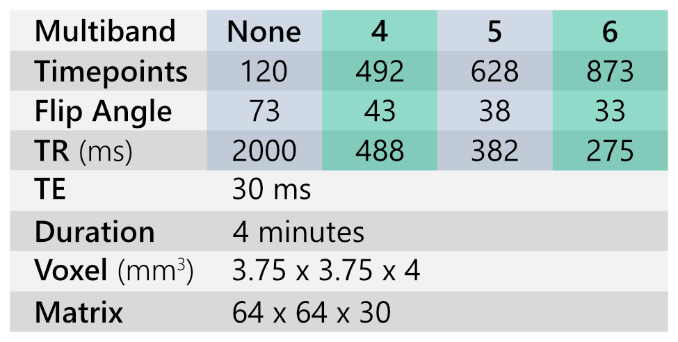
Following institutional review board approval of protocol and written informed consent, seven healthy volunteers (ages: 22–57, mean: 30) were scanned on a C3T scanner using a 32-channel head coil (Nova Medical). A rhyming-task protocol consisting of 6x40 second blocks was employed based on recommendations of the American Society of Functional Neuroradiology.10 The rhyming-task fMRI scan was repeated using multiband factors of 1 (no acceleration), 4, 5, and 6 in a single session. These multiband factors provided a temporal resolution of 2000, 488, 382, and 275 ms, respectively. The order of the scans was randomized to avoid task habituation bias. The scan parameters are listed in Table1.fMRI processing was performed using Prism Process (Prism Clinic Imaging, Elm Grove, WI) which is FDA-approved for fMRI analysis based on AFNI11. The processing steps included motion correction, spatial smoothing, and computation of filtered t-score maps. The different multiband factors were compared by: (i) computing the 99th percentile of the t-score map, and (ii) visually thresholding the t-score maps to provide the most informative visualization for neurosurgical planning and then noting the threshold value. The visual thresholding was performed by an experienced neuroradiologist with 14-years of experience in clinical fMRI.
Results
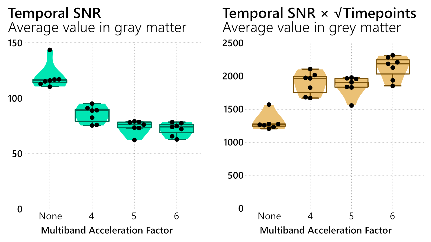
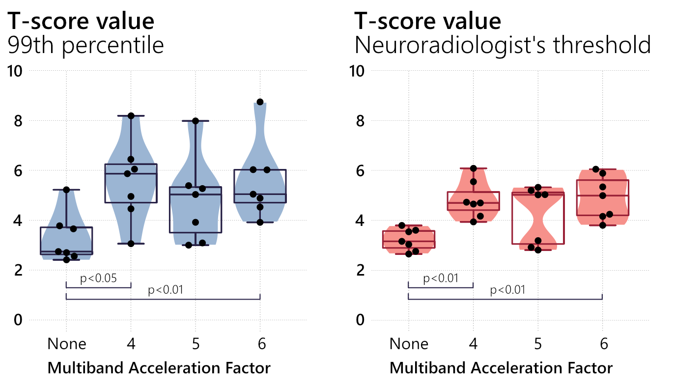
Temporal SNR in gray matter decreased as the multiband (MB) factor increased, but the difference between MB-5 and MB-6 was not substantial (Fig.1). The mean temporal SNR over gray matter volume across all subjects was 119 (MB-1), 85 (MB-4), 74 (MB-5), and 72 (MB-6).The 99th percentile of the t-map was larger at greater MBs, but the difference between MB-1 and MB-5 was not statistically significant (Fig.2-left). Similarly, the t-map threshold used by the neuroradiologist to visualize the language regions was also higher for greater multiband factors, but the difference between MB-1 and MB-5 was not statistically significant. The average t-score threshold across all subjects was 3.2 (MB-1), 4.8 (MB-4), 4.2 (MB-5), and 4.9 (MB-6). Although the mean threshold for MB-5 is larger than MB-1, it should be noted that MB-5 had a bi-modal distribution with negligible improvement for half the subjects when compared to the scan with no acceleration (Fig.2-right).
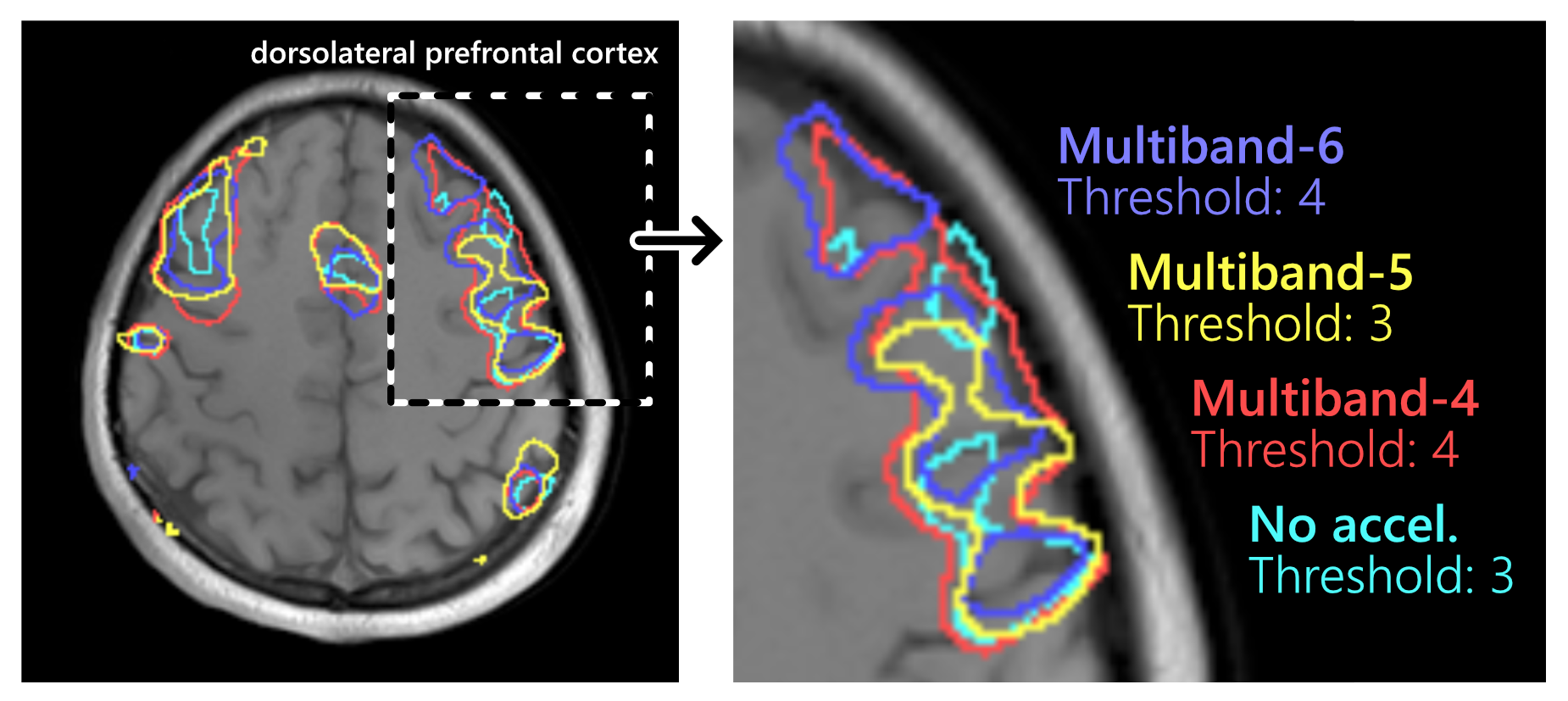
Figure 3 shows thresholded t-maps for a single subject where MB-4 and MB-6 can tolerate a higher threshold of 4 to visualize the language regions while MB-5 required a lower threshold of 3. This is an example of a scenario where MB-4 and MB-6 provided a substantial increase over MB-1 in t-score, while the increase for MB-5 was relatively modest.
Discussion
The relationship between multiband (MB) factor and statistical power was not intuitive, indicating that different MBs must be tested to design an optimized protocol. The t-scores were greatest with MB-4 and MB-6 and the difference between them was not statistically significant, indicating that the improved temporal resolution provided by MB-6 over MB-4 might be counteracted by its lower SNR. MB-5, on the other hand, had an inconsistent trend and it is unclear why it performed poorly compared to MB-4 and MB-6. A potential reason could be due to the coil geometry providing different g-factor penalties at different MB.Additional multiband factors were not explored in the current study because adding them would have lengthened the scanning session and could risk subject fatigue. In-plane acceleration was not utilized to isolate the impact of multiband factor but the combination of multiband with in-plane acceleration may be worth exploring in the future.
Conclusion
Multiband factors of 4 and 6 may be used in rhyming-task fMRI on a C3T scanner to provide maps with higher t-scores, allowing clinicians to use higher t-score threshold to suppress statistical noise when viewing the functional activation maps for pre-surgical planning.Acknowledgements
This work was supported by NIH U01 EB024450 and NIH U01 EB026979.References
1. Setsompop K, Gagoski BA, Polimeni JR, Witzel T, Wedeen VJ, Wald LL. Blipped-controlled aliasing in parallel imaging for simultaneous multislice echo planar imaging with reduced g-factor penalty. Magn Reson Med. 2012 May;67(5):1210-24. doi: 10.1002/mrm.23097.
2. Geng, X., Gu, H., Yang, Y., & Li-Hai, T. (2014). Multiband EPI in brain functional mapping – an fMRI study with rhyme judgment tasks. Proc. Intl. Soc. Mag. Reson. Med. 22.
3. Todd, N., Josephs, O., Zeidman, P., Flandin, G., Moeller, S., & Weiskopf, N. (2017). Functional sensitivity of 2D simultaneous multi-slice echo-planar imaging: Effects of acceleration on g-factor and physiological noise. Frontiers in Neuroscience, 11(MAR), 1–14. https://doi.org/10.3389/fnins.2017.00158
4. Demetriou, L., Kowalczyk, O. S., Tyson, G., Bello, T., Newbould, R. D., & Wall, M. B. (2018). A comprehensive evaluation of increasing temporal resolution with multiband-accelerated protocols and effects on statistical outcome measures in fMRI. NeuroImage, 176(April), 404–416. https://doi.org/10.1016/j.neuroimage.2018.05.011
5. McDowell, A. R., & Carmichael, D. W. (2019). Optimal repetition time reduction for single subject event-related functional magnetic resonance imaging. Magnetic Resonance in Medicine, 81(3), 1890–1897. https://doi.org/10.1002/mrm.27498
6. Mark, I. T., Black, D. F., DeLone, D. R., Passe, T. J., Witte, R. J., Little, J. T., Ho, M. L., Fagan, A. J., Parney, I. F., Burns, T. C., & Welker, K. M. (2020). Higher temporal resolution multiband fMRI provides improved presurgical language maps. Neuroradiology. https://doi.org/10.1007/s00234-020-02569-8
7. Risk, B. B., Murden, R. J., Wu, J., Nebel, M. B., Venkataraman, A., Zhang, Z., & Qiu, D. (2021). Which multiband factor should you choose for your resting-state fMRI study? NeuroImage, 234(March), 117965. https://doi.org/10.1016/j.neuroimage.2021.117965
8. Foo, T. K. F., Laskaris, E., Vermilyea, M., Xu, M., Thompson, P., Conte, G., Epps, C., Immer, C., Lee, S., Tan, E. T., Graziani, D., Mathieu, J., Hardy, C. J., Schenck, J. F., Fiveland, E., Stautner, W., Ricci, J., Piel, J., Park, K., … Bernstein, M. A. (2018). Lightweight, compact, and high‐performance 3 <scp>T MR</scp> system for imaging the brain and extremities. Magnetic Resonance in Medicine, 80(5), 2232–2245. https://doi.org/10.1002/mrm.27175
9. Kang, D., Jo, H. J., In, M.-H., Yarach, U., Meyer, N. K., Bardwell Speltz, L. J., Gray, E. M., Trzasko, J. D., Huston III, J., Bernstein, M. A., & Shu, Y. (2020). The benefit of high-performance gradients on echo planar imaging for BOLD-based resting-state functional MRI. Physics in Medicine & Biology, 65(23), 235024. https://doi.org/10.1088/1361-6560/abb2ec
10. Black, D. F., Vachha, B., Mian, A., Faro, S. H., Maheshwari, M., Sair, H. I., Petrella, J. R., Pillai, J. J., & Welker, K. (2017). American society of functional neuroradiology-recommended fMRI paradigm algorithms for presurgical language assessment. American Journal of Neuroradiology, 38(10), E65–E73. https://doi.org/10.3174/ajnr.A5345
11. Cox RW (1996). AFNI: software for analysis and visualization of functional magnetic resonance neuroimages. Comput Biomed Res 29(3):162-173. doi:10.1006/cbmr.1996.0014
12. Smith, S. M., Beckmann, C. F., Andersson, J., Auerbach, E. J., Bijsterbosch, J., Douaud, G., Duff, E., Feinberg, D. A., Griffanti, L., Harms, M. P., Kelly, M., Laumann, T., Miller, K. L., Moeller, S., Petersen, S., Power, J., Salimi-Khorshidi, G., Snyder, A. Z., Vu, A. T., … Glasser, M. F. (2013). Resting-state fMRI in the Human Connectome Project. NeuroImage, 80, 144–168. https://doi.org/10.1016/j.neuroimage.2013.05.039
Figures