2109
Altered rich club organization and dynamic structure-function relationship in treatment-naïve new-onset juvenile myoclonic epilepsy1Department of Magnetic Resonance, Lanzhou University Second Hospital, Lanzhou, China, 2Department of Biomedical Engineering, Zhejiang University, Zhejiang, China
Synopsis
Neuroimaging studies have shown that juvenile myoclonic epilepsy (JME) is characterized by impaired brain networks. However, few studies have investigated the potential disruptions in the rich club organization. Our study described the alterations of anatomical rich club organization and abnormalities of dynamic SC-FC coupling in treatment-naïve newly diagnosed JME. The results showed that the anatomical rich club organization was disrupted in the patient group, along with decreased connectivity strength among rich club hub nodes. The aberrant dynamic SC-FC coupling of rich club organization suggests a selective influence of interconnected network core in JME at the early phase of the disease.
Background/Purposes
Juvenile myoclonic epilepsy (JME) is the most common generalized genetic epilepsy syndrome, accounting for up to 10% of all epilepsies. Although the underlying neural substrates for JME remain elusive, recent neuroimaging studies have conceptualized JME as a disorder of brain network dysfunction, providing a new insight for understanding the underlying pathophysiology and etiology of this disorder. However, to our knowledge, no prior study has explored the time-varying characteristics of SC-FC coupling in JME, and it is still unclear how anatomical connectivity constraint supports dynamic functional interaction in the rich club organization. This study aims to investigate the changes of rich club organization and its dynamic SC-FC coupling in treatment-naïve newly diagnosed patients with JME.Materials and Methods
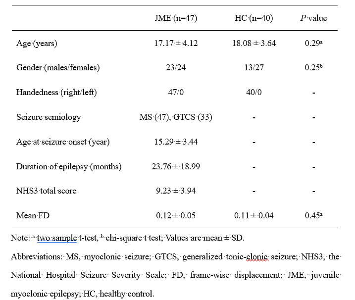
We investigated structural and dynamic functional brain networks, respectively, using diffusion-weighted imaging and resting-state functional magnetic resonance imaging data in 47 treatment-naïve new-onset patients with JME and 40 matched healthy controls. We focused on the anatomical rich club organization, as well as its structural connectivity and functional connectivity (SC-FC) coupling, in different dynamic states. We further evaluated dynamic functional network efficiency and its association with SC-FC coupling.Results:
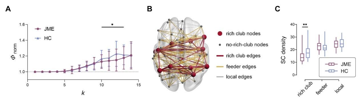
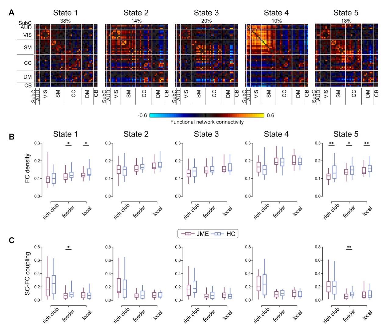
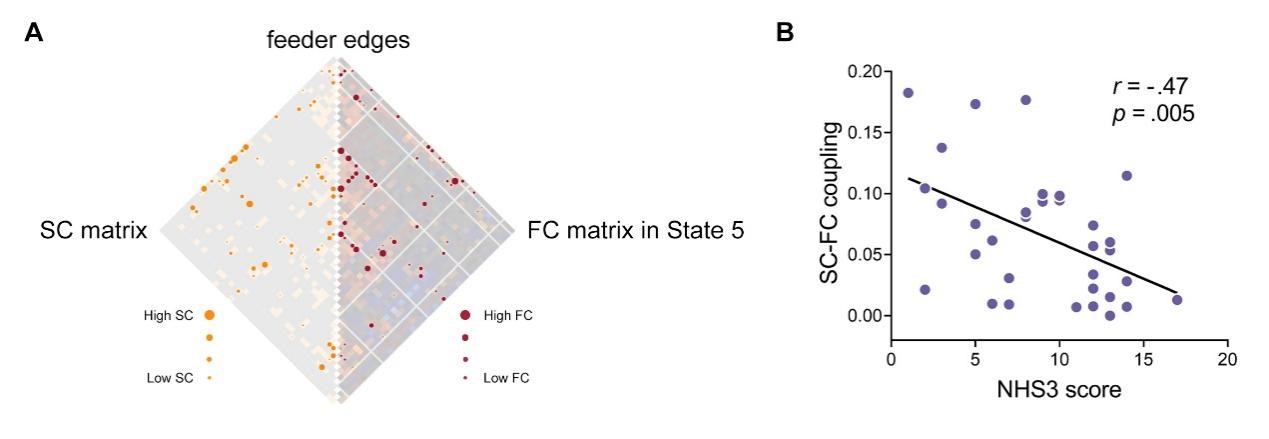
Disrupted organization of anatomical rich club, along with decreased connectivity strength, were found in the patient group . Furthermore, reduced SC-FC coupling in rich club organization of the patients was found in two functionally independent states, i.e., the functional segregation state (State 1) and the strong somatomotor-cognitive control interaction state (State 5); and the later was significantly associated with disease severity. In addition, the relationships between SC-FC coupling of rich club hub’s connections and functional network efficiency in State 1, which had been established in healthy controls, were absent in patients.Conclusions
In this study we performed a combination analysis of SC and dynamic FC, for the first time, to examine the abnormalities in the rich club organization and its dynamic SC-FC coupling in treatment-naïve newly diagnosed JME. Beyond previous studies reporting altered the rich club system and structure-function coupling in generalized epilepsy, we not only found significant rich club organization deficits but also state-specific SC-FC coupling alterations in patients with JME. These results suggest the rich club disorganization and its disrupted role in dynamic structure-function coupling for JME patients at the early phase of the disease, which may contribute to the cognitive deficits such as impaired executive function in patients during development, advancing our understanding of the neurobiology.Acknowledgements
This work was supported by the National Natural Science Foundation of China (No.82160326) and Cuiying Scientific and Technological Innovation Program of Lanzhou University Second Hospital(No. CY2018-QN03).
References
1. Scheffer IE, Berkovic S, Capovilla G, et al. ILAE classification of the epilepsies: Position paper of the ILAE Commission for Classification and Terminology. Epilepsia 2017;58:512-521.
2. Jiang S, Luo C, Gong J, et al. Aberrant Thalamocortical Connectivity in Juvenile Myoclonic Epilepsy. International journal of neural systems 2018;28.
3. Ur Ozcelik E, Kurt E, Sirin NG, et al. Functional connectivity disturbances of ascending reticular activating system and posterior thalamus in juvenile myoclonic epilepsy in relation with photosensitivity: A resting-state fMRI study. Epilepsy research 2021;171:106569.
4. Zhang Z, Liu G, Zheng W, Shi J, Liu H, Sun Y. Altered dynamic effective connectivity of the default mode network in newly diagnosed drug-naive juvenile myoclonic epilepsy. NeuroImage Clinical 2020;28:102431.
5.Lee HJ, Park KM. Structural and functional connectivity in newly diagnosed juvenile myoclonic epilepsy. Acta Neurol Scand 2019;139:469-475.
Figures