2097
Model validation of a 500 MHz Inductive Resonator for 11.7 T Brain MRI
Natalia Gudino1, Jacco A de Zwart2, Peter Van Gelderen2, Stephen J Dodd1, Jeff H Duyn2, and Joe Murphy-Boesch2
1LFMI, NIH, Bethesda, MD, United States, 2NIH, Bethesda, MD, United States
1LFMI, NIH, Bethesda, MD, United States, 2NIH, Bethesda, MD, United States
Synopsis
To evaluate safety of a 500 MHz inductive birdcage resonator for human brain MRI at 11.7 T, we compared phantom SAR measurements with predictions based on simulations. On a head-shaped phantom with realistic permittivity and conductivity, good correspondence was achieved in heating distributions when irradiating the phantom with 85 W RF power. The ability to predict SAR at 500MHz with simulation is critical step towards in-vivo MRI at 11.7 T.
Introduction
With the goal of improving sensitivity and contrast for MRI of human brain, we are currently developing transmit and receive RF hardware for the 11.7 T head-only MRI system being developed at our institution. As part of this, we are performing RF safety assessment of any novel hardware. A critical point in this assessment is the accuracy of the coil model used to simulate transmit B1 and associated electrical field. In this work, we present electromagnetic field simulations, benchtop measurements and power tests performed with different coil-phantom setups to validate the model of a 500 MHz inductive birdcage transmit-receive head coil1,2. Towards comprehensive safety analysis, we plan to extend our electromagnetic field simulations with realistic human brain models (virtual family3) at various position inside the coil.Methods
A detailed electrical model of the coil was developed in FDTD-based EM simulation software (Remcom, XFdtd 7.9) as illustrated in Figure 1a and b. Optimization of the model and mesh were performed through several iterations based of bench measurements of scattering parameters using the physical hardware. The expected symmetry in the scattering parameter matrix of the coil model was tested first with a head-size sphere. After this first validation, simulations were run with a model of a head-shaped phantom filled with a polyvinylpyrrolidone-based solution4 having a conductivity and permittivity of average brain tissue at 500 MHz (ε=48.42 and σ=0.63 S/m, https://amri.ninds.nih.gov/phantomrecipe.html). A first experimental power validation of the model was performed with the quadrature ports of the coil connected to 2 independent Tx channels of a 11.7 T animal system (11.7T Bruker Avance Neo Console). Total average power at the coil input was 82 W. Based on simulated SAR maps, optical temperature probes were placed at three different locations inside the head phantom centered inside the coil. SAR at each location was estimated from the slope of the temperature curve and compared with the value obtained in simulation at that location. A second experimental power validation was performed using MRI thermometry at 3 T. In this setup, each port of the resonator was connected to a 50 W broadband RF power amplifier (LZY, 20-512 MHz, Mini-Circuits) through a 90° hybrid for quadrature excitation. The head resonator and phantom were located inside the scanner bore at isocenter. A sliding support allowed moving the coil out the integrated body coil to image the phantom for MR thermometry5. Gel capsules were placed at the edge of the resonator to determine phantom placement relative to the coil using imaging. Three oil tubes were placed around the phantom to obtain a reference for post-processing correction of frequency drifts. A baseline MRI acquisition was performed (1-min temporal, 4 mm spatial resolution) followed by cycles of 2- or 5-minute 500 MHz RF excitation of total average power of 85 W, interleaved with cool-down periods. MRI thermometry was performed in between and after heating cycles. Temperature was also monitored using 2 optical fiber probes. For this experiment, 2% agarose was added to the polyvinylpyrrolidone-based solution to reduce heat convection.Results
Simulated and measured frequency responses (S-parameter measurement) were in good agreement. Figure 1c shows as example the coupling between couplers C1 and C3 for all resonance modes. Figure 1d shows the homogenous mode measured on the bench and simulation space with an RF probe located at the center of the resonator and only C1 and C3 exciting the linear mode. In the first power validation test, we were able to readily predict the location of hot and cold spots based on simulated SAR maps. Figure 2a shows simulated sagittal SAR maps and approximate locations of temperature probes inside phantom (Figure 2b). Figure 2c shows temperature curves for the 5-minute heating and cooling cycle. Figure 2d shows the initial slopes during the heating cycle used for SAR estimation. Measured and simulated heating patterns across multiple axial slices with 16 mm separation are shown in Figure 3a. Probe location could be assessed accurately, as they were visible in the MR images. Linear regression of temperature curves at 2 locations during the first part of an RF heating cycle is shown in Figure 3b.Measured and simulated SAR ratios at these probe locations, for both RF power tests, are shown in Figure 3c.Discussion
Preliminary simulated and experimental data are presented for 500 MHz coil model validation. Model adjustment was done manually with values of lumped elements set within 10 % of values implemented in hardware. Simulated reflection coefficient amplitudes were within 8.5 % of measured values. Co-simulation methods could be implemented for finer model adjustment as previously presented by others6-8. To determine absolute measured SAR, we will confirm PVP solution heat capacity estimation with measurements. MR thermometry data will be validated using an increased number of temperature probes. This validation workflow was designed based on the hardware frequency response and electrical field distribution across a head phantom. Currently, validation of the B1 distribution is not possible since our 11.7 T system is not at field yet. Further workflow optimization will allow us to complete validation of this single channel transmitter and extend to validation of multi-channel transmitters.Acknowledgements
References
- Murphy-Boesch J, Dodd S, van Gelderen P, et al. Proc. ISMRM 2011; 19: 3986.
- Murphy-Boesch J, Duan Qi, Dodd S, et al. Proc ISMRM 2014; 22: 4803.
- Christ A, Kainz W, Hahn EG , et al. Phys Med Biol. 2010 Jan 21;55(2):N23-38
- Ianniello C, de Zwart JA, Duan Q, et al. Magn Reson Med. 2018 Jul;80(1):413-419.
- De Zwart JA, Vimeux FC, Delalande C, et al. Magn Reson Med. 1999 Jul;42(1):53-9.
- Restivo M, Raaijmakers A, van den Berg C, et al. Magn Reson Med. 2017;77:2040–2047.
- Kozlov M, Turner R. J Magn Reson. 2009;200:147–152.
- Sadeghi-Tarakameh A, DelaBarre L, Lagore RL, et al. Magn Reson Med. 2020 Jul;84(1):484-496.
Figures

Figure 1: (a-d) Simulated versus measured performance of transmit-receive volume resonator. (a) Physical 500 MHz Tx/Rx coil resonator and (b) its 3D model, driven by 4 inductive couplers C1-C4 located azimuthally 90 degrees apart. Zoomed sections are shown in the right of panels (a) and (b). Frequency response measurement: (c) Measure and simulated coupling between C1 and C3 (d) and frequency response detected with a probe located at the center of the resonator while simultaneously driving the same pair of couplers.
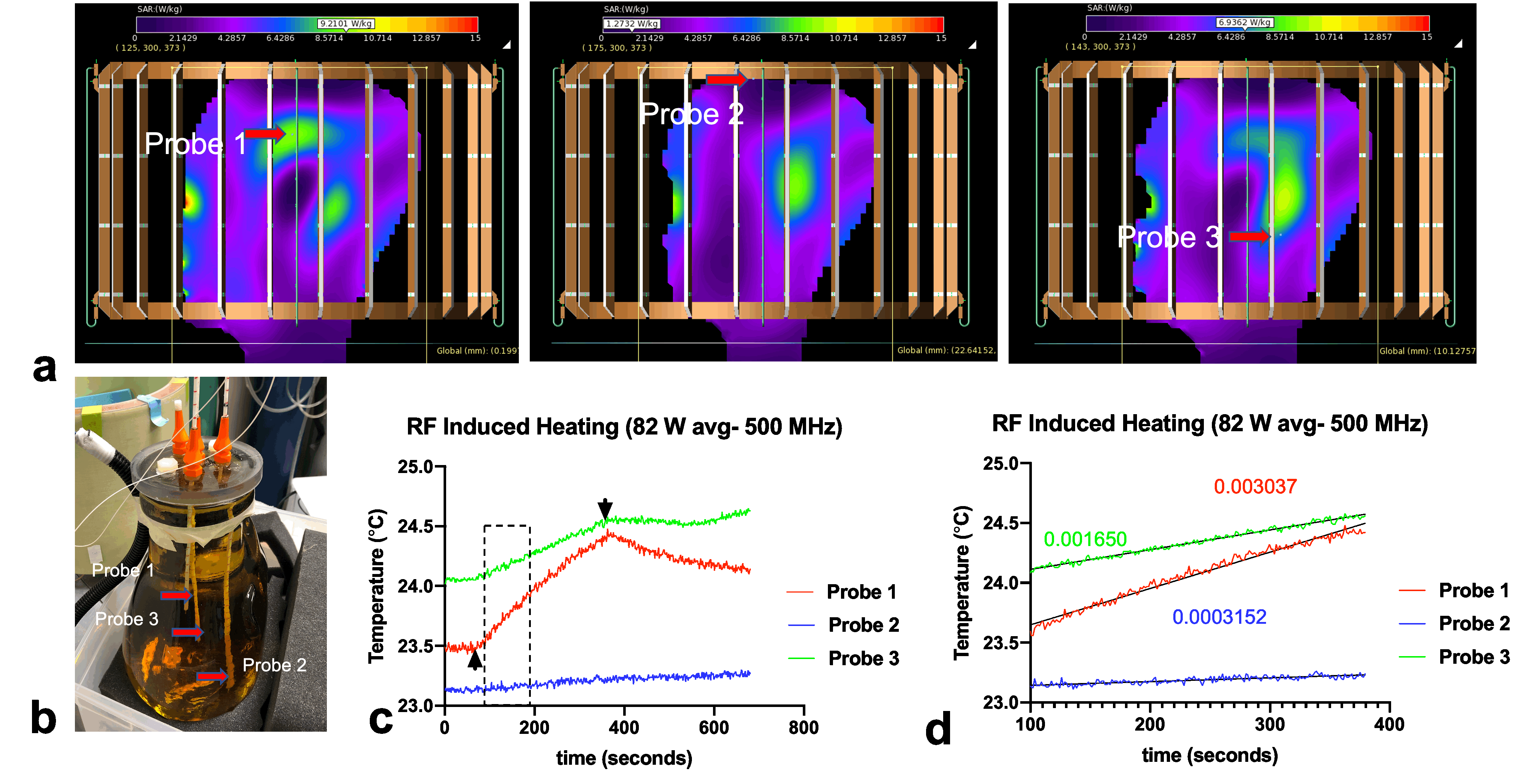
Figure 2: (a) Simulated SAR map in the brain phantom model (average brain permittivity and conductivity). (b) Picture of the PVP phantom with temperature probes inserted in 3 different locations selected from simulated SAR maps. (c) Temperature measurement during RF transmission (82 W net available power) and cool down cycles. (d) Temperature curves and estimated slope values (°C/seconds) during the first part of the RF transmission cycle.
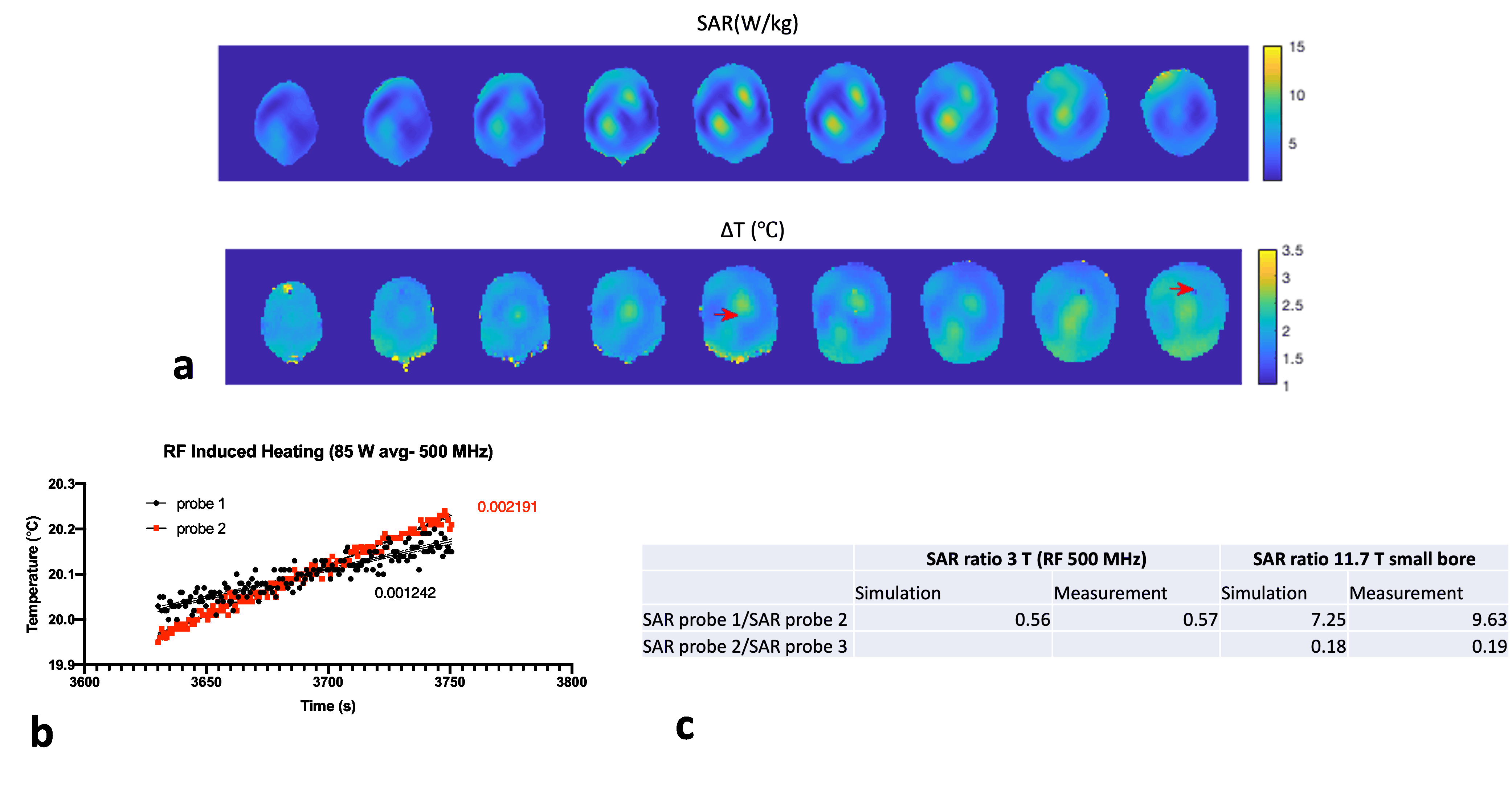
Figure 3: (a) Simulated SAR (top) and experimental MR thermometry maps (bottom) of 9 axial slices with 16 mm separation for a 5-min, 85 W heating experiment. (b) Linear regression of temperature curves and estimated slopes (°C/seconds) during the first part of the RF transmission cycle. Approximate locations of the 2 temperature probes are indicated with red arrows on the thermometry maps (c) Simulated and measured SAR ratio at temperature probe locations for both RF power experiments.
DOI: https://doi.org/10.58530/2022/2097