2092
Parallel Transmit DANTE-SPACE for improved black-blood signal suppression at 7 Tesla1Wellcome Centre for Integrative Neuroimaging, FMRIB, Nuffield Department of Clinical Neurosciences, University of Oxford, Oxford, United Kingdom
Synopsis
DANTE-SPACE can be used for visualizing intracranial vessel walls at 7T, where the flow-sensitive signal attenuation of the DANTE preparation simultaneously suppresses signal from the internal blood and external CSF. However, the efficacy of DANTE is reduced for blood moving upstream through the neck due to limited B1+ coverage at 7T. Here, EPG simulations are used to estimate the effect of this limited B1+ coverage on the achieved contrasts. A pTx-based approach which uses separate RF shims for the DANTE-preparation and the SPACE readout to improve the attenuation of blood moving through the neck is proposed, and validated in vivo.
Introduction
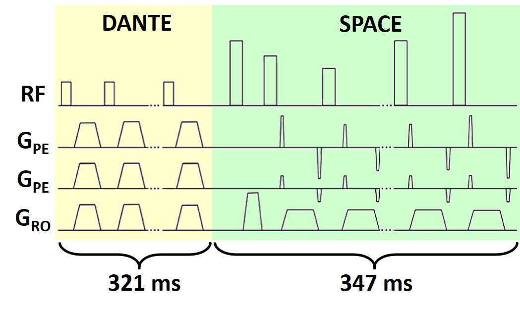
Vessel wall imaging (VWI) can be used to assess the pathology of intracranial vessel walls. For the assessment of intravascular pathology in the vessels above and around the Circle of Willis, such as the middle cerebral arteries (MCAs), the lumen signal needs to be suppressed while retaining signal from the vessel wall. A previous study presented the DANTE-SPACE sequence at 7T1 (Figure 1), which achieves delineation of the MCA walls by employing an optimized variable-flip-angle turbo spin-echo sequence (SPACE2) with additional DANTE-preparation3 for flow-sensitive signal suppression of both the CSF and the lumen. However, assessment of intravascular pathology and calculation of vessel wall thickness using black-blood sequences such as DANTE-SPACE can be hindered by remaining unsuppressed blood signal.In this work, a previously introduced framework for the simulation of DANTE-SPACE4 was used to identify and quantify the cause of this unsuppressed blood signal. Based on the simulation results, a parallel transmission (pTx)5 implementation of DANTE-SPACE is proposed to reduce the effects of limited B1+ coverage in the neck at 7T, and thereby obtain improved blood suppression.
Methods
DANTE-SPACE was simulated using the extended phase graph (EPG) framework6,7. For accurate representation of the blood signal, the simulations included pulsatile flow velocity variations8, estimated flow trajectories, and B1+ variations in different parts of the vasculature based on in vivo B1+ maps. Constant B1+ signal was assumed for the vessel walls.To characterize the limitations when using single-channel transmit coils, B1+ flip angle maps were acquired using the 3D-DREAM sequence9 from four volunteers on a 1Tx/32Rx system. To assess the potential B1+ improvements when using pTx, additional channel-by-channel relative B1+ maps were acquired in 3 volunteers for all 8 transmit channels using an 8Tx/32Rx coil. Those individual maps were combined using circular polarization (CP)-mode, as well as using an optimized radiofrequency (RF) shim aimed at maximizing the minimum B1+ values in the neck.
DANTE-SPACE signal was both simulated and acquired (from 3 healthy volunteers) using the acquisition parameters used in Ref. (1), adopting the optimized DANTE-preparation proposed in Ref. (4) for improved contrast and reduced SAR. These parameters include 200 DANTE pulses of 9º, and a SPACE readout consisting of 75 pulses with an echo spacing of 4.6ms. The TR for each readout was 2.64s. The scan time was 11 minutes for data acquired at 0.5×0.5×1.0 mm3 using a GRAPPA acceleration factor of 4. pTx DANTE-SPACE data were acquired using an 8Tx/32Rx coil on a Siemens Magnetom 7T scanner (Erlangen, Germany) under an institutional ethical agreement. Rigid-body image registration was used where necessary.
Results
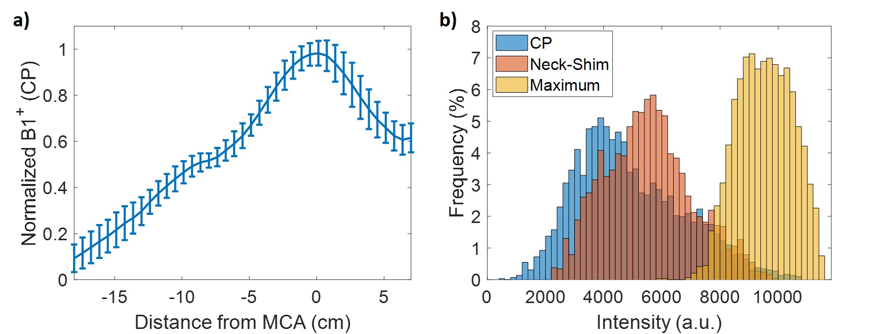
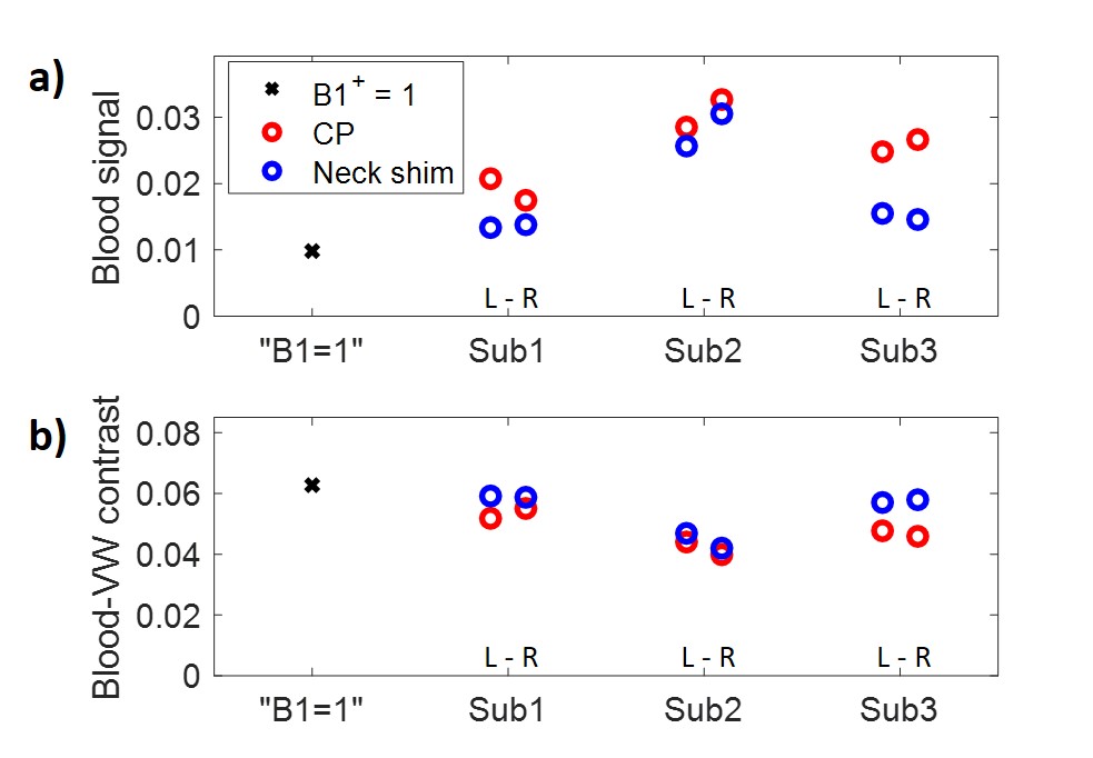
Figure 2a shows measured B1+ profiles at 7T from four volunteers. Figure 2b shows the possible B1+ improvement in the neck when using pTx with a neck-specific RF shim, based on masked voxels in the neck of 3 volunteers.Figure 3 shows the simulated blood signal and blood/vessel wall contrast when running pTx DANTE-SPACE with both DANTE and SPACE in CP-mode (red circles), and when running a neck-specific pTx RF shim for DANTE and CP-mode for SPACE (blue circles). On average, the blood signal is expected to decrease by 26±15% when a neck-shim is used during DANTE-preparation.
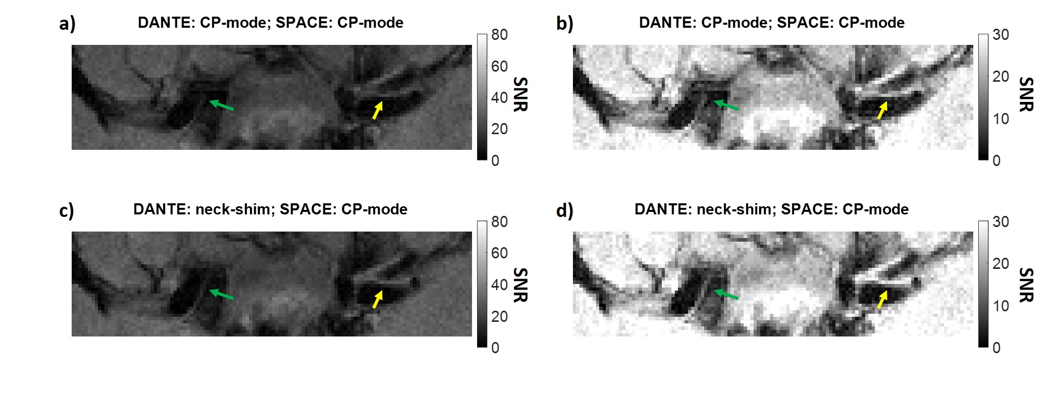
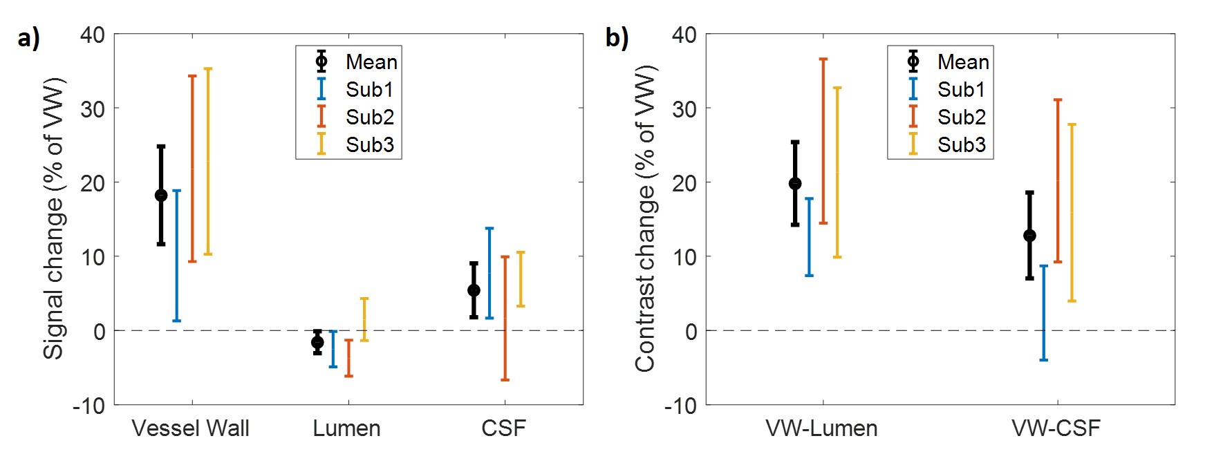
Figure 4 shows a single-slice segment acquired both with and without a neck-specific RF shim for the DANTE-preparation. Figure 5 shows a comparison of the resulting signal in the lumen, vessel wall, and CSF for multiple slices in all three volunteers, with values relative to the vessel wall signal in CP. Across the three subjects, the lumen signal is found to decrease (by 7±6% of the CP-mode lumen signal), while the vessel wall and CSF signal both increase.
Discussion
DANTE-SPACE simulations (Figure 3) show a reduced vessel wall/blood contrast as a result of the reduced B1+ coverage in the neck of a 1Tx system at 7T, which reduces the efficiency of the DANTE-preparation on blood spins flowing into the MCAs from the neck. If the B1+ coverage is increased during DANTE-preparation by using a pTx shim tailored for maximizing the minimum signal in the neck, the simulations show improved suppression of the blood signal.The reduction in blood signal using a neck-specific shim is also visible in vivo (Figures 4-5), while the signal from both the vessel walls and the CSF increases. Those signal increases can be a result of reduced B1+ outside the neck when using the neck-specific shim, resulting in reduced DANTE-suppression of the vessel walls and CSF. Although this was not a target during this work, the increased vessel wall signal further increases the vessel wall/lumen contrast, while also providing increased vessel wall-CSF contrast despite the higher CSF signal. Future work will look into increasing this effect using RF shims that aim to increase the B1+ in the neck while simultaneously suppressing the B1+ around the vessels of interest. Future work will also study the potential of using dielectric pads10 around the neck for additional improvement of the B1+ in the neck.
Conclusion
Suppression of blood signal in DANTE-SPACE at 7T is less effective due to reduced B1+ coverage in the neck. Using a neck-specific pTx shim during the DANTE-preparation reduces this effect and improves vessel wall contrast.Acknowledgements
MdB receives financial support from Siemens Healthineers and the Dunhill Medical Trust. JK is supported by an iCASE stipend award in collaboration with Siemens Healthineers. PJ receives support from the Dunhill Medical Trust and the NIHR Oxford Biomedical Research Centre. The Wellcome Centre for Integrative Neuroimaging is supported by core funding from the Wellcome Trust (203139/Z/16/Z).
References
1. Viessmann O, Li L, Benjamin P, Jezzard P. T2-Weighted intracranial vessel wall imaging at 7 Tesla using a DANTE-prepared variable flip angle turbo spin echo readout (DANTE-SPACE). Magnetic Resonance in Medicine. 2017;77(2):655-663. doi:10.1002/mrm.26152
2. Mugler JP. Optimized three-dimensional fast-spin-echo MRI. Journal of Magnetic Resonance Imaging. 2014;39(4):745-767. doi:10.1002/jmri.24542
3. Li L, Miller KL, Jezzard P. DANTE-prepared pulse trains: A novel approach to motion-sensitized and motion-suppressed quantitative magnetic resonance imaging. Magnetic Resonance in Medicine. 2012;68(5):1423-1438. doi:10.1002/mrm.24142
4. de Buck MHS, Hess AT, Jezzard P. Optimized DANTE preparation for intracranial DANTE-SPACE vessel wall imaging at 7T. Proceedings of the 29th Annual Meeting of ISMRM. Published online 2021:4201.
5. Padormo F, Beqiri A, Hajnal JV, Malik SJ. Parallel transmission for ultrahigh-field imaging. NMR in Biomedicine. 2016;29(9):1145-1161. doi:10.1002/nbm.3313
6. Hennig J. Multiecho imaging sequences with low refocusing flip angles. Journal of Magnetic Resonance (1969). 1988;78(3):397-407. doi:10.1016/0022-2364(88)90128-X
7. Weigel M. Extended phase graphs: Dephasing, RF pulses, and echoes - Pure and simple. Journal of Magnetic Resonance Imaging. 2015;41(2):266-295. doi:10.1002/jmri.24619
8. Augst AD, Barratt DC, Hughes AD, Thom SAM, Xu XY. Various issues relating to computational fluid dynamics simulations of carotid bifurcation flow based on models reconstructed from three-dimensional ultrasound images. Journal of Engineering in Medicine. 2003;217(5):393-403. doi:10.1243/095441103770802568
9. Ehses P, Brenner D, Stirnberg R, Pracht ED, Stöcker T. Whole-brain B1-mapping using three-dimensional DREAM. Magnetic Resonance in Medicine. 2019;82(3):924-934. doi:10.1002/mrm.27773
10. Teeuwisse WM, Brink WM, Webb AG. Quantitative assessment of the effects of high-permittivity pads in 7 Tesla MRI of the brain. Magnetic Resonance in Medicine. 2012;67(5):1285-1293. doi:10.1002/mrm.23108
Figures