2091
Traveling pulses visit 7T Terra sites: Getting ready for Parallel Transmission in routine use1Paris-Saclay University, CEA, CNRS, BAOBAB, NeuroSpin, Gif-sur-yvette, France, 2Siemens Healthcare SAS, Saint-Denis, France, 3German Center for Neurodegenerative Diseases (DZNE), Bonn, Germany, 4Montreal Neurological Institute, McGill University, Montreal, QC, Canada, 5Siemens Healthineers Ltd, Seoul, Korea, Republic of, 6Techna Institute & Koerner Scientist in MR Imaging, University Health Network, Toronto, ON, Canada, 7Center for Neuroscience Imaging Research, Institute for Basic Science & Department of Biomedical Engineering, Sungkyunkwan University, Suwon, Korea, Republic of, 8CHU Poitiers, Poitiers, France, 9LRCOM I3M, University and University Hospital of Poitiers, Poitiers, France, 10Laboratory of Applied Mathematics, UMR CNRS 7348, University of Poitiers, Poitiers, France
Synopsis
Parallel transmission universal pulses (UPs) consist of pre-optimized RF pulse solutions mitigating the RF field inhomogeneity problem for a given RF coil. Optimized offline on a database of representative field maps, they are designed to be robust to intersubject variability and spare the user a cumbersome online calibration. Initially designed for the MAGNETOM 7T Classic system from Siemens Healthineers, with the RF coil being strictly identical, this abstract describes the set of transformations on the pulses necessary to meet the MAGNETOM Terra specifications as well as successful in vivo results already achieved at four Terra sites using the same UPs.
Purpose
Despite recent progress, the use of parallel transmission (pTx) to mitigate the RF field inhomogeneity problem at UHF has remained marginal due to a cumbersome calibration procedure requiring subject-specific field mapping and pulse design, furthermore requiring expertise. Universal pulses (UPs) were proposed to circumvent this problem by providing calibration-free solutions. These “traveling” pulses were shown to be immune to inter-site variability1 among ten sites equipped with MAGNETOM 7T Classic. For broader dissemination and impact, the purpose of this work was to transform the pulses to meet the new specifications of the more widespread MAGNETOM Terra (Siemens Healthineers, Erlangen, Germany) and to verify again inter-site robustness.Methods
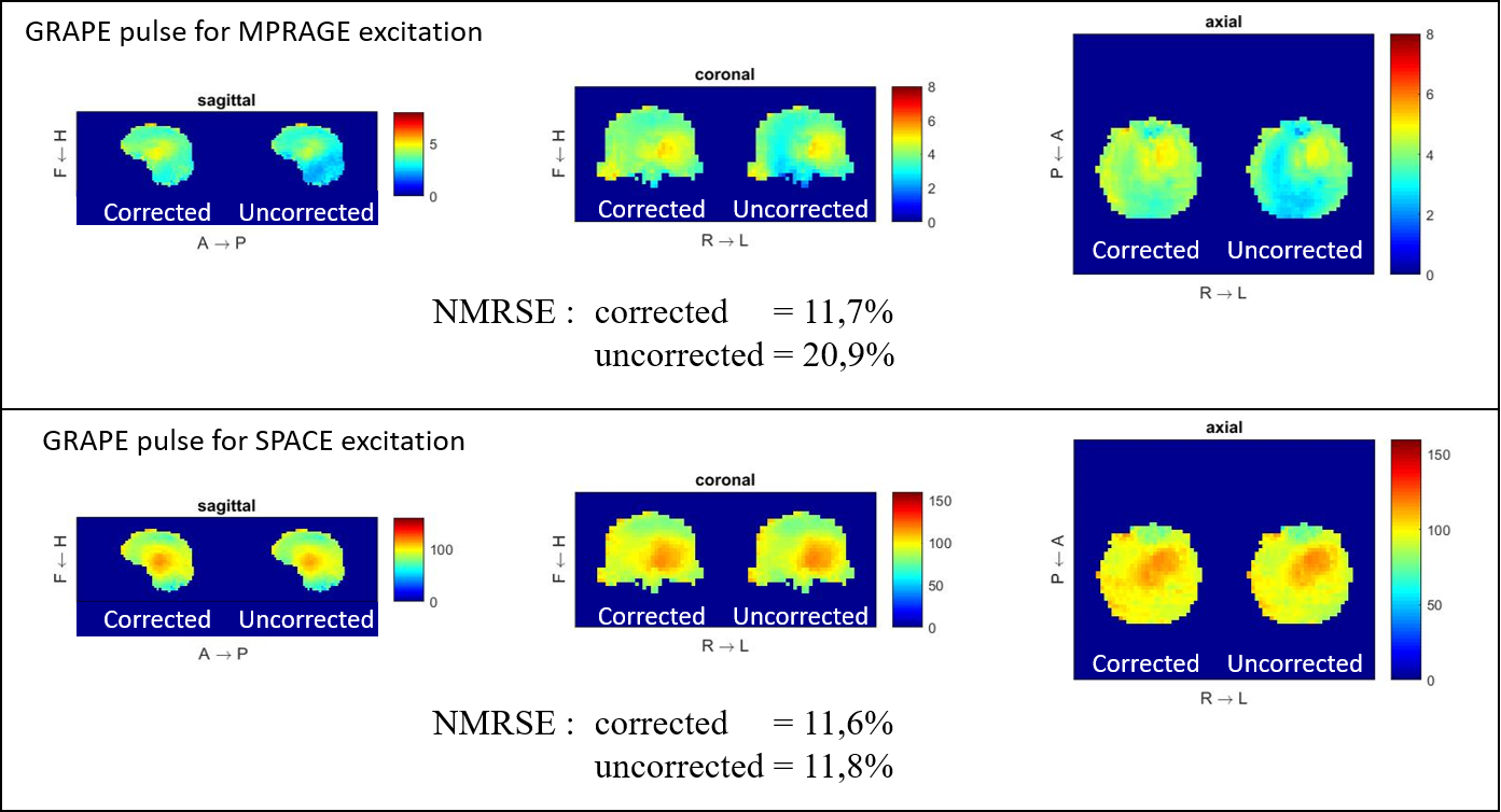
Universal pulses were designed based on a database of 20 B1+ and B0 field maps acquired at NeuroSpin (CEA Saclay, France) and DZNE (Bonn, Germany) on whole-body investigational MAGNETOM 7T scanners, both equipped with the same SC72 gradient coil, as well as a 8Tx/32Rx RF coil (Nova Medical, Wilmington, MA, USA). The UPs embedded in the PASTEUR2 package are non-selective kT-point3 and GRAPE4 pulses, and incorporate small excitation, inversion and refocusing pulses which were inserted in the MP(2)RAGE, GRE3D, vFA-TSE (FLAIR5, DIR) sequences. The following transformations were performed on the PASTEUR pulses to meet the MAGNETOM Terra specifications. CP mode phases: on the MAGNETOM 7T Classic, the coil file applies a CP mode by default with 45 deg increments implemented within the Siemens SyngoMR VB17 software. This is no longer the case on the MAGNETOM Terra, so the phase shifts were added accordingly to the RF waveforms. Voltage definitions: voltages are defined at the coil plug and TALES respectively for the 7T Classic and Terra. There is 1.61 dB power attenuation between the two so that the pulse voltages were scaled up accordingly. Channels permutation: RF waveforms were permuted according to 1$$$\rightarrow$$$8, 2$$$\rightarrow$$$1, 3$$$\rightarrow$$$2 etc., to correspond to the new labeling convention on the Terra. Table position: the patient bed is believed to be located 1.6 cm higher in the A-P direction with respect to isocenter. This affects RF pulse performance, as shown in Figure 1, whenever the Y gradient is used. Fortunately, this offset can be corrected analytically by modulating the phase of the RF across all channels according to the integral of the gradient waveform, thereby recovering the same exact performance. The different pulses were integrated into the PASTEUR sequences and disseminated to four different Terra sites for testing. For comparison, brain images were also acquired from the same subjects at each site using the Circularly-Polarized (CP) mode with the same coil (TrueForm mode). All MR sequences run under the local SAR supervision system provided by the manufacturer for pTx imaging. Chosen parametrizations were always compatible with UHF clinical research standards.Results
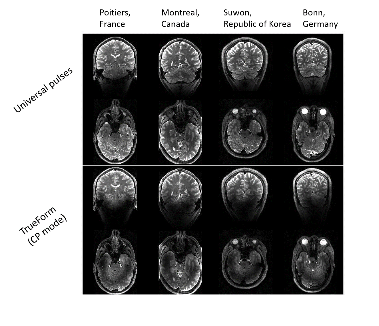
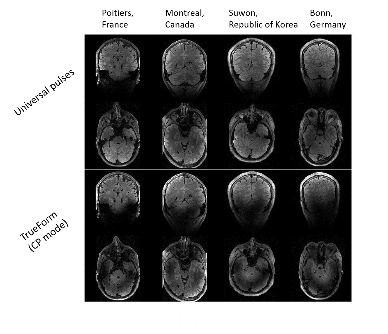
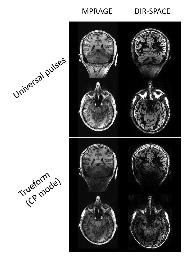
UP versus CP comparisons acquired at Poitiers (France), Montreal (Canada), Suwon (Republic of Korea) and Bonn (Germany) are shown in Figure 2 for the vFA-TSE (SPACE) sequence and in Figure 3 for the T2-prepared FLAIR sequence. Figure 4 shows the UP versus CP comparison for MPRAGE and DIR-SPACE sequences acquired at Poitiers (France). The excitation, inversion and refocusing pulses mitigate to a great extent the RF field inhomogeneity problem while sparing the user any calibration or calculation procedure.Discussion/Conclusion
Here we have shown that the UPs designed using a database of pTx field maps acquired on the MAGNETOM 7T Classic can be transformed to mitigate the RF field inhomogeneity problem on the MAGNETOM Terra. The proposed 3D MR sequences incorporating the UPs can be applied directly, without subject specific calibration, to collect high quality whole brain structural T1w and T2w images at 7T. The sequences and pulses are part of the PASTEUR package, which is available to Terra users upon C2P request. Additionally to the four testing sites reported here, nearly 30 healthy volunteers from around a total of ten sites have been scanned so far without encountering any failure. When analyzed, images were extremely well received by local radiologists. The traveling pulses are COVID-free and are willing to continue to travel along their journey toward being widely accepted as a calibration-free, B1+ mitigation solution that can benefit a large array of neuroimaging applications at ultrahigh field. More work is under way to complete the package with additional sequences (such as 3D-EPI and 3D slab-selective pulses) incorporating UP solutions.Acknowledgements
This project has received funding from the European Union’s Horizon 2020 research and innovation program under grant agreement No 885876 (AROMA project). The authors also thank their collaborators in London (King’s college and UCL), Charlestown (MGH), Minneapolis (CMRR), Leipzig (MPI), Trondheim (NTNU) and Rehovot (WIS) for testing the UP sequences.References
[1] X. Wu, V. Gras, A. Vignaud, F. Mauconduit, M. Boland, T. Stoecker, K. Ugurbil, N. Boulant. The travelling pulses: multicenter evaluation of universal pulses at 7T. In proceedings of the 27th international meeting of the ISMRM, Paris, France, p 1133.
[2] V. Gras, F. Mauconduit, A. Vignaud, C. Le Ster, L. Leroi, A. Amadon, E. Pracht, M. Boland, R. Stirnberg, Rüdiger, T. Stöcker, B. Poser, C. Wiggins, X. Wu, K. Ugurbil, N. Boulant. PASTeUR: Package of Anatomical Sequences using parallel Transmission UniveRsal kT-point pulses. In proceedings of the 28th international meeting of the ISMRM, Montreal, Canada, p 4626.
[3] Cloos MA, Boulant N, Luong M, Ferrand G, Giacomini E, Le Bihan D, Amadon A. kT-points: Short three-dimensional tailored RF pulses for flip-angle homogenization over an extended volume. Magn Reson Med 2012;67:72–80.
[4] L. Van Damme, F. Mauconduit, T. Chambrion, N. Boulant, V. Gras. Universal nonselective excitation and refocusing pulses with improved robustness to off-resonance for Magnetic Resonance Imaging at 7 Tesla with parallel transmission. MRM 85: 678-693 (2021).
[5] V. Gras, E. Pracht, F. Mauconduit, D. Le Bihan, T. Stöcker, N. Boulant. Robust nonadiabatic T2 preparation using universal parallel-transmit k T -point pulses for 3D FLAIR imaging at 7 T. MRM 81: 3202-3208 (2019).
Figures