2054
ROI-focused Non-Rigid Groupwise Registration approach for Motion Correction in ASL Renal Blood Flow Imaging
Anne Oyarzun1, Rebeca Echeverria-Chasco2,3, Paloma L. Martin Moreno4, Nuria Garcia-Fernandez4, Gorka Bastarrika2,3, María A. Fernández-Seara1,3, and Arantxa Villanueva1,3,5
1Electrical, Electronics and Communications Engineering Department, Universidad Pública de Navarra, Pamplona, Spain, 2Radiology, Clínica Universidad de Navarra, Pamplona, Spain, 3IdiSNA, Instituto de Investigación Sanitaria de Navarra, Pamplona, Spain, 4Nephrology, Clínica Universidad de Navarra, Pamplona, Spain, 5ISC, Instituto de Smart Cities, Pamplona, Spain
1Electrical, Electronics and Communications Engineering Department, Universidad Pública de Navarra, Pamplona, Spain, 2Radiology, Clínica Universidad de Navarra, Pamplona, Spain, 3IdiSNA, Instituto de Investigación Sanitaria de Navarra, Pamplona, Spain, 4Nephrology, Clínica Universidad de Navarra, Pamplona, Spain, 5ISC, Instituto de Smart Cities, Pamplona, Spain
Synopsis
Motion correction methods are a prerequisite in multiple-image registration tasks. We implemented a non-rigid groupwise registration method and ROI-focused non-rigid groupwise registration method for renal pCASL images. We evaluated the temporal signal variation in the renal cortex after motion correction using both methods. Motion correction technique shows statistically significant improvement on the tSNR and ROI-focused method performs statistically better.
INTRODUCTION
Arterial spin labeling (ASL) allows evaluation and quantification of Renal Blood Flow (RBF). Quantitative RBF values are obtained through a mathematical model applied to ASL perfusion maps, that are computed by subtraction of control and label images. The sensitivity of RBF measuring is hampered by subject motion, and even with optimal background suppression, motion will still result in misregistration of label and control images [1]. Pairwise registration method only considers two point sets and requires a reference point set to be chosen. In contrast, groupwise image registration aims to match correspondent points across a set of images simultaneously [2] and offers the possibility of focusing the registration within a region of interest (ROI). The goal of this work was to evaluate whether non-rigid mask-based groupwise registration was a successful approach for motion correction for ASL renal blood flow imaging.METHODS
Subjects: 18 renal transplanted patients (mean age ± standard deviation (SD), 53.74±14.65 years) participated in this study. This study was approved by the Ethics Research Committee of the University of Navarra. Written informed consent was obtained from all subjects before MRI evaluation. Inclusion criteria: adults, MRI compatible and clinically stable, considered as patients with eGFR>50 ml/min/1.73m2 that were transplanted more than a year before the study. Subjects were recruited by their referring nephrologist.MRI protocol: ASL-MRI scans were performed on a 3T Skyra (Siemens, Erlangen, Germany) using an 18-channel body-array coil. Perfusion images were acquired using a pseudo continuous arterial spin labeling (PCASL) sequence with background suppression (BS) and spin-echo echoplanar (SE-EPI) readout [5]. Pre-saturation pulses were applied before the labeling pulses, and BS pulses were optimized to suppress the static signal to 10%. The imaging plane was coronal-oblique or coronal-sagittal. Sequence parameters are shown in Table 1.
Dataset: For the registration, data from 17 patients was used. One patient was discarded due to low image contrast. For each patient, each data consisted of an M0-image and 50 (25 control and 25 labels) pCASL images and 3 slices.
Registration: The ASL and M0 images were registered using non-rigid groupwise registration based on BSplineStackTransform transform and PCA2 metric [3]. The method aligns volumes on a slice-wise basis. We use reduced dimension BS-spline interpolator, stochastic gradient descent optimizer and 200 iterations. We compared the registration method without focusing on ROI (NRR) using RandomCoordinate image sampler and within a ROI (ROI-focused registration, RR) using RandomSparse image sampler. The ROI in each slice of the volume was manually marked and subsequently dilated to encompass whole renal area (Figure 1A). The registration was implemented in Elastix [4] on Intel(R) Core(TM) i5-7500 CPU.
Post-processing: After image registration, PWI maps were extracted by subtracting registered control and label images. Temporal SNR (tSNR) was computed as the ratio of the mean to the temporal standard deviation, as a measure of signal stability. Outliers were discarded when the ASL signal was more than 2 standard deviations (SD) away from the global mean [5]. Manually defined and subsequently eroded ROI on the cortex was used to measure the tSNR along ASL pairs.
RESULTS
Figure 1 depicts the workflow of the study. Motion correction techniques (NRR and RR) show statistically significant improvement (p<0.025) on the tSNR (Figure 2). No statistical difference was found between two registration approaches in terms of temporal signal variation of the images (p>0.025). However, our dataset presents high inter-subject variability, to which groupwise registration method is highly dependent on. For that reason, for those tSNR samples higher than mean tSNR, RR method shows statistically significant difference (p<0.025) on tSNR mean, compared to NRR method, indicative of a more successful image alignment. For both methods average runtime was about 11 minutes.DISCUSSION & CONCLUSION
Our results demonstrate the applicability of groupwise registration as a single optimization procedure and PCA2 groupwise dissimilarity measure for motion correction in ASL renal flow imaging. Besides, the implementation of ROI-focused registration shows significant improvement on image alignment on successfully corrected PWIs. Inter-subject image variability and image contrast play an important role in registration performance.Acknowledgements
Project PC181-182 RM-RENAL supported by the Department of University, Innovation and Digital Transformation (Government of Navarra).References
- Assessment of Renal Perfusion in Transplanted Kidney Patients Using Pseudo-Continuous Arterial Spin Labeling with Multiple Post-Labeling Delays - ScienceDirect. https://www.sciencedirect.com/science/article/pii/S0720048X20303892.
- Crum, W. R., Hartkens, T. & Hill, D. L. G. Non-rigid image registration: theory and practice. Br. J. Radiol. 77 Spec No 2, S140-153 (2004).
- Huizinga, W. et al. PCA-based groupwise image registration for quantitative MRI. Med. Image Anal. 29, (2015).
- Klein, S., Staring, M., Murphy, K., Viergever, M. A. & Pluim, J. P. W. elastix: a toolbox for intensity-based medical image registration. IEEE Trans. Med. Imaging 29, 196–205 (2010).
- Echeverria-Chasco, R. et al. Optimization of pseudo-continuous arterial spin labeling for renal perfusion imaging. Magn. Reson. Med. 85, 1507–1521 (2021).
Figures
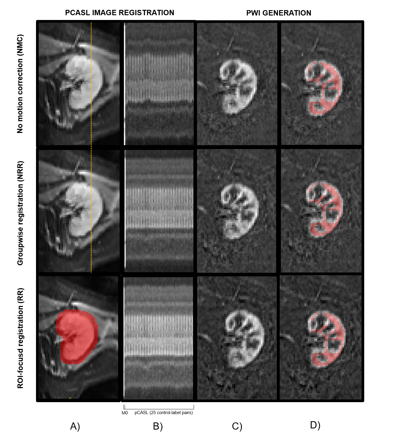
Figure 1.-
Example
images of before and after segmentation (using both groupwise registration
methods). A) M0 image. RR process uses a renal ROI to focus the registration
process. B) Image series representing the sagittal intersection of the image
for M0 and 25 control-label pCASL pairs. C) Mean Perfusion Weighted Images (PWI)
for each method. D) Superimposed cortex mask on Mean PWI.
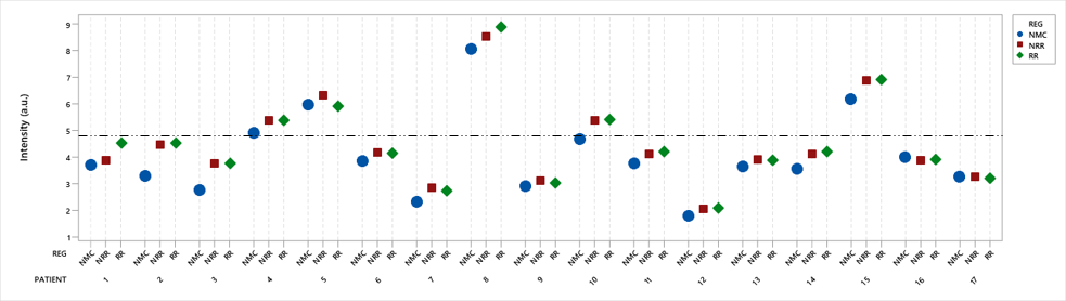
Figure 2.- Slice-wise mean tSNR
values for No Motion Correction (NMC), No-ROI-focused registration (NRR) and ROI-focused
registration (RR). Horizontal line represents mean tSNR.
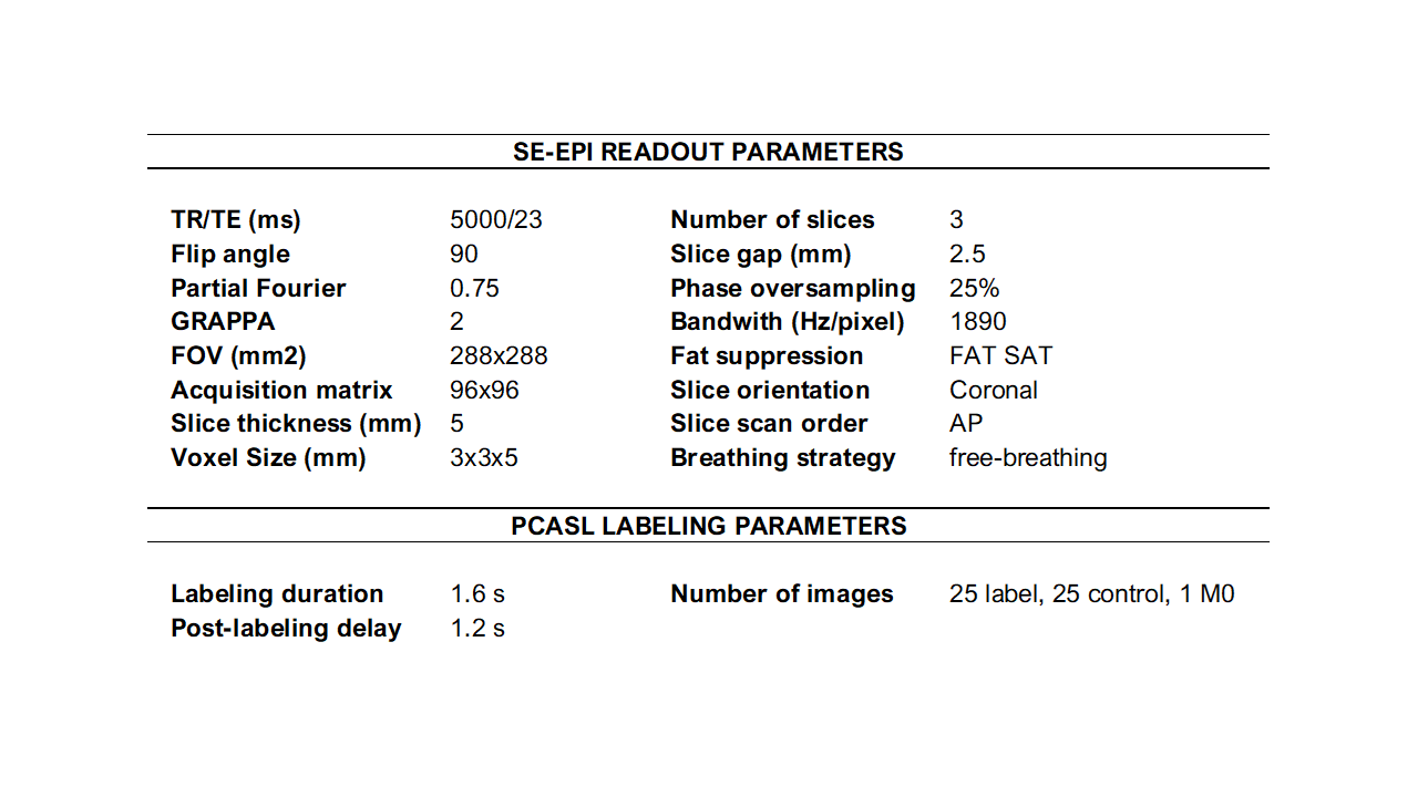
Table 1.- ASL-MRI Sequence parameters.
DOI: https://doi.org/10.58530/2022/2054