2041
Gastric emptying whilst upright
Hannah Askill 1, Emily Gaudoin1, Dipendra Jayantilal Mistry1, Olivier Mougin1, Caroline Hoad1,2, Hayfa Sharif1,2, Luca Marciani1,2, and Penny A Gowland1,2
1Sir Peter Mansfield Imaging Centre,, University of Nottingham, Nottingham, United Kingdom, 2Nottingham Biomedical Research Centre, Nottingham, United Kingdom
1Sir Peter Mansfield Imaging Centre,, University of Nottingham, Nottingham, United Kingdom, 2Nottingham Biomedical Research Centre, Nottingham, United Kingdom
Synopsis
Gastric accommodation and gastric tone are key measure in functional diseases such as gastroparesis, dyspepsia, and even reflux. Whilst gastric accommodation has been assessed using conventional MRI such measures will have been be perturbed by the subject lying supine. Gastric tone is either assessed using a barostat or pressure sensors. This work investigate the gastric response to a meal in the upright position with the aim of analysing the data to study accommodation and markers of tone in future.
Introduction
Gastric accommodation and gastric tone [1] are key measure in functional diseases such as gastroparesis, dyspepsia, and even reflux. Whilst gastric accommodation has been assessed using conventional MRI [2], gastric tone is either assessed using a barostat (balloon inflated with gas in the stomach to controlled pressure [1]) or pressure sensors, including MRI compatible sensors [3]. Previous work has indicated that for simple meals gastric function can be similar in the supine and upright positions. However it is clear that the shape of the stomach will be affected by gravity, and we postulate that the weight of the gastric contents will change the shape of the stomach in the upright position in a way that will provide an indication of gastric tone. In this work we present initial data studying the gastric emptying of two different liquid meals expected to have very different effects on gastric tone using upright MRI.Methods
Ethics approval for a feeding study was obtained from the University of Nottingham Medical School Ethics Committee. Two subjects have been scanned so far. The subjects arrived fasted at about 9 am and were scanned seated (80o) in an open 0.5T ASG MRI scanner. Multislice HASTE scans (10 slices with 20 mm gap to cover abdomen, FOV 31mm) and multislice balanced fast field echos (FOV 31mm, TR 7 TE 3.5, FA 80,) were acquired repeatedly as the subjects drank 500 mL of chilled water and then until stomach emptied completely. The rate of acquisition of the data was reduced by the operator as the stomach emptied. This procedure was then repeated as the volunteer drank 500 mL of yoghurt drink (The Collective Kefir Mango Cultured Drink, Sainsburys; 7.8g fat, 33g carbohydrate, 14g protein total). From the acquired images, the volume of the gastric contents and gastric lumen was outlined was measured at regular intervals during the emptying using MIPAV (https://mipav.cit.nih.gov/), however for preliminary analysis only alternate slices were measured.Results
Figure 1 shows an example HASTE images when the stomach is filled for both meals and both subjects. The difference in accommodation between the two subjects is very apparent.Figure 2 shows the variation in the volume of the gastric contents with time for both subjects. (Subject 2 drank the water slowly which is why the volume initially rose).All curves have been fitted to an exponential which approximated a straight line for the nutrient meals.
Discussion
We have developed a protocol for measuring gastric emptying seated in an open low field MRI scanner. The water and nutrient meals produces the expected gastric emptying profiles, with the nutrient meal emptying much slower. The shape of the stomach will be influence by gastric tone, the weight of the gastric contents and the intra-abdominal pressure. Gas volumes varied during the study, probably due to physiological reasons and errors in determining the position of the stomach wall in this preliminary analysis. Future work will focus on comparing the area of the gut wall to the weight in the stomach as a potential marker of gastric tone in both upright and supine positions.Acknowledgements
No acknowledgement found.References
1. Gregersen and Christensen, Neurogastro. Mot. 12, 501-8, 2000.
2. Coleman et al, Aliment Pharmacol ter, 18, 1039-48, 2003.
3. Kwiatek et al, Am. J. Physiol, Gastro Liver Physiol, 297, G1894-2901, 2009.
Figures
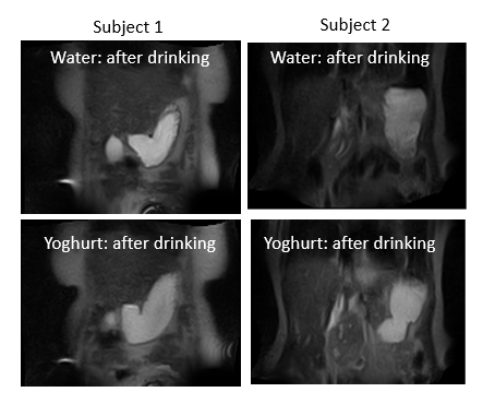
Images from both subjects just after each drink had been consumed, showing the differences in the gastric accommodation between both subjects.
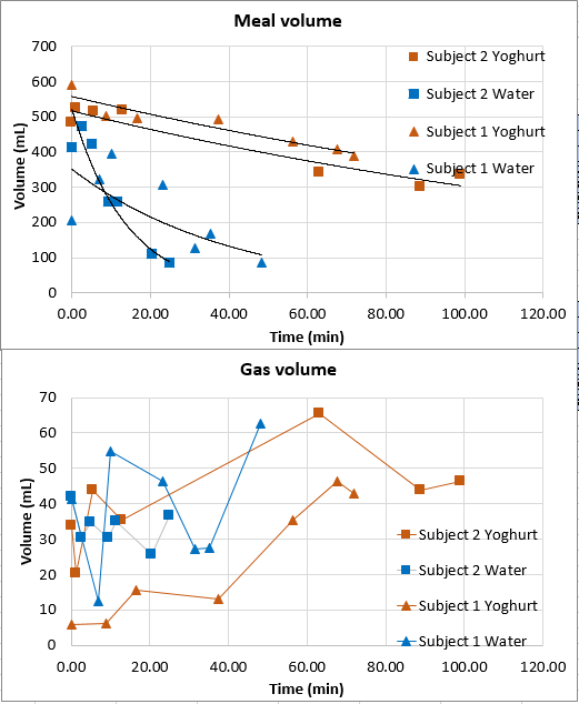
Gastric emptying curves for both subjects and both meals. For the meal data exponential fits are shown (which approximate to linear for the nutrient yoghurt meals). For the gas data the data points are simply joined for clarity.
DOI: https://doi.org/10.58530/2022/2041