2027
Independent component analysis of rsfMRI data in pedophilic disorder: A Swedish case control study
Linnéa Andersson1, Christoffer Rahm2, Benny Liberg2, Maria Ljungberg1,3, and Isabella M. Björkman-Burtscher4,5
1Department of Medical Physics and Biomedical Engineering, Sahlgrenska University Hospital, Gothenburg, Sweden, 2Department of Internal Medicine, Karolinska Institute, Stockholm, Sweden, 3Department of Medical Radiation Sciences, Institute of Clinical Sciences, Sahlgrenska Academy, University of Gothenburg, Gothenburg, Sweden, 4Department of Radiology, Institute of Clinical Sciences, Sahlgrenska Academy, University of Gothenburg, Gothenburg, Sweden, 5Department of Radiology, Sahlgrenska University Hospital, Gothenburg, Sweden
1Department of Medical Physics and Biomedical Engineering, Sahlgrenska University Hospital, Gothenburg, Sweden, 2Department of Internal Medicine, Karolinska Institute, Stockholm, Sweden, 3Department of Medical Radiation Sciences, Institute of Clinical Sciences, Sahlgrenska Academy, University of Gothenburg, Gothenburg, Sweden, 4Department of Radiology, Institute of Clinical Sciences, Sahlgrenska Academy, University of Gothenburg, Gothenburg, Sweden, 5Department of Radiology, Sahlgrenska University Hospital, Gothenburg, Sweden
Synopsis
Fifty percent of all child sexual abuse (CSA) perpetrators are individuals with pedophilic disorder. The neural underpinnings of pedophilic disorder are unknown. We conducted a case-control study aiming to investigate functional connectivity alterations using independent component analysis on resting state fMRI data in search of biomarkers that can be used for preventing CSA. The results suggest there are functional connectivity alterations in several resting state networks in individuals with pedophilic disorder.
Introduction
Child sexual abuse (CSA) causes extensive suffering for the victims. A risk factor in CSA offenders is pedophilic disorder (PD). The neurocognitive underpinnings of PD are still unknown. It is of great importance to find biomarkers that can serve as tools in risk assessments and treatment monitoring, and by extension prevent CSA. Previous findings suggest PD is related to neuroanatomical alterations (e.g. in white matter and cortical surface area)1,2 as well as functional alterations3,4. These findings suggest there is potential in finding functional connectivity biomarkers using resting state (rs)fMRI. However, the number of imaging studies and in particular rsfMRI studies performed on this group is limited.The aim of this study was to investigate differences in functional connectivity between individuals with PD and healthy controls in an exploratory manner using rsfMRI in search of biomarkers for PD.
Methods
ParticipantsStudy participants included 46 help-seeking, self-identified men with PD, age 18-66 years, and an age matched control group of 55 healthy controls. The PD cohort was recruited from Preventell, a helpline for unwanted sexuality, linked to Anova at the Karolinska University Hospital, Stockholm, Sweden. The National Swedish Ethics Review Appeals Board approved the study protocol (Ö 26-2014) which is part of the Priotab (Pedophilia at risk – investigations of treatment and biomarkers) trial (Eudra-CT 2014---000647---32).
MRI Image Acquisition
Imaging was performed on a Siemens Prisma 3 T scanner. A T1-weighted(w) MPRAGE scan for registration and normalization to standard space (resolution=1 x 1 x 1 mm3, TR=1900 ms, TE=2.52 ms) and a rsfMRI scan with 180 volumes (resolution=3 x 3 x 3 mm3, TR=2500 ms, TE=34 ms, flip angle 90°) were acquired.
rsfMRI preprocessing and analysis
FMRIB Software Library (FSL) v6.05 and ICA-AROMA6 was used in pre-processing of image data. The T1w image was brain extracted using BET and segmentation of CSF and white matter performed using FAST. Preprocessing of functional data were performed using preprocessing steps implemented in FSLs FEAT v6.00. Preprocessing steps used were motion correction, slice timing correction, smoothing (4 mm FWHM) and calculation of registration parameters to T1w image using the BBR-algorithm followed by non-linear normalization to MNI152 2 mm space. rsfMRI data was then denoised in native space using ICA-AROMA and CSF and white matter signal regressed out using fsl_regfilt. A temporal highpass filter (0.01 Hz cutoff) was applied and rsfMRI data was then transformed to MNI152 2 mm space.
Multi-subject temporal concatenation Independent Component Analysis (ICA) was performed using MELODIC v3.15 (FSL) with number of output components restricted to 30. This was followed by dual regression v0.6 (FSL). Group comparisons were carried out with a two-sample t-test using randomise v2.9 (FSL), with non-parametric permutation testing (5000 permutations) and statistic thresholding performed with threshold-free cluster enhancement and family-wise error (FWE) rate <0.05. Age was included as a regressor of no interest.
Results
The ICA analysis resulted in 23 components classified as resting state networks. The remaining 7 components were classified as noise/physiological noise.Differences in functional connectivity were found in 8 components (figure 1). The components were identified as medial visual network, sensorimotor network, lateral visual network, part of cerebellar network, auditory network, task-positive network, motor network and an unidentified network. Differences were found in the healthy controls > PD contrast. No differences were found in the PD > healthy controls contrast. The volume of the regions with differences are reported in table 1 together with MNI152 coordinates, probabilistic lobar structure (MNI structural atlas) and probabilistic cortical structure (Harvard-Oxford cortical atlas) of the voxel with minimum p-value found in each component.
Discussion
The present study demonstrates a decrease in functional connectivity in the PD group compared to healthy controls in 8 of 23 resting state network components generated with group ICA. Some of the alterations overlap spatially with areas of alterations found in a previous rsfMRI study using ICA to evaluate differences between PD offenders and a control group consisting of non-PD non-offenders and non-sexual offenders3. The overlaps are localized in the alteration areas of the lateral visual network, sensorimotor network, and cerebellar network. To our knowledge, very few studies have evaluated differences between PD and healthy controls using ICA and we acknowledge that further studies are needed to confirm our results. The results from our study can, however, be valuable for formulation of hypotheses in future studies exploring potential functional connectivity biomarkers in PD.Conclusion
Findings in this study suggest there are differences in functional connectivity between PD and healthy controls. These findings can be valuable in future studies aiming to find PD biomarkers useful for preventing CSA.Acknowledgements
Staff at Anova clinicReferences
1. Abé C, Adebahr R, Liberg B, et al. Brain structure and clinical profile point to neurodevelopmental factors involved in pedophilic disorder. Acta Psychiatr Scand. 2021;143(4):363-374.2. Cantor JM, Kabani N, Christensen BK, et al. Cerebral white matter deficiencies in pedophilic men. J Psychiatr Res. 2008;42(3):167-183.
3. Cantor JM, Lafaille SJ, Hannah J, et al. Independent Component Analysis of Resting-State Functional Magnetic Resonance Imaging in Pedophiles. J Sex Med. 2016;13(10):1546-1554.
4. Poeppl TB, Eickhoff SB, Fox PT, et al. Connectivity and functional profiling of abnormal brain structures in pedophilia. Hum Brain Mapp. 2015;36(6):2374-2386.
5. Jenkinson M, Beckmann CF, Behrens TE, et al. FSL. Neuroimage. 2012;62(2):782-790.
6. Pruim RHR, Mennes M, van Rooij D, et al. ICA-AROMA: A robust ICA-based strategy for removing motion artifacts from fMRI data. Neuroimage. 2015;112:267-277.
Figures
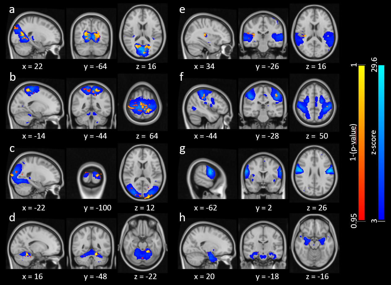
Figure 1:
Components with differences in the PD > healthy control contrast. Blue show
component maps generated with group ICA, thresholded at z=3. Red-yellow show
where differences were found within those components. The components were
identified as a) medial visual network, b) sensorimotor network, c) lateral
visual network, d) part of cerebellar network, e) auditory network, f)
task-positive network, g) motor network and h) an unidentified network.
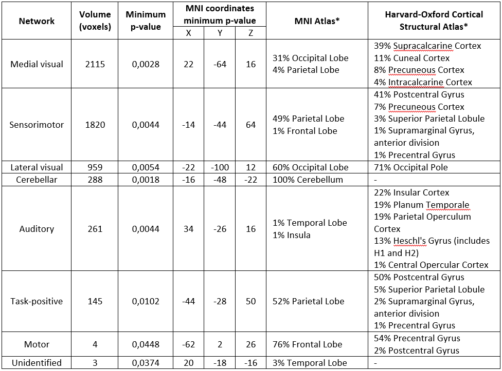
Table 1:
Identified resting state networks where differences were found in the PD >
healthy control contrast. *probabilistic lobar structure (MNI structural atlas)
and probabilistic cortical structure (Harvard-Oxford cortical atlas) of the
voxel with minimum p-value found in each component.
DOI: https://doi.org/10.58530/2022/2027