2015
A concurrent tDCS-MEGA PRESS MRS study to investigate polarity dependent effects on neurometabolites1Human Magnetic Resonance Center, Institute for Applied Life Sciences, University of Massachusetts Amherst, Amherst, MA, United States, 2Biomedical Engineering, University of Massachusetts Amherst, Amherst, MA, United States, 3University of Massachusetts Medical School - Baystate Medical Center, Springfield, MA, United States, 4University of Massachusetts Medical School, Worcester, MA, United States
Synopsis
Up- or downregulation of neurotransmitter/receptor concentration have been seen when transcranial direct current stimulation (tDCS) and proton Magnetic Resonance Spectroscopy (MRS) were combined in concurrent experiments; however, results have been mixed and not reliable across studies. In this study, we investigated dose and polarity effects of tDCS in an MRS voxel centered over the anterior cingulate/prefrontal region using three dose levels of tDCS with stimulation electrode placed over left a supraorbital area and return electrode over right mastoid bone. Dose and polarity dependent modulation of GABA and Glx was observed in a single subject study.
Introduction
Transcranial direct current stimulation (tDCS) is a non-invasive brain stimulation method capable of modulating neuronal activity in targeted brain regions1,2. tDCS is applied with two or more electrodes placed over the scalp and passing a small electrical current between these electrodes. Non-invasive measurement of Glx (Glutamate+Glutamine), the major excitatory neurotransmitter, and GABA, the inhibitory neurotransmitter using proton (1H) MRS, is of interest for studying the mechanisms behind various brain disorders3,4. The majority of tDCS-MRS studies have applied stimulation to the motor cortex, making it challenging to interpret the findings across different brain regions5,6. These studies have shown little to no effect of tDCS on neurotransmitters. Reasons for these minimal changes are considered to be (1) movement from in and out of the scanner between MRS scans and (2) low dose of the tDCS. In this study, we are conducting a concurrent tDCS-MRS experiment to investigate how the change in dose and polarity of tDCS modulates neuro metabolites in the anterior cingulate cortex (ACC).Materials and Methods
One healthy volunteer (age 30 years) participated in this study. The study was approved by the institutional review board. MRI imaging was performed on a 3T Siemens Skyra system using a 32-channel head receive coil. The tDCS stimulation was applied using MR compatible Neuroconn DCMC multichannel stimulator with two rubber electrodes which were placed on the scalp/skin (primary electrode over left supraorbital area and return electrode over the right mastoid bone). MRI/ MRS: Structural MRI data were acquired using a T1-weighted 3D MPRAGE sequence with sagittal acquisition, resolution 1.0 × 1.0 × 2.0 mm3; TR/TE= 1490/3.36 ms; flip angle = 9°, matrix = 256 × 256; field of view = 256 × 256mm2. MEGA-PRESS7 spectra were acquired from a 3 × 3 × 3 cm3 voxel. The voxel was centered over interhemispheric fissure and covered ACC from both the left and right hemispheres. MEGA-PRESS data were then acquired with the following parameters: TR/TE = 2000/ 68ms; 128 averages; 1024 data points; spectral width = 2000 Hz; editing pulse frequencies set to 1.9 ppm and 7.5 ppm for editing of GABA. Water-unsuppressed MEGA-PRESS data were acquired with one average for optimal co-localization of the unsuppressed water signal. The complete spectroscopy protocol was around 45 minutes, with total of five MRS scans of 9 minutes each. tDCS stimulation was concurrently applied while recording third MRS scan with five epochs labeled as OFF1, OFF2, ON, OFF3, and OFF4. Subject participated in five stimulation sessions separated by 48 hours or more. In each session, different stimulation was applied with electrode polarity at the left supraorbital area being either no-stimulation, Anodal 2.5 mA, Anodal 5 mA, Cathodal 2.5 mA, or Cathodal 5mA. Processing: MEGA-PRESS data were processed using the MATLAB-based toolbox Gannet 3.18. The Gannet Co-register and Gannet Segment modules call SPM 12 to determine the tissue volume fractions of GM, WM, and CSF9. Gannet analysis provides GABA and Glx neurometabolite concentrations in the MRS voxel as well as the fit error against the model function used to calculate these concentrations. Neurometabolites concentrations were calculated as change from the baseline of each session where first MRS (OFF1) was considered as the baseline.Results
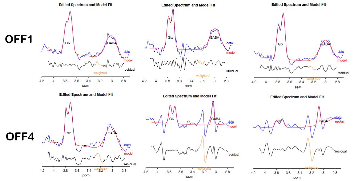
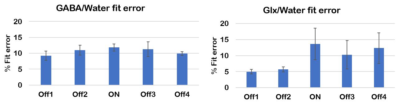
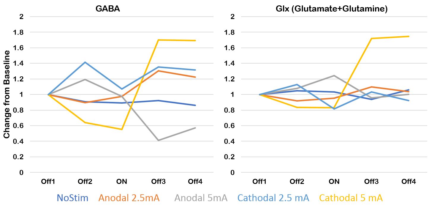
Figure 1 shows the schematic diagram to demonstrate the voxel placement, GM, WM, CSF percentage, along with a bar graph. The MRS spectrum of OFF1 and OFF4 from baseline, anodal 5mA, and cathodal 5mA processed by Gannet software were shown in Figure 2. The fit error of GABA/water and Glx/water were shown in Figure 3. Figures 4 shows the changes in GABA and Glx vs. tDCS epoch with different polarities. Subject did not have any tolerability issues or significant adverse effects with the high dose tDCS stimulation.Discussion and Conclusions
We have observed the differential effects of tDCS in GABA and Glx with different doses compared to baseline. MEGA-PRESS was able to detect and quantify GABA with improved fitting compared to PRESS10. We did not see any changes in the GABA and Glx during the no stimulation. During the stimulation (ON), change in GABA was less than 20% for both anodal while it reduced for cathodal 5mA. Similarly, the Glx change was minimal (less than 20%) for all active stimulations, with a maximum change observed with anodal 5mA. In the post-stimulation (OFF3 and OFF4), the effect of dose in anodal tDCS on the GABA was in the opposite direction showing a slight increase with Anodal 2.5mA and a decrease of more than 50% with Anodal 5mA. In contrast, GABA concentrations increased during the post-stimulation period with increasing dose levels when cathodal stimulation was applied. Further, there was almost no effect of stimulation on Glx except with cathodal 5mA which caused an increase of ~80% post-stimulation. In conclusion, we have developed a model paradigm to examine the modulation of neurotransmitter-receptors GABA (inhibitory) and Glx (excitatory). GABA behaved as expected with high dose stimulation: Andal stimulation leads to a decrease, while cathodal stimulation leads to an increase. Ongoing study will aim to replicate these findings in more subjects.Acknowledgements
This research was supported by an NIH Brain Initiative grant (RO1MH111874). Dr. Schlaug also acknowledges support from U01NS102353. We are thankful for and very much appreciate the generous support that Klaus Schellhorn from neuroConn has provided to us with making a state-of-the-art multi-channel MR DC stimulator available to us and his helpful suggestions with setting up the concurrent tDCS-MR experiments and being available for any troubleshooting over the years. We would also like to thank Elena Bliss, our MR technologist, for her support with all of the complicated and lengthy concurrent tDCS-MRS acquisitions.References
1. Nitsche MA, Paulus W. Excitability changes induced in the human motor cortex by weak transcranial direct current stimulation. J Physiol. 2000;527(3):633–639.
2. Shinde AB, Lerud KD, Munsch F, Alsop DC, Schlaug G. Effects of tDCS Dose and Electrode Montage on regional cerebral blood flow and motor behavior. NeuroImage. 2021 Aug 15;237:118144.
3. van Nuland AJM, den Ouden HEM, Zach H, Dirkx MFM, van Asten JJA, Scheenen TWJ, Toni I, Cools R, Helmich RC (2020) GABAergic changes in the thalamocortical circuit in Parkinson’s disease. Hum Brain Mapp 41(4):1017–1029.
4. Huang D, Liu D, Yin J, Qian T, Shrestha S, Ni H. Glutamate-glutamine and GABA in brain of normal aged and patients with cognitive impairment. Eur Radiol. 2017 Jul;27(7):2698-2705.
5. Patel HJ, Romanzetti S, Pellicano A, Nitsche MA, Reetz K, Binkofski F. Proton magnetic resonance spectroscopy of the motor cortex reveals long term GABA change following anodal transcranial direct current stimulation. Scientific Reports 2019; 9, 2807.
6. Kim S, Stephenson MC, Morris PG, Jackson SR (2014) tDCS-induced alterations in GABA concentration within primary motor cortex predict motor learning and motor memory: a 7 T magnetic resonance spectroscopy study. Neuroimage 99, 237–243.
7. M Mescher, H Merkle, J Kirsch, M Garwood and R Gruetter NMR Biomed, 11 (1998), pp. 266-272.
8. RA Edden, NA Puts, AD Harris, PB Barker and CJ Evans J Magn Reson Imaging, 40 (2014), pp. 1445-1452.
9. KJ Friston Elsevier/Academic Press, Amsterdam; Boston. (2007).
10. Nagarajan R, Shinde A, Gunduz ME, Schlaug G. Polarity Dependent Modulation of the Motor Region Using tDCS: A Proton MR Spectroscopy Study. Abstract accepted at the ISMRM, Abstract, 2234, 2021.
Figures