1962
Single-image superresolution of hyperpolarized 13C spectroscopic images1Memorial Sloan Kettering Cancer Center, New York, NY, United States
Synopsis
Hyperpolarized 13C spectroscopic images are acquired at a low spatial resolution, making it necessary to apply superresolution techniques to the metabolite maps prior to fusion with the proton anatomic image for visualization of metabolite biodistribution. In this work, we demonstrate great improvement in image quality for preclinical and clinical images when the metabolite map is upsampled by high spatial frequency transfer from magnetic resonance images of thermally polarized 13C phantoms to the invivo metabolic image.
INTRODUCTION
The low gyromagnetic ratio of 13C means hyperpolarized (HP) 13C imaging is conducted in the coil-dominated region which favors the use of low spatial resolution spectroscopic acquisition methods, such as Chemical Shift (CSI) or Echo-Planar Spectroscopic (EPSI) imaging, to obtain images with high signal-to-noise ratio (SNR). Fortuitously, it also means knowledge of the coil’s spatial resolution transfer properties can be used to improve superresolution (SR), regardless of the noise in the biological sample. While deep learning (DL) is currently the most popular method for SR, it is impractical to acquire the many high-resolution training images required for DL, because of the long spectroscopic scan times. Instead, we propose the use of example-based super-resolution (EBSR) which requires only a single image of a 13C phantom.METHODS
Reconstructing a high-resolution image, x, from an observed low-resolution image, y, can be done with the model:$$y = Hx + n$$ (1)
where H represents a linear degradation operation including downsampling and blurring, and n is the image acquisition noise. Based on a principle, known as cross-scale invariance, that certain image features are preserved regardless of spatial resolution, researchers have proposed a solution of the form(2):
$$\widehat{x} = {arg\,min}_{x}\parallel y - Hx\parallel + \lambda \phi(x\mid y)$$ (2)
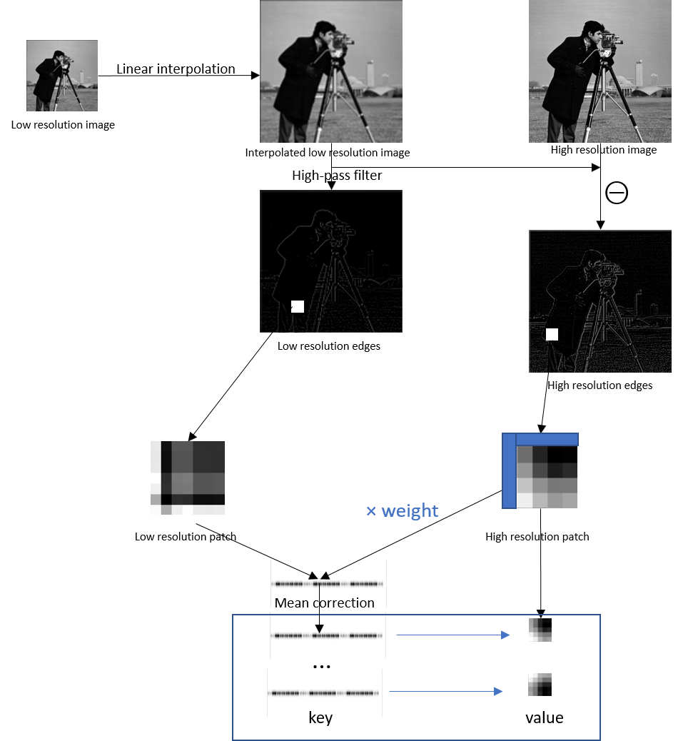
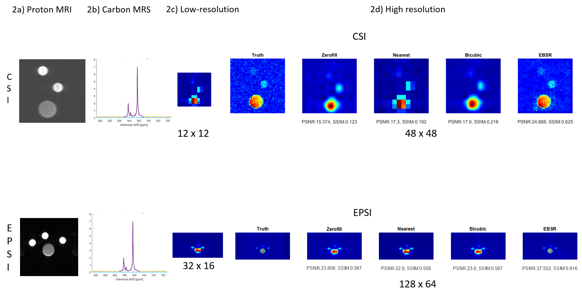
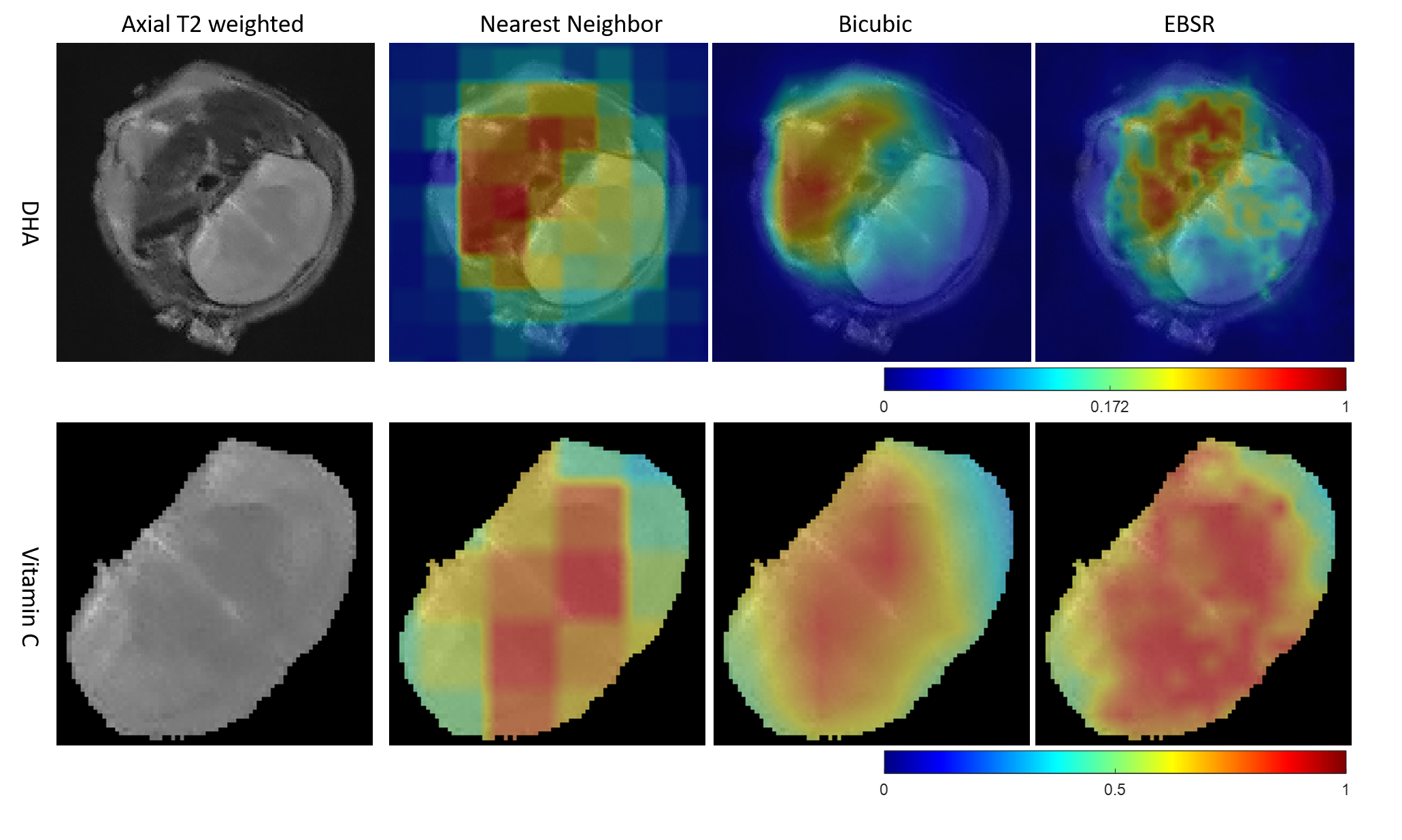
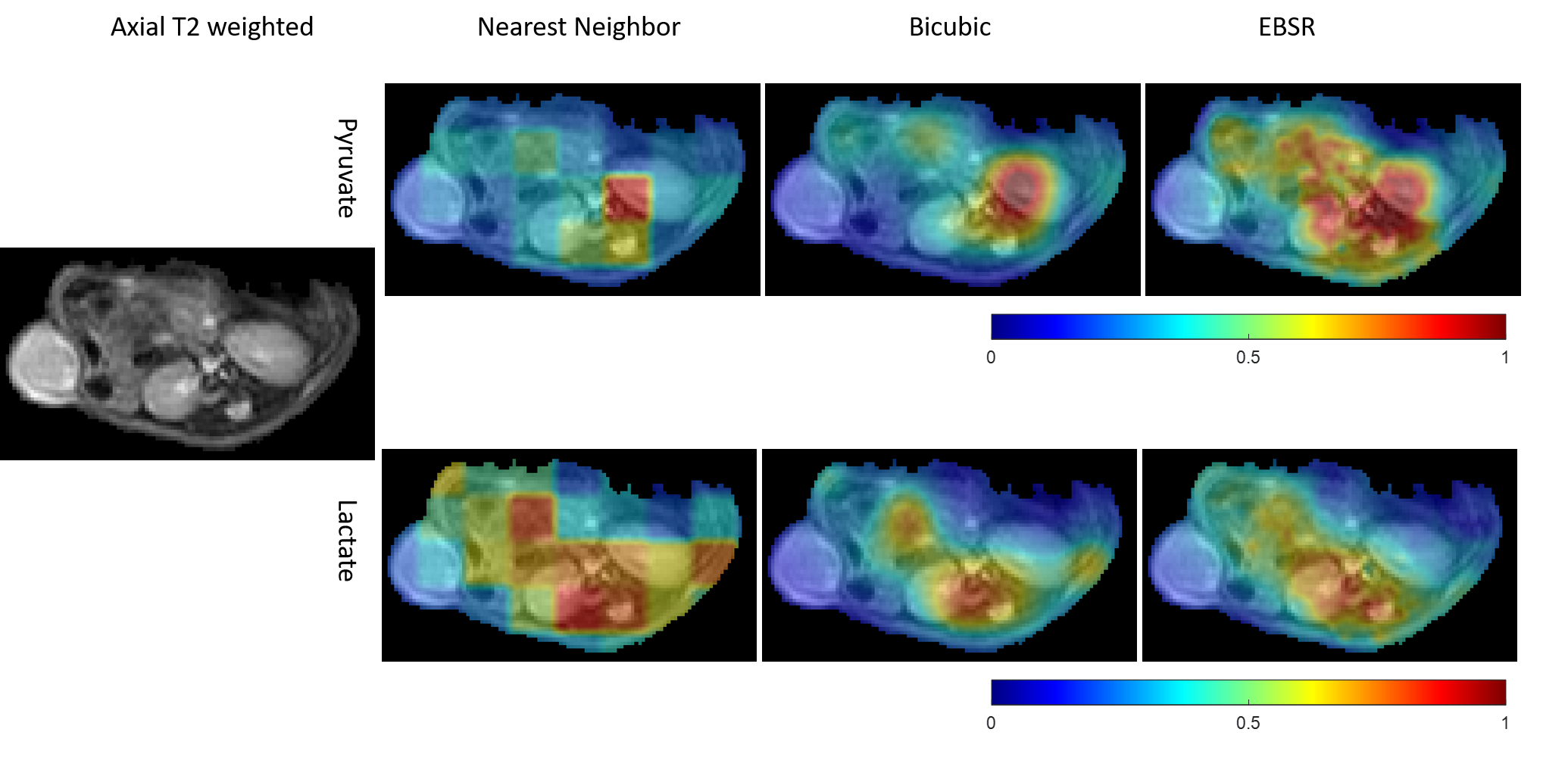
where $$$\phi(x\mid y)$$$ is regularization incorporating prior knowledge of the relationship between one or more low- and high-resolution training image pairs of a phantom that is similar to the invivo metabolite distribution by virtue of being a 13C chemical in a compartment. The phantom images are acquired with the same sequence acquisition parameters used for invivo imaging, but with a larger number of signal averages for increased SNR. This use of exvivo images for upsampling invivo images is justified because, in HP 13C imaging, the injected substrate and its metabolites are directly imaged. EBSR may be implemented as a database of image patches, for example as a KdTree data structure and nearest neighbor search algorithm in MATLAB(Fig. 1). Two preclinical experiments involving CSI of [1-13C] dehydroascorbate (DHA) metabolism in the mouse brain and EPSI of [1-13C] pyruvate metabolism in the mouse kidney, and one clinical experiment on EPSI imaging of [1-13C] pyruvate metabolism in metastatic human brain cancer were used to test this method. Training data was acquired from thermally polarized 13C phantoms (Fig. 2). The FIDs from the data acquisitions were Fourier-transformed, baseline, and phase-corrected to obtain spatially localized maps of 13C spectra(3). Intensity maps of metabolites were generated by integrating the magnitude of the spectra for the range corresponding to metabolite’s chemical shift plus or minus one. The EBSR method described above was applied with training parameters of $$$\lambda$$$ = 0.5, a low-resolution patch size of 7 x 7 pixels, and a high-resolution patch size of 5 x 5 pixels(Fig. 1). The peak signal-to-noise ratio (PSNR) and structural similarity index matrix (SSIM) between the natively acquired high-resolution image and the sinc, nearest neighbor, and bicubic and EBSR interpolations of the low-resolution image were compared.
RESULTS
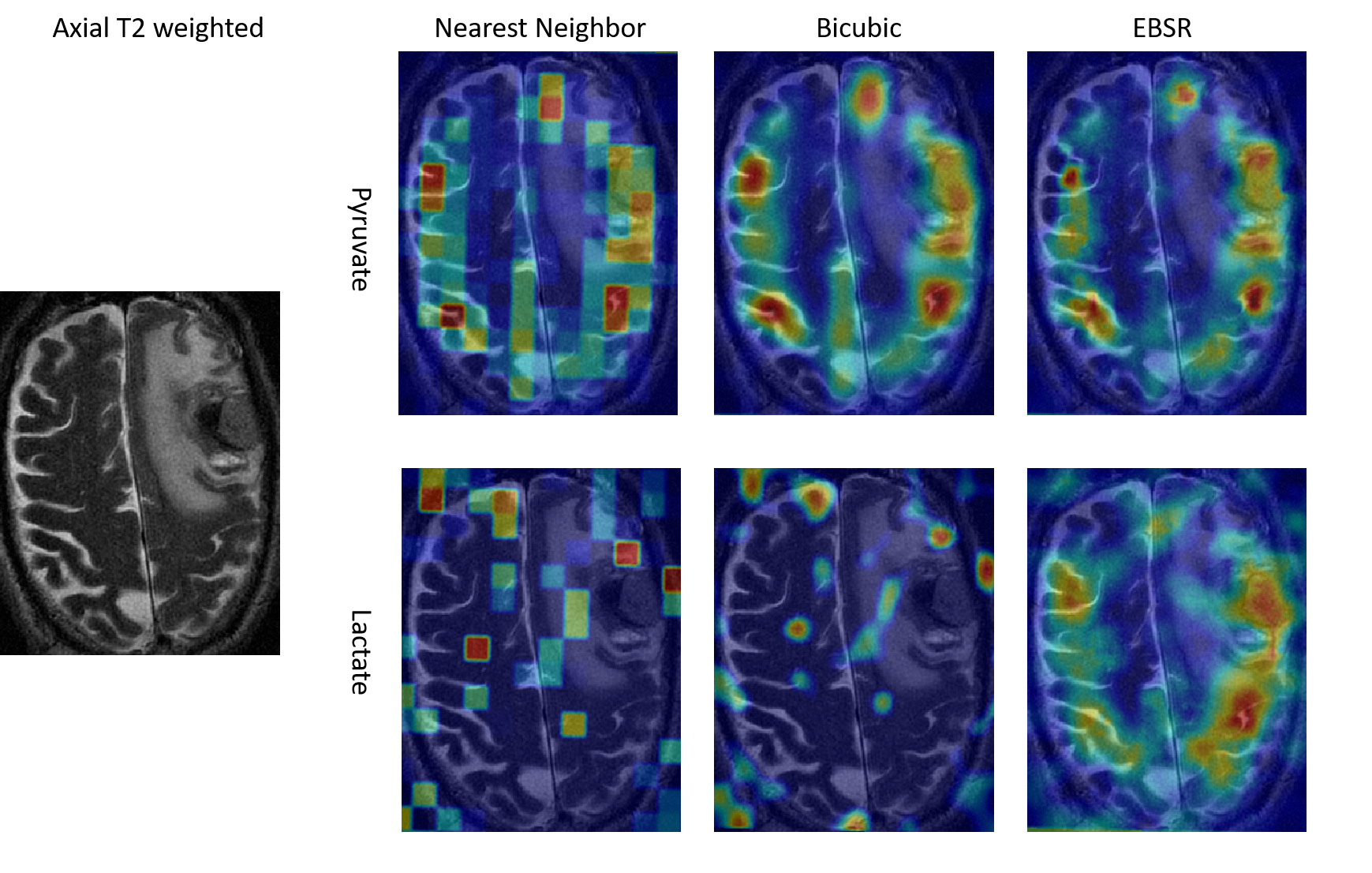
After training with a single phantom image, EBSR was able to successfully upsample low-resolution training and test phantom images. The upsampled EBSR images had the highest values of PSNR and SSIM computed against the natively acquired high-resolution image compared to upsampled images that use sinc, nearest neighbor and bicubic interpolation (Fig 2). Compared to bicubic interpolation, EBSR shows a more plausible metabolite biodistribution in the mouse brain (Fig. 3), kidneys (Fig. 4), and for human metastatic brain cancer (Fig. 5)DISCUSSION
We have demonstrated a reduction in blurring and noise present in low-resolution spectroscopic HP 13C images by using a patch example-based superresolution technique to transfer high spatial frequency information from thermally polarized 13C phantoms to the invivo image. This approach showed improvements in image quality compared to the use of bicubic interpolation which is the standard interpolation technique in most imaging software. The proposed method is also attractive because it requires only a single high-resolution image of a phantom.Acknowledgements
This work was supported by grants R01CA252037, R01CA237466, and S10OD016422.References
1. Keshari KR, Wilson DM. Chemistry and biochemistry of 13C hyperpolarized magnetic resonance using dynamic nuclear polarization. Chem Soc Rev 2014;43(5):1627-1659.
2. Freeman WT, Jones TR, Pasztor EC. Example-based super-resolution, IEEE Computer Graphics and Applications, Volume: 22, Issue: 2, Mar/Apr 2002.
3. Granlund KL, Tee SS, Vargas HA, Lyashchenko SK, Reznik E, Fine S, Laudone V, Eastham JA, Touijer KA, Reuter VE, Gonen M, Sosa RE, Nicholson D, Guo YW, Chen AP, Tropp J, Robb F, Hricak H, Keshari KR. Hyperpolarized MRI of Human Prostate Cancer Reveals Increased Lactate with Tumor Grade Driven by Monocarboxylate Transporter 1. Cell Metab 2019.
Figures