1936
Diagnostic performance of histological fibrous capsule and pseudocapsule (a false-positive fibrous capsule) in hepatocellular carcinoma1Radiology, Kawasaki Medical School, Kurashiki, Japan, 2Pathology, Kawasaki Medical School, Kurashiki, Japan
Synopsis
We used contrast-enhanced MRI and CT to evaluate the diagnostic potential of histological fibrous capsule and pseudocapsule in HCC. 51.4% (36/70) of HCCs had capsules, and pseudocapsule was found in 35.3% (18/51) of lesions with enhancing capsule imaging. Among the various imaging features, T1WI low signal rim, arterial phase low signal capsule, enhancing capsule on MRI and CT, and tumor margin enlargement sign on HBP were useful in diagnosing fibrous capsules. Among these, tumor margin enlargement sign on HBP showed the highest accuracy for the diagnosis of capsule, and was the only finding that could diagnose pseudocapsule.
INTRODUCTION
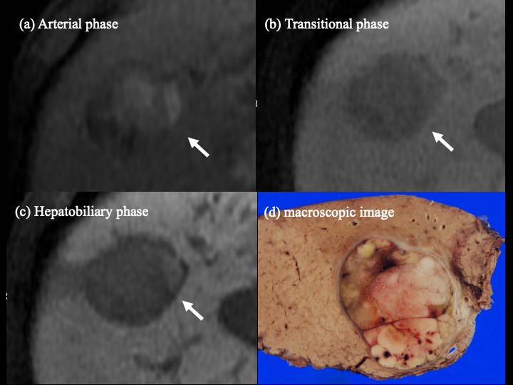
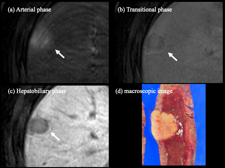
Hepatocellular carcinoma (HCC) is the fourth leading cause of cancer-related mortality, and diagnostic imaging plays a pivotal role in patient care1. Currently, the Liver Imaging Reporting Data System (LI-RADS) is widely used for the diagnosis of HCC2. Nodule size, arterial phase hyperenhancement, washout appearance, enhancing capsule, and threshold growth are major features in determining the diagnostic category. Of these, the diagnosis of fibrous capsule is made by observing the enhancing capsule, which is the contrasted rim around the tumor on dynamic contrast-enhanced CT or MRI. However, this diagnosis is often difficult because some HCCs do not have a fibrous capsule despite the presence of an enhancing capsule3. It is called a pseudocapsule because the peritumoral fibrosis and prominent sinusoids around the tumor are contrasted and look as if they are enhancing capsules. Gd-EOB-DTPA contrast-enhanced MRI has a higher lesion detection ability than conventional extracellular contrast agents, but it is sometimes difficult to evaluate the enhancing capsule. Intriguingly, in Gd-EOB-DTPA contrast-enhanced MRI, an increase in the size of the nodal margins is often observed from the transitional phase (TP) to the hepatobiliary phase (HBP). This imaging feature (we call it "tumor margin enlargement sign on HBP") has not been reported so far and may be important to diagnose histological fibrous capsule or pseudocapsule. The purpose of this study was to evaluate the diagnostic performance of various imaging features, including the tumor margin enlargement sign on HBP, for the histological fibrous capsule and pseudocapsule.METHODS
Surgically resected 70 HCCs between March 2008 and August 2020 were included in the study. A 64-row multidetector CT scan was used for contrast imaging (precontrast, arterial, portal, and equilibrium phases). 3-T scanner MRI was used to obtain T1-weighted images (T1WI), T2-weighted images (T2WI), contrast imaging (pre-contrast, arterial, portal, and transitional phases), and hepatobiliary phase. The images were evaluated by consensus reading of two radiologists, and the presence or absence of a T2WI low signal rim, T1WI low signal rim, arterial phase low signal capsule, low signal rim inside corona enhancement, enhancing capsule on MRI and CT, increase in size from TP to HBP, and HBP hypointense rim were recorded. The radiologists evaluated these various findings in a comprehensive manner to determine the final diagnosis on MRI and CT. Histopathological evaluation was performed using HE-stained specimens from the largest circumferential area of the tumor to determine if there was fibrous capsule of more than two-thirds of the tumor circumference. All statistical tests were performed using SPSS Statistics 24 software (SPSS, Chicago, IL, USA). Comparisons between groups were made using the χ2 test or Fisher's exact test for the association between each imaging finding and histological coverage. In cases where contrast-enhancing capsules were clearly observed in MRI and CT, intergroup comparisons were made for each imaging features between groups with and without fibrous capsules.RESULTS
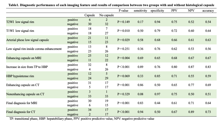
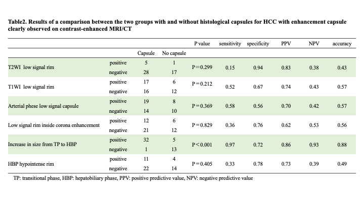
Among the 70 HCCs, 36 HCCs (51.4%) had a histological fibrous capsule, and pseudocapsule was found in 35.3% (18/51) of cases with enhancing capsule on contrast-enhanced MRI and CT. The sensitivity, specificity, positive predictive value, negative predictive value, and accuracy of multiparametric MRI and dynamic contrast-enhanced CT for the diagnosis of histological fibrous capsules were 0.83, 0.44, 0.61, 0.71, 0.64, and 0.94, 0.50, 0.67, 0.89, 0.73, respectively. The results of the comparison between the two groups for each imaging finding showed that T1WI low signal rim (P = 0.010), arterial phase low signal capsule (P = 0.029), enhancing capsule on MRI (P = 0.004) and CT (P = 0.001), and increase in size from TP to HBP (P<0.001) were found to be significantly different. The sensitivity, specificity, positive predictive value, negative predictive value, and accuracy of each imaging finding are shown in Table 1. Among the cases in which the enhancing capsule on contrast-enhanced MRI and CT was clearly observed, the results of the two-group comparison between the group with and without histological capsule showed a significant difference in the increase in size from TP to HBP (P<0.001) (Table2).DISCUSSION
Diagnostic performance of multiparametric MRI and dynamic contrast-enhanced CT for histological fibrous capsule showed high sensitivity and negative predictive value, but not high specificity, probably due to the presence of a certain percentage of HCC with pseudocapsule. Among the various imaging findings, T1WI low signal rim, arterial phase low signal capsule, enhancing capsule on MRI and CT, and increase in size from TP to HBP were useful in diagnosing the capsule. Among them, tumor margin enlargement sign on HBP showed the highest accuracy for the diagnosis of capsule, and was the only finding that could diagnose pseudocapsule. This result may be due to the fact that both the true fibrous capsule and the pseudo-capsule are contrasted in the PVP to TP, but the fibrous capsule without hepatocytes is not contrasted in the HBP and thus slightly increases in size, while the prominent sinusoids and peritumoral fibrosis, which are the cause of the pseudo-capsule, contain some hepatocytes and maintain some contrast effect in HBP and thus remain constant in size.CONCLUSION
Tumor margin enlargement sign on HBP is useful for the diagnosis of histological fibrous capsule and pseudocapsule in HCC.Acknowledgements
No acknowledgement found.References
1. Global Burden of Disease Liver Cancer C, Akinyemiju T, Abera S, Ahmed M, Alam N, Alemayohu MA, et al. The Burden of Primary Liver Cancer and Underlying Etiologies From 1990 to 2015 at the Global, Regional, and National Level: Results From the Global Burden of Disease Study 2015. JAMA Oncol. 2017;3(12):1683-91.
2. Moura Cunha G, Chernyak V, Fowler KJ, Sirlin CB. Up-to-Date Role of CT/MRI LI-RADS in Hepatocellular Carcinoma. J Hepatocell Carcinoma. 2021;8:513-27.
3. Ishigami K, Yoshimitsu K, Nishihara Y, Irie H, Asayama Y, Tajima T, et al. Hepatocellular carcinoma with a pseudocapsule on gadolinium-enhanced MR images: correlation with histopathologic findings. Radiology. 2009;250(2):435-43.
Figures