1930
Application of Machine Learning in Comparison between Multimodal Neuroimaging Markers of Laterality in Temporal Lobe Epilepsy
Alireza Fallahi 1,2, Mohammad Pooyan3, Jafar Mehvari-Habibabadi 4, Narges Hoseini Tabatabaei5, Mohammadreza Ay1,6, and Mohammad-Reza Nazem-Zadeh1,6
1Research Center for Molecular and Cellular Imaging, Tehran University of Medical Sciences, Tehran, Iran (Islamic Republic of), 2Biomedical Engineering, Hamedan University of Technology, Hamedan, Iran (Islamic Republic of), 3Biomedical Engineering, Shahed University, Tehran, Iran (Islamic Republic of), 4Isfahan Neuroscience Research Center, Isfahan University of Medical Sciences, Isfahan, Iran (Islamic Republic of), 5Medical School, Tehran University of Medical Sciences, Tehran, Iran (Islamic Republic of), 6Medical Physics and Biomedical Engineering, Tehran University of Medical Sciences, Tehran, Iran (Islamic Republic of)
1Research Center for Molecular and Cellular Imaging, Tehran University of Medical Sciences, Tehran, Iran (Islamic Republic of), 2Biomedical Engineering, Hamedan University of Technology, Hamedan, Iran (Islamic Republic of), 3Biomedical Engineering, Shahed University, Tehran, Iran (Islamic Republic of), 4Isfahan Neuroscience Research Center, Isfahan University of Medical Sciences, Isfahan, Iran (Islamic Republic of), 5Medical School, Tehran University of Medical Sciences, Tehran, Iran (Islamic Republic of), 6Medical Physics and Biomedical Engineering, Tehran University of Medical Sciences, Tehran, Iran (Islamic Republic of)
Synopsis
Five neuroimaging markers including T1 volume, FLAIR signal intensity, and mean diffusivity in hippocampus, and fractional anisotropy in both posteroinferior cingulum and crus of fornix were used for lateralization of temporal lobe epilepsy (TLE). Support vector machine (SVM) was used as a classifier and for measuring the importance of neuroimaging attributes. The classification results demonstrated that the hippocampal volumetric and mean diffusivity showed the highest correct classification rate and the largest area under the curve (AUC) for the receiver operating characteristic (ROC), thus considered as the most important attributes of TLE laterality among all markers investigated in this study.
Introduction
Temporal lobe epilepsy (TLE) is the most common type of drug-resistant epilepsy, mainly characterized by a history of seizures, and EEG abnormalities in the temporal lobe 1,2. The only standard treatment for these patients is surgery, and the success of the surgery depends on the precise localization of the onset of the seizure. However, preoperative localization of epilepsy especially in situation of non-lesional MRI, is highly dependent on nuclear medicine evaluations or invasive intracranial monitoring, which are non-invasive, time consuming and costly. Recently, magnetic resonance imaging (MR) based techniques have played an important role in accurately identifying the seizures foci in TLE patients who are candidates for surgical treatments. Hippocampal atrophy in T1-weighted images and signal enhancement in fluid attenuated inversion recovery (FLAIR) images are fundamental findings of MR images 3,4. Quantitative MR and machine learning studies of the hippocampus and other subcortical areas that are consented to undergo structural alterations may provide more useful information for diagnosis of the severity and the extent of epileptogenic damage in cases of TLE and lateralizing the associated bran hemisphere. In this study, we evaluated the performance of different neuroimaging markers from multimodal MRI (T1, FLAIR, and diffusion MRI) using support vector machine (SVM) as the classifying method. Results of applied method can detect the relative significance of neuroimaging markers in detection of laterality in individual mTLE patients.Materials and Methods
Thirty-five unilateral patients with left or right mTLE who were candidates for a surgical resection of a medial temporal structure were involved in this study. Based on the presurgical medical records by a multi-disciplinary decision-making team, 21 cases were diagnosed as left-mTLE (LTLE) and 14 cases as right-mTLE (RTLE) (male: female, 19:21 men; age range: 17-54; mean age 30.4 yrs). Hemispheric variation uncertainty (HVU) measures of the established markers of hippocampal T1 volumetry (Hip. Vol) and FLAIR signal intensity (Hip. FLAIR) were estimated based on structural MRI. In addition, HVU measures of mean diffusivity (MD) in the hippocampus (Hip.MD), and fractional anisotropy (FA) in both posteroinferior cingulum (FA in cingulum) and crus of fornix (FA in fornix) were estimated from diffusion MRI images. Each estimated HVU measure was employed as a ground for comparison of the interhemispheric changes in corresponding imaging attribute. To evaluate the reliability of neuroimaging features, SVM applied as a classifier using different subsets of the abovementioned features. Leave-one-out cross-validation approach was used for validating the performance of SVM classification. For each imaging marker, a single subject data was used for test, and the remaining data was used for training. The performance of the classifier was assessed by the classification accuracy and receiver operating characteristic (ROC) curves. The overall accuracy, and the accuracy for identification of left and right TLE cases are calculated based on Eq (1), where TL, FL, TR and FR are the number of true left, false left, true right and false right cases assigned by the classifier, respectively. Overall Accuracy Rate = (TL+TR) / (TL+ FL+ TR+ FR) (1) True Left Accuracy Rate= TL / (TL+ FR) True Right Accuracy Rate=TR / (TR + FL)Results
Fig 1 shows performance of mTLE lateralization in detail for the proposed biomarker, evaluated by the leave-one-out cross validation. As it shows, Hip. Vol and Hip.MD markers together lateralized 95% of patients in left TLE group and 64% of patients in right LE group with overall 82.9% accuracy. Hip. Vol and Hip. FLAIR attributes together lateralized 80% of patients in left TLE group and 57% of patients in right LE group with overall 71.4% accuracy. Using FA in fornix and FA In cingulum lateralization achieved 76% of patients in left TLE group and 42% of patients in right LE group with overall 62.9% accuracy. Using Hip. Vol, Hip FLAIR and Hip.MD together lateralized 90% of patients in left TLE group and 57% of patients in right LE group with overall 77.1% accuracy. Combination of all attributes lateralized 100% of patients in left TLE group and 57% of patients in right LE group with overall 82.9% accuracy. The ROC curves of the SVM classifier using all feature and also Hip.Vol and Hip.MD (best results) are shown in Fig. 2. The AUC of the ROC for the SVM classifiers based on all feature and Hip.Vol and Hip.MD was calculated as 83% and 89%, respectively.Conclusion
In this study, a SVM classification technique was customized for a lateralization task, as well as measuring the importance of imaging attributes for this task. The results demonstrated that the hippocampal volumetric and mean diffusivity would have the same correct classification rate and the best AUC among all imaging markers investigated in this study. The classification accuracy also showed that the fractional anisotropy in cingulum and in fornix crus were the least influencing markers of seizure laterality. This finding is concordant with a previous work focusing on distinguishing between left and right mTLE patients 5.Acknowledgements
We must acknowledge the contribution of the Iranian National Brain Mapping Lab (NBNL) for MRI data acquisition throughout this project. This work was partially funded and supported by Iran’s National Elites Foundation, National Institute for Medical Research Development (Grant No. 971683), and Cognitive Sciences & Technologies Council (Grant No. 6431), between 2017 and 2021.References
1. Engel J (2001) Mesial Temporal Lobe Epilepsy: What Have We Learned? The Neuroscientist 7:340–352. https://doi.org/10.1177/107385840100700410 2. Téllez-Zenteno JF, Hernández-Ronquillo L (2012) A Review of the Epidemiology of Temporal Lobe Epilepsy. Epilepsy Research and Treatment 2012:1–5. https://doi.org/10.1155/2012/630853 3. Jafari-Khouzani K, Elisevich K, Wasade VS, Soltanian-Zadeh H (2018) Contribution of Quantitative Amygdalar MR FLAIR Signal Analysis for Lateralization of Mesial Temporal Lobe Epilepsy. Journal of Neuroimaging 28:666–675. https://doi.org/10.1111/jon.12549 4. Mahmoudi F, Elisevich K, Bagher-Ebadian H, Nazem-Zadeh MR, Davoodi-Bojd E, Schwalb JM, Kaur M, Soltanian-Zadeh H (2018) Correction: Data mining MR image features of Select structures for lateralization of mesial Temporal lobe epilepsy (PLoS ONE (2018)13:8 (e0199137) DOI: 10.1371/journal.pone.0199137). PLoS ONE 13:209866. https://doi.org/10.1371/journal.pone.0209866 5. Sanjari Moghaddam H, Rahmani F, Aarabi MH, Nazem-Zadeh M-R, Davoodi-Bojd E, Soltanian-Zadeh H (2019) White matter microstructural differences between right and left mesial temporal lobe epilepsy. Acta Neurologica Belgica. https://doi.org/10.1007/s13760-019-01074-xFigures
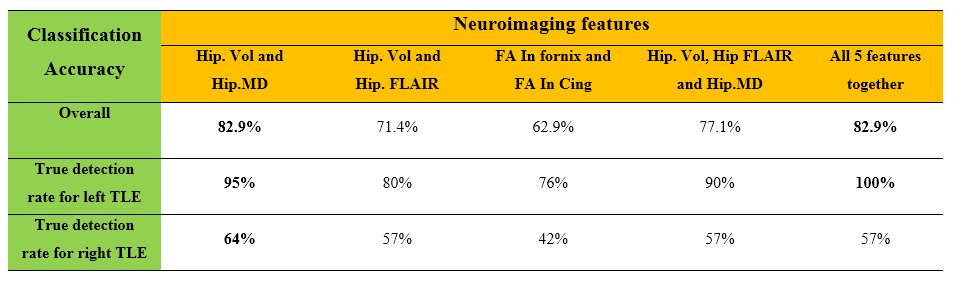
Fig.1: Result of SVM classification accuracy for
different number of neuroimaging features.
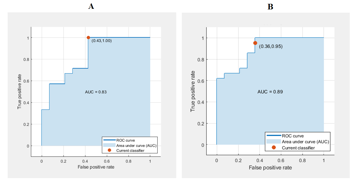
Fig.
2. The receiver operating characteristic curve (ROC) for the
classification of patients with R-TLE and L-TLE using neuroimaging attributes.
A: The area under the curve (AUC) was 83% when all feature were used for the
classification. B: The AUC was 89% when Hip. Vol and Hip.MD together were used
for the classification.
DOI: https://doi.org/10.58530/2022/1930