1924
Diffusion-weighted MRI-based Virtual Elastography is feasibility for the evaluate of Rolandic epilepsy1Departemnt of diagnostic Radiology, The First Affiliated Hospital of Xi'an Jiaotong University, Xi'an, China, 2The First Affiliated Hospital of Xi'an Jiaotong University, Xi'an, China, 3Departemnt of Neurosurgery, The First Affiliated Hospital of Xi'an Jiaotong University, Xi'an, China, 4Departemnt of Pediatrics, The First Affiliated Hospital of Xi'an Jiaotong University, Xi'an, China, 5GE Healthcare, MR Research China, Beijing, Beijing, China
Synopsis
Conventional MRI is difficult to discover the subtle changes caused by epilepsy. Virtual magnetic resonance elastography (vMRE) is based on diffusion MRI, which is sensitive for variety of physiologic and pathologic conditions. This study use vMRE to evaluated left Rolandic epilepsy. We found that left precentral gyrus, left postcentral gryus and left superior temporal gyrus is more stiffer than controls. The longer the seizure duration the stiffer of the brain tissues. vMRE has the potential to noninvasive evaluate epilepsy.
Introduction
Epilepsy is the most common serious neurological disorder, which affects 65 million people worldwide.1 Compared with adults children are at risk for epilepsy.2 Epilepsy in children is harmful to developmental programmes and resulting in inadequate construction of cortical networks.3 Even though antiepileptic drugs have been used but have limited therapeutic benefits. There are approximately one third of patients remains medical treatment failing.1 Surgery is an important approach in drug-refractory epilepsy. Magnetic resonance imaging(MRI)is the first choice for evaluating patients before operations.Methods
The local Institutional review board approved this study and written informed consents were obtained from parents of the children. Subjects Children diagnosed with RE according diagnostic criteria defined by the International League Against Epilepsy4.Subjects who were confirmed to have a history of intracranial infection,cerebral trauma and abnormalities of brain parenchyma on MRI were excluded.Controls were matched for gender, postnatal age at MRI scan, gestational age (GA) and without any MRI abnormality. MRI Protocols Each MRI session was finished on a 3.0T scanner(Signa HDxt,GE Healthcare, Milwaukee, WI) with an 8-channel head coil. Data acquistition included three-dimensional fast spoiled gradient-echo T1-weighted sequence(TR/TE, 10.2ms/4.6ms;NEX, 1;isotropic 1×1×1mm3;FOV, 24cm) and transverse fast spin-echo T2-weighted sequence(TR/TE, 4200ms/116ms;NEX, 1.5; matrix,320×320;thickness,4mm;FOV,24cm),followed by DKI(b values = 0, 50, 200, 500, 1000, 2000, 2500 s/mm2; 18 gradient directions per nonzero b value; NEX = 1; repetition time/echo time = 11000/91.7 ms; slice thickness = 4 mm; field of view = 240 × 240 mm2; acquisition matrix =172 × 172; the acquisition voxel size = 1.4 × 1.4 × 4 mm3). ASL (4.0-mm-thick, TR=4500ms, TE=10ms, T1=700ms, T2=1900ms, PLD=1500ms, FOV=230×230mm, phase matrix/frequency matrix = 96/96, resolution = 2.0 × 2.0 mm). Data and statistical analysis Analyses were performed in Matlab (MathWorks, Natick, Massachusetts),FMRIB software library (FSL; http://www.fmrib.ox.ac.uk/fsl) and SPSS 19.0 (Chicago,IL,USA). Lower b-value (Slow, b value = 200 s/mm2) and those of the higher b-value (Shigh, b value = 1000 s/mm2) were used to estimate the virtual shear stiffness [5,6].Virtual shear stiffness = a·ln (Slow/Shigh) + b. The scaling (a) and the shift (b) factors were separately set to −9.8 and 14 according to the previous calibration studies.5,6 We compared stiffness measures and cerebral blood flow (CBF) between ipsilateral and contralateral to epileptogenesis in RE patients. Meanwhile, we also compared stiffness measures and CBF between RE and controls. Wilcoxon signed-rank tests and paired t-test were used for the group difference between RE group and the match controls. Results were considered statistically significant if p<0.05.Results
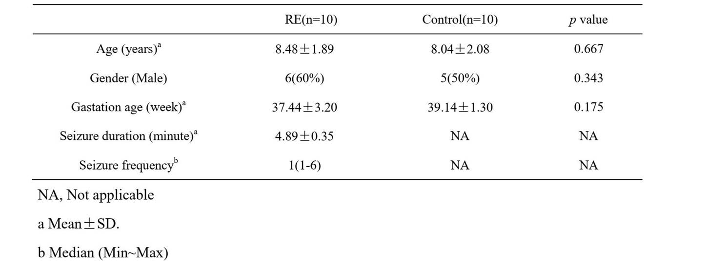
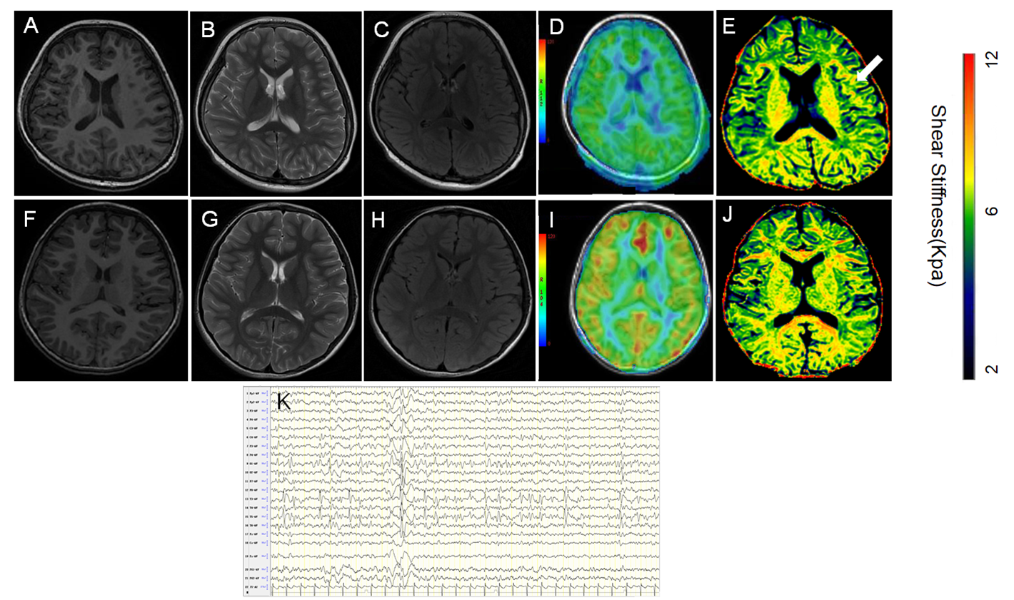
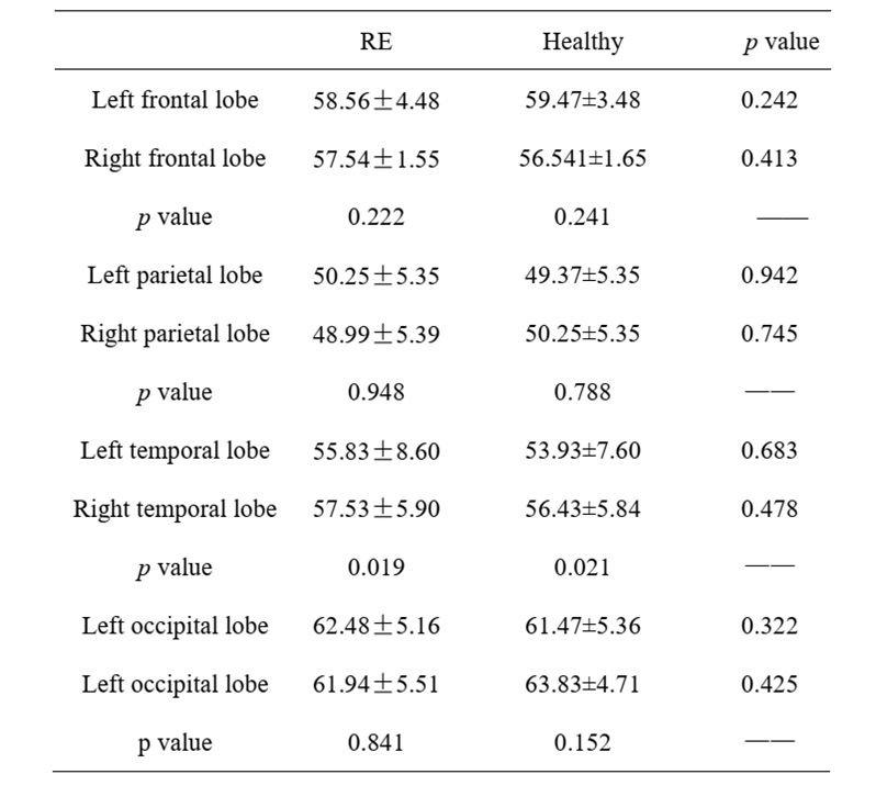
A total of 10 left REs and 10 match controls were enrolled. No significant difference in GA, ages, gender betweent REs and controls (Table 1).Compared with contralateral side and controls,a higher stiffness in left precentral gyrus, left postcentral gryus and left superior temporal gyrus in REs (Table 2). Stiffness significantly improved the ability to differentiate the two groups (Fig 1).
Stiffness correlated with seizure duration (p=0.013, r=0.745). There was no significant difference of CBF in both RE patients and controls (Table 3).
Discussion
This study quantitatively demonstrate that Rolandic areas’ stiffness measured with vMRE is altered in patients with RE compared with healthy participants. This may due to seizure activity might lead to sclerosis of tissue7. The loss of cell and reactive gliosis is the characteristic of sclerosis of tissue. It is reported that reactive glial cells are more stiffer than normal cells, which may influence vMRE measures8. The longer longer the seizure duration the more serious reactive gliosis is.Conclusion
Epileptic seizures can induce changes in stiffness. Stiffness were correlated with the seizure duration. The quantitative analysis of stiffness on MRI might be a biomarker to find the epileptic focus. vMRE might be a useful approach in assessing patient with epilepsy.Acknowledgements
This study was supported by National Natural Science Foundation of China (81901516, 81901823, 81971581, 81771810 and 51706178), Shaanxi Provincial Innovation Team (2019TD-018), National Key Research and Development Program of China (2016YFC0100300), the 2011 New Century Excellent Talent Support Plan of the Ministry of Education, China (NCET-11-0438), the Project Funded by China Postdoctoral Science Foundation (No. 2019M653659), Foundation of the First Affiliated Hospital of Xi’an Jiaotong University (No. 2018MS-09),the Natural Science Basic Research Plan in Shaanxi Province of China (No. 2019JQ-198), the Key Research and Development Program of Shaanxi (2020SF-118).References
1. Moshé SL, Perucca E, Ryvlin P, et al. Epilepsy: new advances. Lancet,2015, 385(9971) :884-98.
2. Ben-Ari Y, Holmes, GL. Effects of seizures on developmental processes in the immature brain. Lancet Neurol, 2006;5(12) :1055-63
3. Scheffer IE, Berkovic S, Capovilla G, et al. ILAE classification of the epilepsies: position paper of the ILAE commission for classification and terminology. Epilepsia, 2017;58(4):512–21.
4. Kwan P, Brodie, MJ. Early identification of refractory epilepsy. N Engl J Med, 2000, 342(5):314-9.
5. Le Bihan D, Ichikawa S, Motosugi U. Diffusion and intravoxel incoherent motion mr imaging-based virtual elastography: a hypothesis-generating study in the liver. Radiology. 2017, 285(2):609–19.
6. Lagerstrand K, Gaedes N, Eriksson S, et al. Virtual magnetic resonance elastography has the feasibility to evaluate preoperative pituitary adenoma consistency. Pituitary, 2021, 24(4):530–41.
7. Chan HW, Pressler R, Uff C, et al . A novel technique of detecting MRI-negative lesion in focal symptomatic epilepsy: Intraoperative ShearWave Elastography. Epilepsia, 2014, 55(4): 30–3.
8. Lu YB, Iandiev I, Körber, N, Körber, N, et al. Reactive glial cells: increased stiffness correlates with increased intermediate filament expression. FASEB J, 2011,25(2):624–631.
Figures