1910
Longitudinal decrement of cerebral blood flow in high-impact sports1Radiology, Stanford University, Stanford, CA, United States, 2Medical Biophysics, University of Toronto, Toronto, ON, Canada, 3Stanford Center for Clinical Research, Stanford, CA, United States, 4Neurosurgery, Stanford University, Stanford, CA, United States, 5Bioengineering, Stanford University, Stanford, CA, United States
Synopsis
Longitudinal changes (over four years) of cerebral blood flow (CBF) using arterial spin labeling MRI were investigated in a population of high-contact sport football college athletes and were compared to low-contact cohort of volleyball athletes. A linear-mixed-effects model was applied to assess CBF (normalized to the cerebellum) by sport (football vs. volleyball), time from baseline MRI, and the interaction between sport and time. Longitudinal analysis showed a prospective decline in perfusion in football compared to volleyball. Fourteen football players experienced an in-study concussion; in contrast to the longitudinal findings, football players exhibited acutely a mild increase in occipital lobe CBF.
Introduction
Repetitive head trauma is common in high-contact sports such as football. Repeated head impacts may carry significant risks; increasing evidence has suggested persistent neurological impairments1, cognitive and memory decline, even in the absence of concussive events2, and higher risk for neurodegenerative diseases. CBF is sensitive to neurological impairments and functional changes3, as well as acute and chronic effects of concussion4. Emerging evidence suggest that CBF is also sensitive to effects of sub-concussive impacts (high-velocity impacts that do not cause a clinical concussion) during a season of high-contact sports5. The goal of this work is to investigate whether exposure to high-contact sports causes longitudinal changes of CBF over a 4-year period.Method
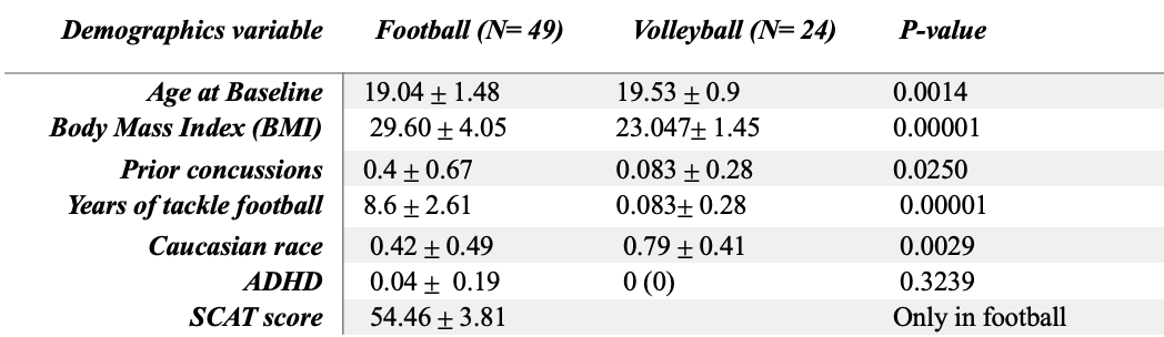
Subjects. We enrolled 65 football and 34 volleyball college athletes. Athletes were scanned at the start of each season, within 1-5 days after concussion, and after their last season of sport participation. In football, a Standardized Concussion Assessment Tool (SCAT) evaluation was acquired in players pre-first-season. For our longitudinal analysis, we only included athletes who had at least two scans spaced at least six months apart, excluding post-concussive scans (which were analyzed separately). Collectively, 50 football and 24 volleyball athletes were included for longitudinal analysis (Table 1, demographics variables were compared at baseline using Wilcoxon-rank-sum-test), and 14 football subjects who had within-study post-concussion MRI.Imaging data acquisition and analysis. Imaging was conducted on one 3T GE-MR750 system, with an 8-channel receive head coil. T1-weighted images (inversion recovery fast spoiled gradient echo) were collected with TR=7.9ms, TE=3.1ms, NEX=1, 1mm isotropic, FOV=240cm. Arterial spin labeling was undertaken using a 3D pseudo-continuous ASL (PCASL) to measure regional CBF, with 24cm FOV, NEX=3, TR=2, labeling duration of 1.45s, post-label-delay 2.025s, 2x2x4 mm and a simultaneous proton density (PD) image. Segmentation of T1 images was performed using the FreeSurfer 5.3 longitudinal pipeline as previously reported6. The ASL image was co-registered to Freesurfer space using a boundary-based cost function, and this was blindly evaluated for quality. Absolute CBF was then computed using the ASL and PD images using white-paper recommendations described in Alsop 20157. Regional median CBF was computed in each Freesurfer segmented region. To account for intrasubject variability in global CBF, we computed relative CBF (rCBF) by normalizing CBF values with each subject’s median cerebellar CBF8.
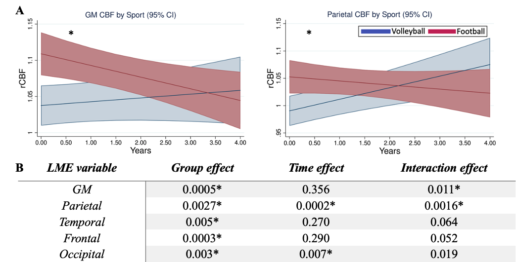
Statistical analysis. A linear-mixed-effect (LME) model was applied in Stata-15 to investigate the longitudinal CBF changes in each region (results were corrected for multiple-comparisons9). Fixed effects in the LME model included sport, time, and their interaction. There was a small but significant difference between the cohorts in BMI, age at baseline, and Caucasian race (Table 1); however, none of these significantly contributed to the LME model by likelihood-ratio-test. A similar LME model was used exclusively within football to test whether impact risk associated with football players position (HITsp), baseline SCAT score, cumulative years playing football prior to the study, or the presence of prior concussion had any association with CBF alterations. To examine acute effects of in-study concussions, a Wilcoxon-signed-rank test was used to compare relative CBF values pre- and post-concussion.
Results
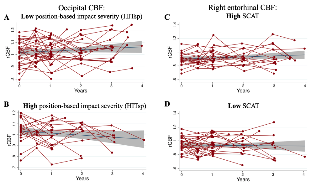
There was significant group by time interaction effect in gray matter (GM): GM rCBF declined over time in football compared to volleyball athletes (Fig.1.A). At a more granular level, similar interactions were observed in the parietal lobe, as well as bilateral precuneus, and left para and post-central regions. Statistically significant sport differences with higher baseline rCBF in football were observed at the global level of whole-brain GM, and multiple lobes including temporal, frontal, occipital and parietal (Fig.1.B). In addition, previously we reported on cortical thickness and volume alterations in the same cohort6, here we examined contributions of cortical-volume to rCBF changes; our findings did not show any change of rCBF effects when adding cortical-volume into the model (p=0.375), and cortical-volume was not associated with rCBF changes (p=0.205).Regarding within football analyses, football players with higher impact risk measured at baseline (high HITsp), had decreased occipital rCBF over time (interaction effect: p=0.004) (Fig.2.B). Football players with higher SCAT score (better performance) at baseline had increased rCBF in right entorhinal cortex over time (interaction effect: p=0.002) (Fig.2.C). Football players with a prior history of concussions had less of a decrease in the left postcentral region over years vs. players without concussion history (interaction effect: p=0.0006). There were no significant contributions of years of prior football experience in rCBF changes (p= 0.76).
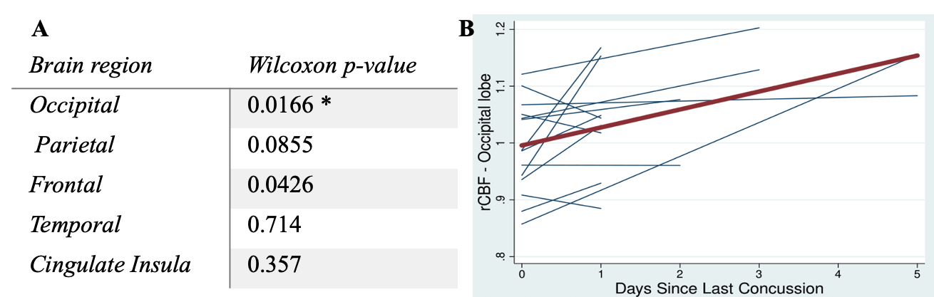
Among 14 football players who had a within-study concussion, an increase in the occipital lobe rCBF immediately after concussion was observed compared to baseline (Fig.3).
Discussion and Conclusion
Our longitudinal analysis demonstrated a prospective decline in GM relative perfusion in football players compared to a control cohort, suggesting exposure to cumulative impacts may cause a decrease in rCBF. We did not find any relationship between longitudinal rCBF alterations and previously reported delayed cortical thinning in football, suggesting this is an independent effect and possibly a different underlying physiologic mechanism. Football athletes had higher rCBF at baseline compared to volleyball. In addition, we observed a transient increase in occipital rCBF after concussion. rCBF may reflect dynamic changes that could be indicative of long-term physiologic consequences.Acknowledgements
This research was conducted with funding from the Radiology Society of North America, the American Society for Neuroradiology, General Electric Healthcare, and the Pac-12 Conference’s Student- Athlete Health and Well-Being Initiative.References
1. Bazarian et al. PloS one 9.4 (2014): e94734.
2. Strain et al. JAMA neurology 72.7 (2015): 773-780.
3. Ogoh. The Journal of Physiological Sciences 67.3 (2017): 345-351.
4. Churchill et al. Neurology 93.21 (2019): e1980-e1992.
5. Slobounov et al. Neuroimage: clinical 14 (2017): 708-718.
6. Mills et al. Neuroimage 217 (2020): 116864.
7. Alsop et al. Magnetic resonance in medicine 73.1 (2015): 102-116.
8. Wang et al. Brain imaging and behavior 13.5 (2019): 1375-1385.
9. Li and Ji. Heredity 95.3 (2005): 221-227.
Figures