1867
Quantitative Reduced Field-of-view Imaging using 3D Tailored Inner Volume Excitation and Pattern Recognition1Department of Radiology, University of Michigan, Ann Arbor, MI, United States, 22Department of Biomedical Engineering, University of Michigan, Ann Arbor, MI, United States
Synopsis
The goal of this study is to achieve small field-of-view (FOV) T1 and T2 mapping using 3D tailored inner volume (IV) excitation and pattern recognition based on MR Fingerprinting.
Introduction
The goal of this study is to achieve small field-of-view (FOV) T1 and T2 mapping using 3D tailored inner volume (IV) excitation and pattern recognition based on MR Fingerprinting1. Reduced FOV imaging using a localized, inner volume excitation can be used to reduce acquisition time or increase spatial resolution by reducing the FOV encoding burden. A recently proposed auto-differentiable Bloch equation simulation approach allows for joint optimization of RF and gradient waveforms for large-tip flip angles2, potentially enabling inner volume excitation for various applications. However, the signal from the outer volume (from unwanted residual excitation outside the IV) could still alias into the inner volume, especially in the presence of B0 field inhomogeneities.Using the concepts of signal encoding and pattern recognition from MRF, it may be possible to impart signals from the inner volume and the outer volume with distinguishable signal evolutions by exciting them with different flip angles and RF phases. By matching the signals from both volumes to the pre-calculated dictionary containing expected signals, improved reduced FOV quantitative mapping can be achieved in a reduced scan time.
Methods
A 3D inner volume inversion pulse (IV pulse) was designed to excite a 80*80*50 mm3 region using the auto-differentiable Bloch simulation approach implemented in Pytorch2. RF and gradient waveforms were initialized using the extended kt-points method3 with a dwell time of 10 ms. The optimization was performed on a 32 × 32 × 30 voxel grid with a FOV of 400 × 400 × 155 mm3 using a B0 map acquired in the brain of a health subject. B0 field map was acquired with a spoiled gradient echo sequence with delta echo time of 0.5 ms. The optimization of the RF and gradient waveforms were constrained to respect the scanner hardware limitations (peak B1 of 20 mT, peak gradient strength of 15 mT/m, and peak slew rate of 170 mT/m/ms).The designed RF pulse was inserted into a gradient-echo spoiled sequence that acquired 1000 time points with sinusoidally varying flip angles from 5°~ 55° following the IV inversion pulse. Each time point was acquired using one interleave of a uniform density spiral which requires 48 interleaves to fully sample the 400 x 400 matrix. The acquisition time for one slice is 12 seconds. All acquired image data were reconstructed using NUFFT5.
A dictionary was simulated using the Bloch equation based on the acquisition parameters with a range of T1 (20~5000 ms) and T2 (10~500 ms) values. A pattern-matching algorithm was applied to match the acquired signal to the dictionary. All studies were performed on a 3T scanner (MAGNETOM Vida, Siemens AG Healthcare, Erlangen, Germany). Both standard MRF data and MRF with the IV pulse for reduced FOV mapping were collected in a healthy subject.
Results
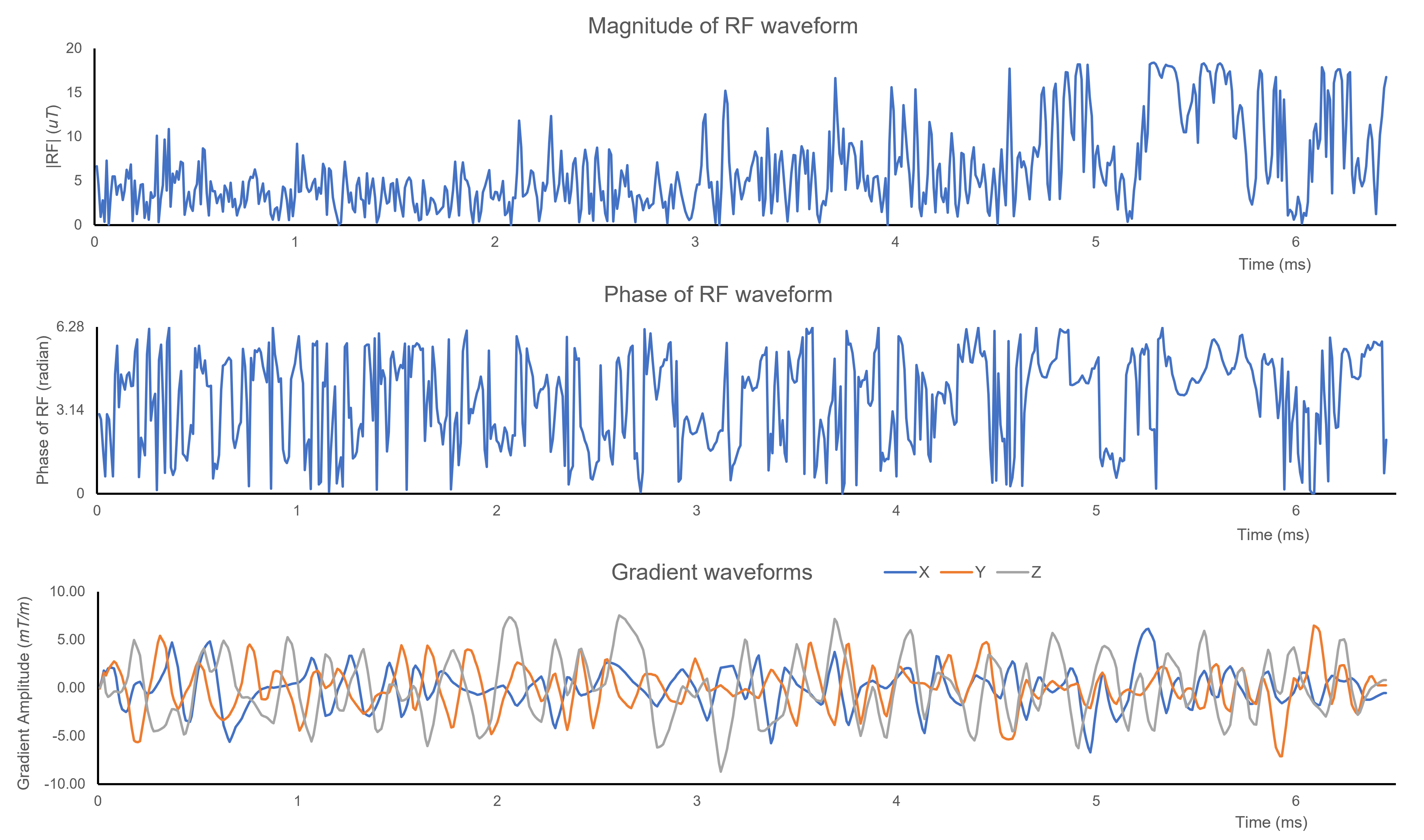
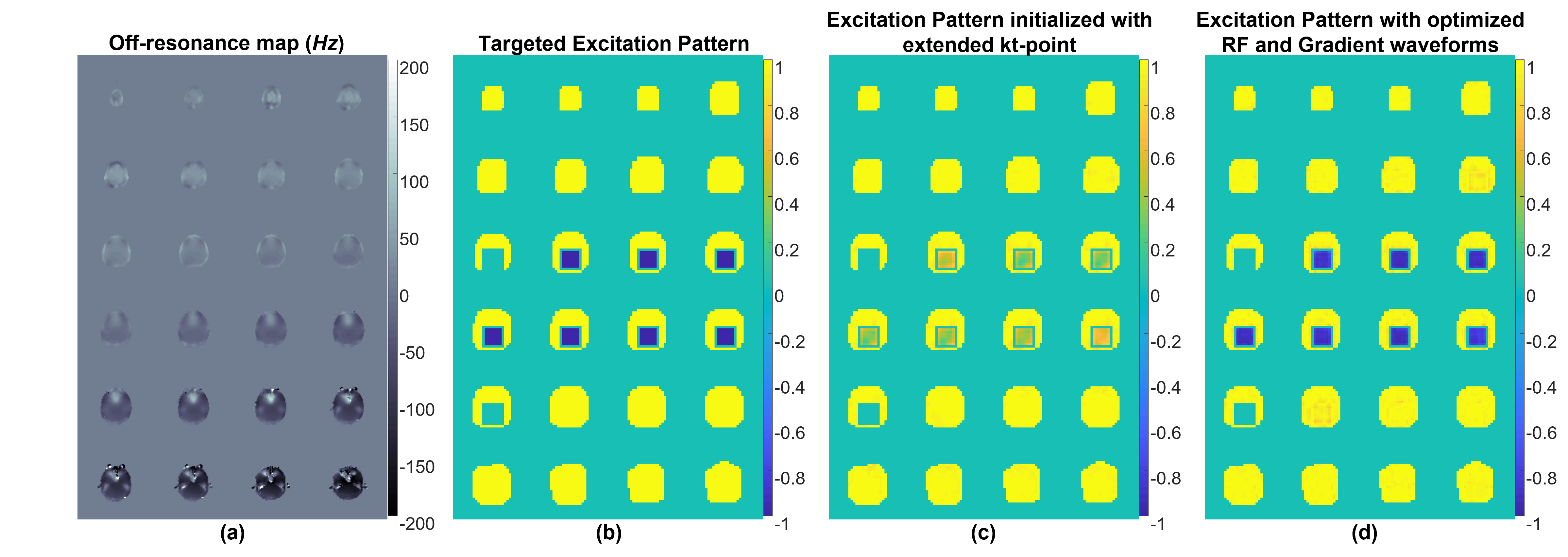
Figure 1 shows one example of the designed RF and gradient waveforms. The duration of the RF that excites a 80*80*50 mm3 region of interest with the larger 400 × 400 × 155 mm3 FOV is 6.45 msFigure 2 shows a B0 field map measured in the brain (a), which was used in the optimization of the RF and gradient waveforms for the targeted excitation pattern (b). The simulated excitation pattern initialized using extended k-t point is shown in Figure 2c, and the simulated excitation pattern generated with the optimized RF and gradient waveforms is shown in Figure 2d.
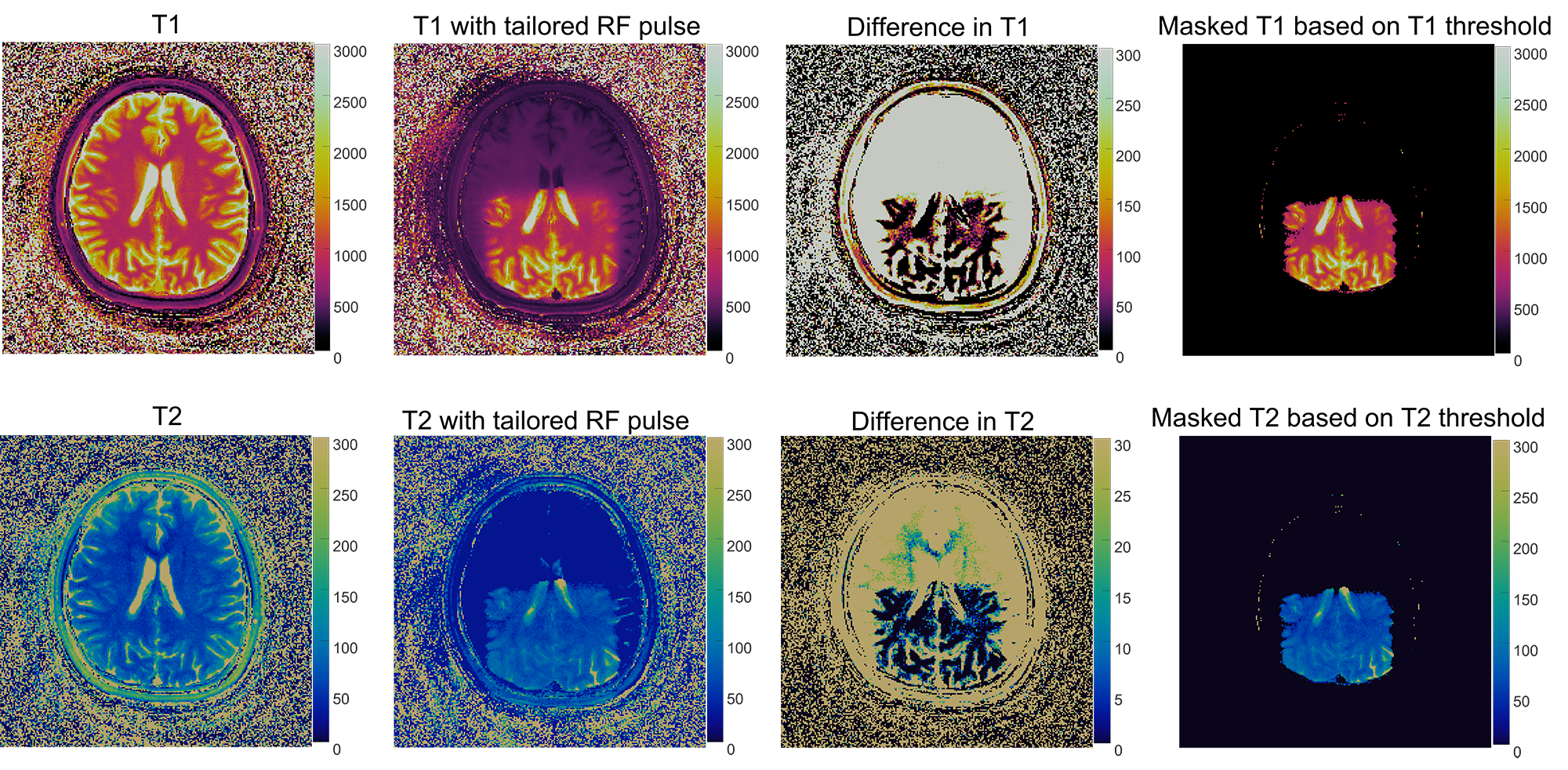
Figure 3 shows T1 and T2 maps excited with normal full FOV RF pulses and with the 3D tailored RF pulse. The difference maps are rescaled by a factor of 10 to amplify the difference, which demonstrates that the T1 and T2 measurements with the 3D tailored RF pulse are close to the results from the conventional MRF. The differences in T1 and T2 maps are mainly confined to the cerebrospinal fluid, which is likely due to physiological fluctuation. A T1 value of 700 ms was used to segment smaller FOV region.
Discussion and Conclusions
We show initial results from a reduced FOV quantitative imaging with a 3D tailored RF pulse. With different RF flip angle and phase patterns between the inner volume and outer volume, the pattern-matching algorithm can retrieve accurate quantitative maps in the inner volume by matching the acquired signal to the signals in the pre-calculated dictionary.Acknowledgements
This work was supported by Siemens Healthcare and NIH grants R37CA263583 and R01CA208236.References
1. Ma D, Gulani V, Seiberlich N, Liu K, Sunshine JL, Duerk JL, Griswold MA. Magnetic resonance fingerprinting. Nature. 2013 Mar 14;495(7440):187–192. PMCID: PMC3602925
2. Joint Design of RF and gradient waveforms via auto-differentiation for 3D tailored excitation in MRI | IEEE Journals & Magazine | IEEE Xplore [Internet]. [cited 2021 Nov 10]. Available from: https://ieeexplore.ieee.org/document/9439482
3. Hao S, Fessler JA, Noll DC, Nielsen J-F. Joint Design of Excitation k-Space Trajectory and RF Pulse for Small-Tip 3D Tailored Excitation in MRI. IEEE Trans Med Imaging. 2016 Feb;35(2):468–479. PMCID: PMC4792784
Figures