1863
Reduction of Acquisition time using RAPID-SI technique: Preliminary in-vitro results and comparison with CSI at Ultra-High-Field.1Carle Foundation Hospital, Center for Clinical Imaging Research, Urbana, IL, United States
Synopsis
It has been shown that RAPID-SI, implemented at 3 Tesla magnet, allows for reduction of acquisition time while providing accurate metabolite information as compared to CSI. To benefit from the higher sensitivity of the Ultra-High-Field (UHF), RAPID-SI was implemented on a Siemens Terra 7T scanner, and phantom MRSI data were acquired, with less averaging, and compared in terms of acquisition time, signal-to-noise ratio (SNR), and data analysis to those collected using CSI method. As expected, and in addition to the acquisition time reduction, proportional to the speed factor R, RAPID-SI provides similar quantification results with similar SNR values, afterward data reconstruction, as compared to CSI.
Introduction
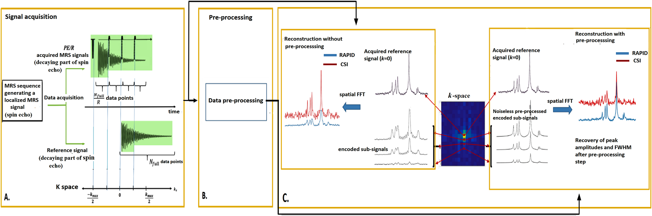
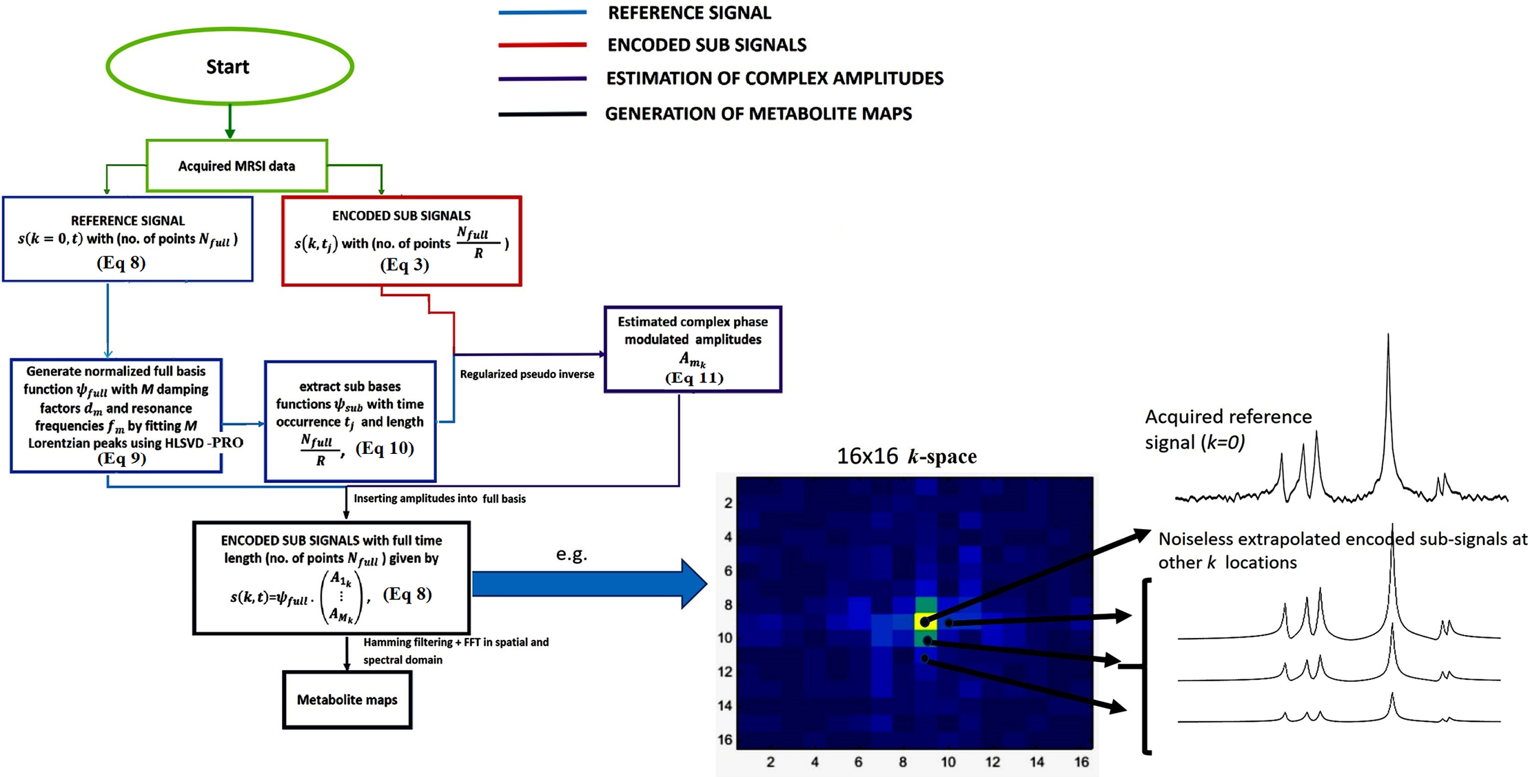
To reduce the long acquisition time in MRSI, a chemical shift imaging technique named Reduction of Acquisition time by Partition of the sIgnal Decay in Spectroscopic Imaging (RAPID-SI), which uses blipped gradients inserted during signal acquisition, has been proposed [1]. In addition to reducing acquisition time in proportion to an acceleration factor R (set equal to the inserted blipped gradients; Figure 1), RAPID-SI has been shown at 3 Tesla to provide comparable quantification results to CSI. However, the partition of the signal into sub-signals for spatial encoding purposes reduces the sensitivity of the acquired data by √R, which is compensated during data reconstruction (Figure 1, 2) [1]. To benefit from the higher sensitivity offered at Ultra-High Field (UHF), RAPID-SI was implemented on a 7 Tesla whole body magnet, and phantom data were acquired and compared to CSI. The results obtained demonstrate that in addition to the expected acquisition time reduction obtained at lower field, RAPID-SI profits from the UHF to reduce signal averaging while providing similar results in term of data analysis and sensitivity to CSI.Methods
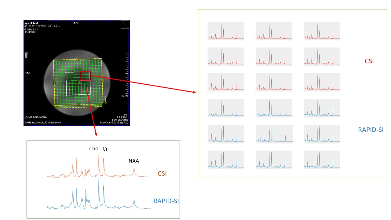
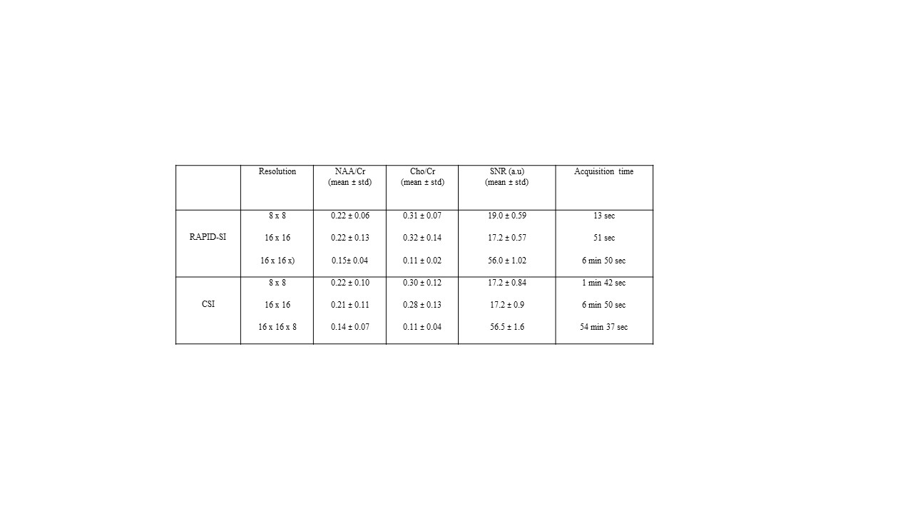
The sequence part of the RAPID-SI technique, displayed in Figure 1A, was implemented on a Siemens Terra 7T scanner, and accelerated in the anterior-posterior (AP) direction by inserting R blipped gradients during data acquisition with classical phase encoding applied along the right-left (RL) direction. The Flow diagram of the data pre-processing part of the technique is presented in Figure 2. The data pre-processing was performed offline in MATLAB. Using the multi-transmit capabilities (pTx) available at the 7 T scanner, 2D (8x8, 16x16) and 3D (16x16x8) RAPID-SI and standard CSI data were acquired from a spherical phantom containing main brain metabolites (e.g: NAA, Creatine, Choline) using the following acquisition parameters: TE/TR=30/1600 msec, BW=2000 Hz, N = 512, avg = 1 (set to 2 at 3 Tesla [2]) , phase encoding steps (PE) = 8 and 16, and number of collected signals (FIDs) along blip (AP) direction = 1 (for PE=8) and 2 (for PE=16). The RAPID acceleration factor was set to R = 8 (AP direction). The FOV dimensions along RL and AP direction = 120 mm, and slice (slab) thickness was set to 10 mm for the 2D and to 120 mm for the 3D.Results and Discussion
As expected, RAPID-SI shortened the acquisition time by the acceleration factor R. Compared to previous 3T studies, this time is further reduced at 7 Tesla by signal averaging reduction. As an example, a 2D 16x16 data set acquired in about 7 min with CSI, was reduced to less than 1 minute with the RAPID-SI with acceleration factor R set to 8 (Table. 1). SNR in RAPID-SI was lower than CSI by approximately the factor √R [1, 2]. However, during data pre-processing (Figs. 1, 2), the SNR recovers to a value closer to that for CSI, as shown in Table 1 and displayed in Figure 3. Data quantification presented here as metabolite ratios (e.g: NAA/Cr and Cho/Cr) reported in Table 1, (absolute metabolite quantification were demonstrated elsewhere [1]), were comparable between of RAPID-SI and CSI. Further in-vivo testing are required to validate this encouraging preliminary phantom results.Conclusion
RAPID-SI reduces acquisition time, while preserving metabolite information, compared to CSI at UHF. To further reduce acquisition time RAPID-SI will be combined with other acceleration methods such as parallel imaging or compressed sensing. In addition, the B1 field inhomogeneity effect manifested at 7T, will be reduced by integrating RAPID-SI approach into a localized MRS sequence employing adiabatic RF pulses (e.g: semi-LASER [3, 4]) with in-vivo validation.Acknowledgements
This work was supported by the Stephens Family Clinical Research Institute, Carle Foundation Hospital, Urbana, IL, USA.References
1. Bhaduri S, Clement P, Achten E, Serrai H. Reduction of Acquisition time using Partition of the sIgnal Decay in Spectroscopic Imaging technique (RAPID-SI). PLOS ONE. 2018; 13(11): e0207015.
2. Bhaduri S., et al. Proc ISMRM-ESMRMB 2018, 1057.
3. Scheenen TW, Heerschap A, Klomp DW. Towards 1H-MRSI of the human brain at 7T with slice-selective adiabatic refocusing pulses. MAGMA. 2008 Mar; 21(1-2):95-101.
4. Scheenen TW, Klomp DW, Wijnen JP, Heerschap A. Short echo time 1H-MRSI of the human brain at 3T with minimal chemical shift displacement errors using adiabatic refocusing pulses. Magn Reson Med. 2008 Jan; 59(1):1-6.
Figures