1853
Improving multimodal imaging with an MR-compatible EEG net: the R-Net-MR-IT1Athinoula A. Martinos Center for Biomedical Engineering, Charlestown, MA, United States, 2Department of Neuroscience, University of Copenhagen, Copenhagen, Denmark, 3Department of Engineering, Boston University, Boston, MA, United States, 4Brain Products GmbH, Gilching, Germany
Synopsis
EEG provides valuable clinical information, but EEG nets produce artifacts in MRI and CT images, preventing these modalities from being combined in typical clinical practice. We tested whether a new MR-compatible EEG net, called the R-Net-MR-IT, could produce high-quality clinical and research images. We assessed image quality in CT and MR images on a phantom with a conventional net, R-Net-MR-IT, and with no net. We then performed the same comparison in humans, as well as fMRI scans. Our results show that the R-Net-MR-IT enables acquisition of high quality CT and MR images, with minimal artifact from EEG hardware.
Introduction
EEG nets are usually removed in both clinical and research settings prior to imaging, in order to maintain high quality images in both CT and MR (1). In previous work, we assessed whether another MR-compatible EEG net could produce high quality images with a limited number of clinical MRI scans (2).Here, we expanded on that previous work by using a novel cap, the R-Net-MR-IT, which uses polymer thick film technology rather than copper wires that can impede MR imaging, and tested its compatibility with CT images, clinical MR sequences, and BOLD fMRI signals. Our results show that this new customized MR-compatible EEG net, the R-Net-MR-IT, produces CT and MR images with reduced artifacts compared to conventional copper nets (Cu-Net), and achieves similar image quality to the No-Net condition.
Methods
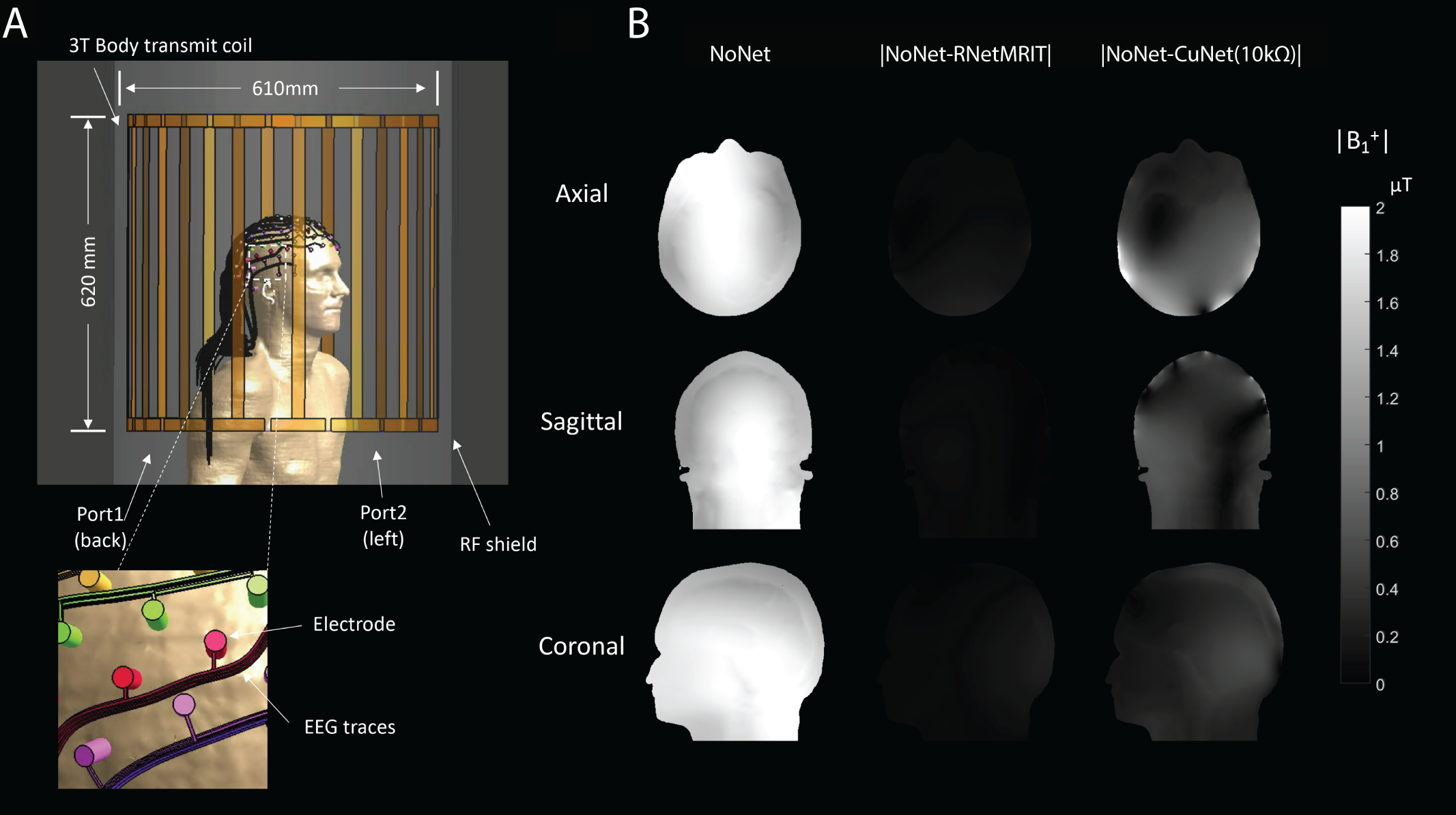
Two MR-compatible EEG nets were used: 1) the R-Net-MR-IT – a novel 128-channel MR-compatible EEG net using high resistance Polymer Thick Film technology (3) and 2) the Cu-Net: a 128-channel MR-conditional Geodesic Sensor Net (MagStim EGI, Eugene, OR) made with conventional copper leads. Three experiments were done to assess the image quality of the R-Net-MR-IT with a CT and Siemens 3T Skyra scanner with a 64-channel head coil array: 1) Sim4Life simulations 2) phantom CT and MR scans and 3) human MR scans.Simulations
Sim4Life (ZMT, Switzerland) was used to estimate the B1+ field distribution in the Duke model (v3.0) in the 3T body transmit coil in the case of No-Net, R-Net-MR-IT (σ: 25.6 S/m), and Cu-Net (σ: 5.8x10ˆ7 S/m) with the trace length of 510 mm. The 128-channel EEG traces were drawn with a similar method to the previous work (4), and the current limiting resistors (10kΩ) were added between electrode and trace in the case of Cu-Net (5).
Phantom Scans
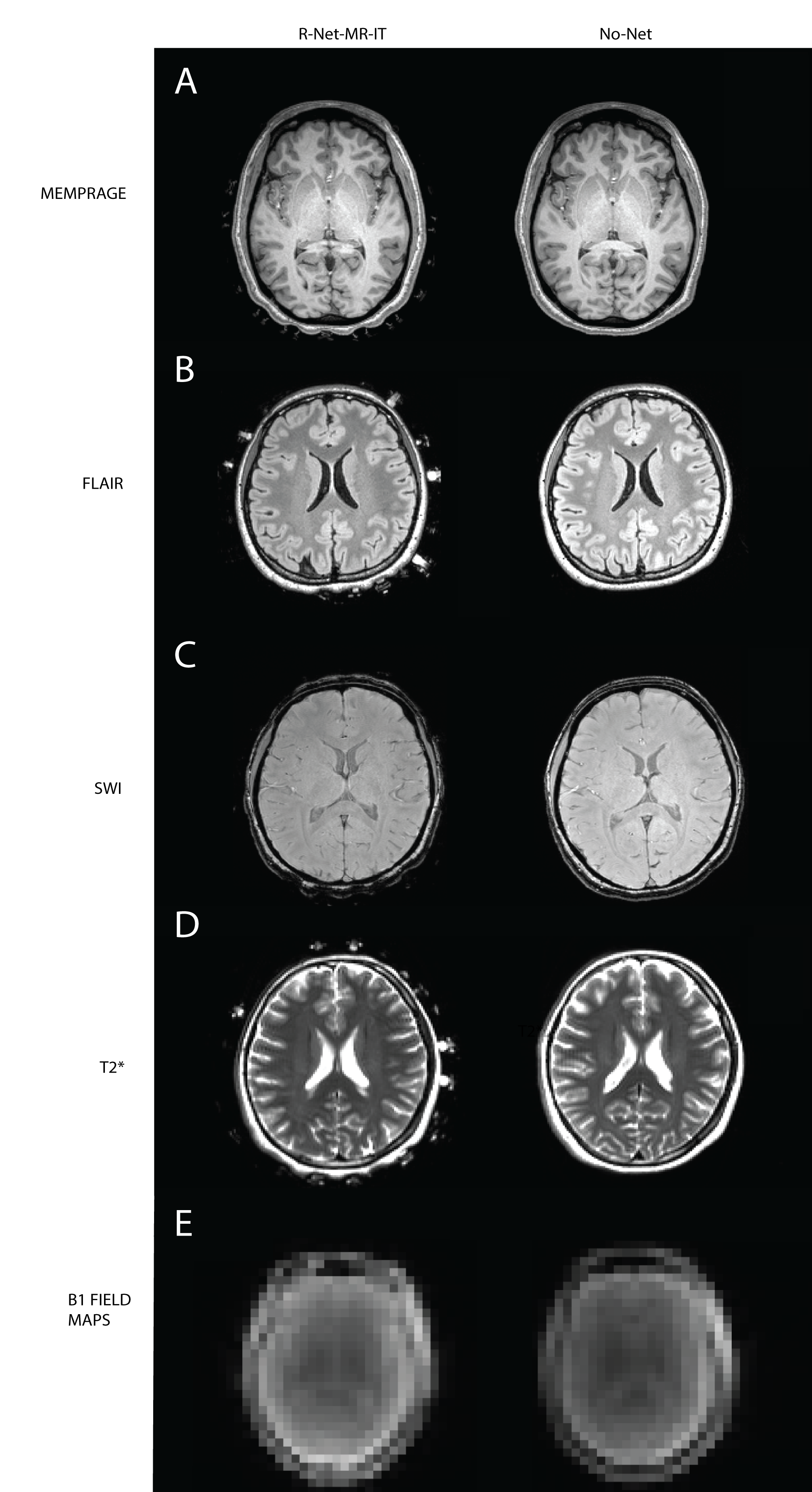
Due to safety concerns, the Cu-Net cannot be used on humans with many clinical MRI sequences, so to examine the differences between the R-Net-MR-IT and the Cu-Net we scanned phantoms in both CT and MR environments. Both phantom scans had three conditions: 1) No-Net 2) R-Net-MR-IT and 3) Cu-Net. The CT Scans compared the three conditions with standard Human Adult Trauma Head scan (SOMATOM Force Scanner). The scans were acquired as follows: a 1.0 mm isotropic resolution single-shot T1 MEMPRAGE, 1.0 mm isotropic resolution FLAIR images (TR=5 s, TE=391 ms, FOV=256 mm), 33 slices with whole brain coverage B1 maps (TR=9 s, TE=1.86 ms, FOV=450 mm, voxel size=7x7x14 mm), 0.9x0.9x1.8 mm resolution susceptibility weighted images (SWI) (TR=30 ms, TE=20 ms, FOV=220 mm) and 1.4x1.4x3.0 mm resolution T2*-weighted images with 31 slices (TR=6 s, TE=95 ms, FOV=450 mm).
Human Scans
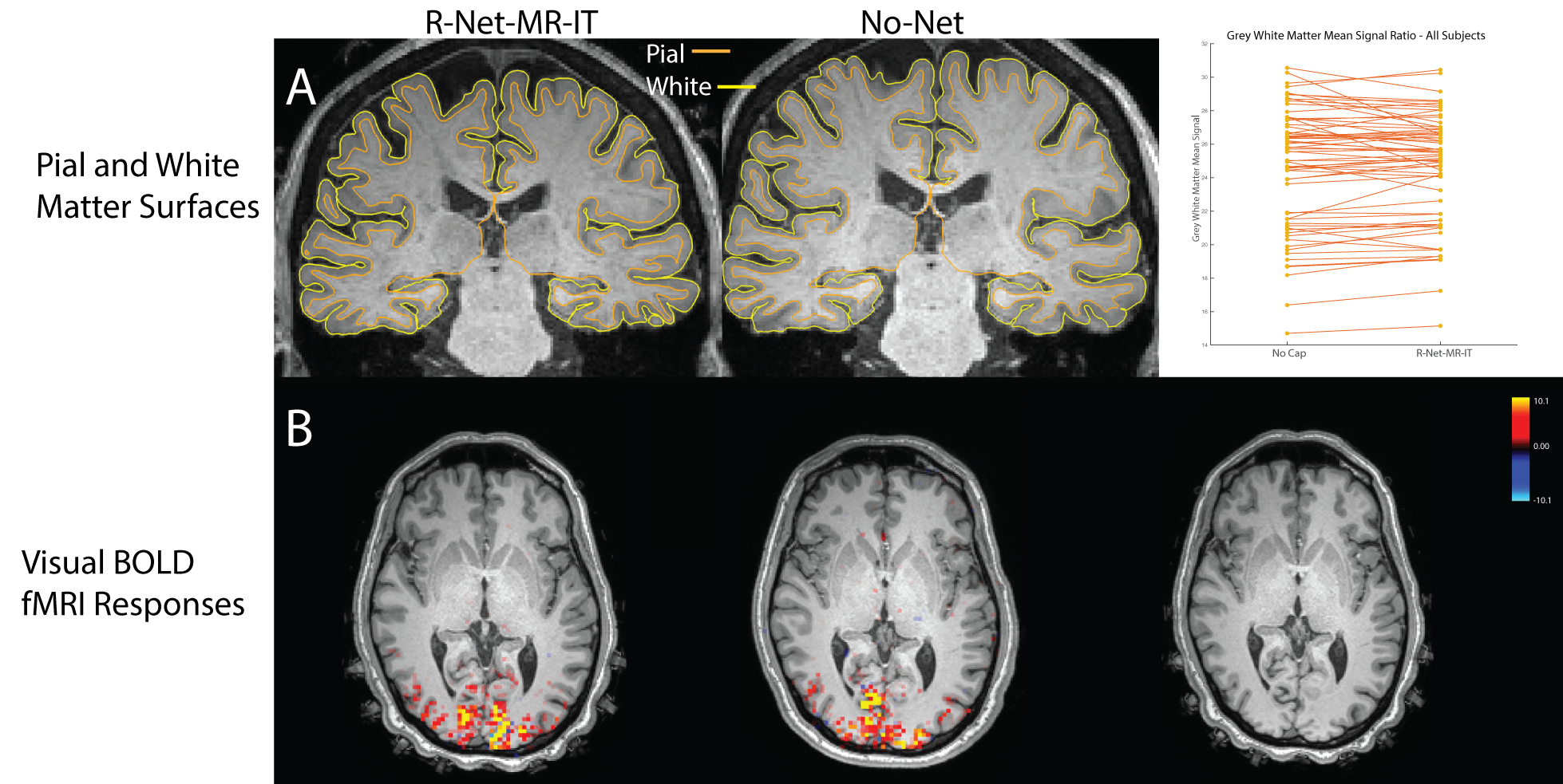
The study was approved by the Institutional Review Board at Massachusetts General Hospital. Five healthy volunteers (mean age=36.2 years, range=22-66) gave informed consent. Participants participated in a two-hour session with two conditions: 1) No-Net and 2) R-Net-MR-IT. Each condition followed the same phantom MR anatomical imaging protocol above. FreeSurfer was used to compute surface-wise gray/white contrast between the T1 MEMPRAGES. Gradient-echo EPI was then acquired with 2.5 mm isotropic resolution with whole-brain coverage (TR=2.71 s, TE=30 ms, FOV=250 mm). To assess functional responses, participants performed a flickering checkerboard block design visual task. Functional data were slice time corrected, and motion corrected. FEAT was used to extract the z-score of functional responses and conditions were compared with a paired t-test.
Results
Qualitative comparison of the CT images showed that the Cu-Net condition had significant artifacts from the copper wires which impeded visualization of the CT image (Fig. 1). While the R-Net-MR-IT electrodes were also visible, no artifact from the leads was visible (Fig. 1b,c).We next examined R-Net-MR-IT performance in the MR environment. The EM simulation results showed that the B1+ field artifact volume using the R-Net-MR-IT was reduced compared to the Cu-Net (Fig. 2). Our phantom scans confirmed these simulation results: the B1 field maps, SWI, FLAIR, T2*, and MEMPRAGE all showed increased dropout in the Cu-Net condition, whereas the R-Net-MR-IT had similar image quality to the No-Net condition (Fig. 3a-c).
In the human imaging, the R-Net-MR-IT again had similar image quality to the No-Net condition in the SWI, FLAIR, T2*, and MEMPRAGE images, as well as B1 field maps (Fig. 4a-e). Qualitative comparison of pial and white matter surfaces showed similar performance, and quantitative evaluation of grey/white matter mean signal intensity ratios showed no significant difference between No-Net and R-Net-MR-IT conditions (Fig. 5a). In the fMRI scans, we found similar activation for the visual cortex in both conditions (Fig. 5b), with no statistically significant difference in activation across conditions (p>0.05).
Conclusion
Our results indicate that with the R-Net-MR-IT, researchers and clinicians can use CT, clinical MRI, and fMRI sequences without sacrificing image quality. Phantom and human comparisons demonstrated that the R-Net-MR-IT provided high image quality with both anatomical and functional sequences. Given the excellent performance of this net, future work could test its potential applications at ultra-high field, where image quality is even more challenging.Acknowledgements
This work was funded by NIH/NIBIB grant R01EB024343 and NIA R01-AG070135. Authors acknowledge to ZMT Zurich MedTech for Sim4Life for Science License. We also thank Dr. John Kirsch and Jonathan Cardona for their help with the MR protocols.
References
1) Chang C, Chen JE. Multimodal EEG-fMRI: advancing insight into large-scale human brain dynamics. Curr Opin Biomed Eng. 2021 Jun;18:100279. doi: 10.1016/j.cobme.2021.100279. Epub 2021 Mar 15. PMID: 34095643; PMCID: PMC8174780.
2) Fultz N, Poulsen C, Lev M, Berman A, Chen JE, Bonmassar G, and Lewis LD (2019). High-quality FLAIR and diffusion imaging with presence of EEG nets (Abstract/Poster). International Society for Magnetic Resonance in Medicine.
3) Poulsen C, Wakeman DG, Atefi SR, Luu P, Konyn A, Bonmassar G. Polymer thick film technology for improved simultaneous dEEG/MRI recording: Safety and MRI data quality. Magn Reson Med. 2017;77(2):895-903. doi:10.1002/mrm.26116
4) Jeong H, Ntolkeras G, Grant PE, Bonmassar G. Numerical Simulation of the Radiofrequency Safety of 128-Channel hd-EEG Nets on a 29-Month-Old Whole-Body Model in a 3 Tesla MRI. IEEE Trans Electromagn Compat 2021:1–9.
5) Krakow K, Allen PJ, Symms MR, Lemieux L, Josephs O, Fish DR. EEG recording during fMRI experiments: Image quality. Hum Brain Mapp 2000;10:10–15.
Figures