1830
Transcranial Focused Ultrasound Suppresses Thalamic Heat Nociceptive Response and Reorganizes Nociceptive Networks in Non-human Primates
Arabinda Mishra1,2, Pai-Feng Yang1,2, Tom Manuel1,3, Allen T Newton1,2, Huiwen Luo1,3, Marshal A Phipps1,2, Michelle Signoa1,3, Jamie L Reed1,2, John C Gore1,2,3, William A Grissom1,3, Charles F Caskey1,2,3, and Li Min Chen1,2
1VUIIS, Vanderbilt University, Nashville, TN, United States, 2Radiology, Vanderbilt University Medical Center, Nashville, TN, United States, 3BME, Vanderbilt University, Nashville, TN, United States
1VUIIS, Vanderbilt University, Nashville, TN, United States, 2Radiology, Vanderbilt University Medical Center, Nashville, TN, United States, 3BME, Vanderbilt University, Nashville, TN, United States
Synopsis
MRI guided transcranial focused ultrasound (MRgFUS) is a neuromodulation tool that can excite or inhibit neural activity with high spatial precision and real-time functional feedback. Our goal is to develop a MRgFUS device for human pain therapy. Here, we studied suppressive effects of FUS on a key hub in the thalamus for transmitting peripheral inputs to cortex for pain perception, and mapped changes in effective functional connectivity of thalamic networks using hierarchical clustering. We show that concurrently delivered FUS of moderate intensity suppressed painful heat responses at the thalamus target and induced reorganization of effective connectivity networks.
Introduction:
Targeted neuromodulation with high spatial precision potentially has significant clinical applications. We previously demonstrated that the concurrent sonication and tactile stimulation of digits using MRI guided transcranial focused ultrasound (MRgFUS) can both excite and inhibit neural activity, causing BOLD signal changes at target and functionally relevant off-target regions in anaesthetized macaque brains [1, 2]. The goals of this work are (1) to demonstrate the suppressive effects of FUS on thermal pain responses in the thalamus and (2) to use psychophysiological interactions (PPI) derived effective connectivity (EC) measures and hierarchical clustering of detected changes to investigate the reorganization of functional networks induced by suppression of thalamic heat pain responses.Method:
MRI studies were performed at 3T (Philips Achieva) on macaque monkeys under isoflurane anesthesia. A 128 channel FUS array delivered 650 kHz pulsed US with fast PRF of 1kHz with a 50% duty cycle for 500ms, slow PRF of 0.5 Hz for 16s. Estimated in vivo pressure was 550 kPa (39% transmission estimate). The thalamic ventroposterior lateral (VPL) nucleus was targeted and validated with MR-ARFI. A pair of flex coils was used for MR-ARFI and fMRI data acquisitions. FMRI data were acquired using single shot GE-EPI sequence (TR = 2s, TE=30ms, voxel size: 1.43x1.43x2mm3). Painful heat stimulation of fingers with and without concurrent FUS were presented in an interleaved manner. Standard fMRI EPI data preprocessing was performed. The EPI data were co-registered to each subject’s anatomic images and then normalized to the macaque template (NMTv2.0) [4] using FSL (flirt, fnirt) for a group (n=4 animals) analysis. Brain regions responding to thermal stimulation were identified using standard GLM analysis. PPI (psychophysiological interactions) regressors [4] based on characteristic temporal variations associated with both stimulus conditions (heat and heat+FUS) were used to evaluate the effective connectivity between the targeted thalamus region and rest of the brain (termed thalamus pain network). We performed objective parcellation of the effective connectivity circuits using hierarchical clustering. The mean temporal dynamics of the ROIs showing significant effective connectivity were the basis for the data driven classification at group level. A direct comparison of effective connectivity based regressor coefficients (beta value) was performed to evaluate the FUS effect in the brain. The changes in the functional organization of the pain regions with and without targeted FUS are used to quantify the network effects of thalamus modulation. The abbreviated cortical regions and re-organization of the clustered groups (networks) shown in the dendrogram (Figure 2) were derived from the NMT2.0 atlas.Results:
Figures 1 A&B show selected coronal views of the fMRI activation maps (thresholded at t > 2.0, p < 0.05, FDR corrected) showing multiple activation foci in the thalamus and cortical regions during painful heat alone (Fig.1A) and painful heat and concurrent sonication of thalamus (Fig.1B). Heat evoked percentage BOLD signal change at the VPL target (VLPdT in table 1, NMTv2.0) was almost abolished when concurrent FUS was delivered (Fig. 1C). Twenty brain regions showed statistically significant effective connectivity measures to the thalamus VPL region (table 1). Classification of these regions in the cortex and targeted thalamus was performed using hierarchical clustering, which unlike other unsupervised machine learning techniques does not require a user defined number of clusters. The multilevel hierarchy clustered the data (ROI mean temporal dynamics) into groups (networks) based on the level most appropriate to describe the dissimilarity between the clusters. During heat pain conditions, the dendrogram clustered the thalamus pain network into four subgroups at cophenetic distance ~1.1 (Fig. 2A). Concurrent FUS suppression of heat pain response at thalamic VPL regrouped the ROIs in the dendrogram (Fig. 2B). Color coded ROIs in Figure 2B depict FUS induced reorganization. A direct comparison of the regression coefficients (beta values) measured for each region associated with both conditions revealed that concurrent FUS significantly (*/**: p < 0.05/0.005, FDR corrected) weakened EF of six ROIs (SI, SII, vlPFC, STGr, STGc, and V4) to thalamus and strengthened DPC’s EF to thalamus (Fig. 3). The mean and standard error of these coefficients are shown in Figure 3. Statistically significant differences were measured using non-parametric Mann Whitney Wilcoxon tests (MWW).Discussion & Conclusion:
A combined use of a 128 channel FUS array that offers steering capacity and real-time MR-ARFI allowed precise FUS modulation of heat pain responses at one thalamus nucleus and its interconnected networks during pain processing. We demonstrated that moderate-intensity FUS suppressed thalamus VPL nucleus’s fMRI response to nociceptive heat stimulation of fingers. Suppression of the thalamus heat pain response also led to thalamus pain network connectivity changes. Hierarchical clustering revealed that heat pain signal reduction by FUS weakened connections between six heat pain cortical regions to thalamus VPC but strengthened connection in one cortical area.Acknowledgements
This work was funded by National Institutes of Health grants R01MH111877, R24MH109105, 1S10OD012297-01A1, 5T32EB014841, and 1F31EB026928, and the Focused Ultrasound Foundation.References
[1] Yang, P.F., et al., Bidirectional and state-dependent modulation of brain activity by transcranial focused ultrasound in non-human primates. Brain Stimul, 2021. 14(2): p. 261-272.[2] Yang, P.F., et al., Neuromodulation of sensory networks in monkey brain by focused ultrasound with MRI guidance and detection. Sci Rep, 2018. 8(1): p. 7993.[3] Jung, B., et al., A comprehensive macaque fMRI pipeline and hierarchical atlas. Neuroimage, 2021. 235: p. 11[4] O'Reilly, J.X., et al., Tools of the trade: psychophysiological interactions and functional connectivity. Soc Cogn Affect Neurosci, 2012. 7(5): p. 604-9.Figures
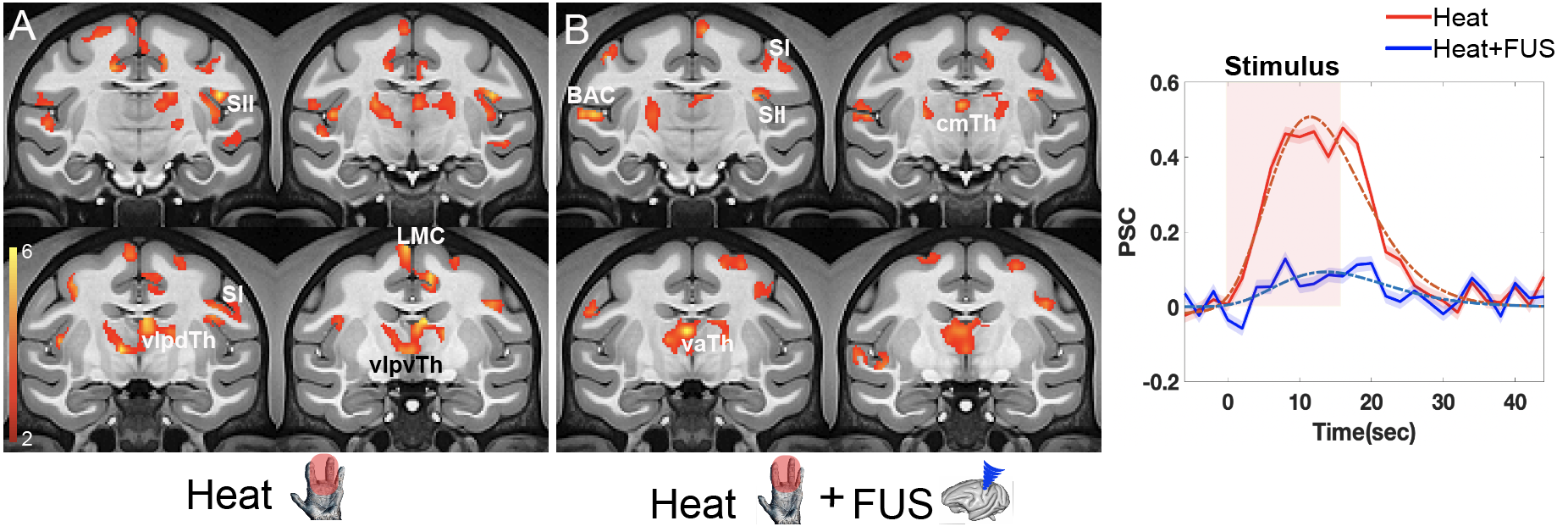
Stimulus driven fMRI activation using thermal (heat) and heat + FUS stimulus. (A) Stimulus evoked activation map at group level (N = 4, 27 runs) overlaid on the coronal slices containing the thalamus area using NMT2.0 atlas t > 2, p < 0.05 (FDR corrected). (B) Activation map showing stimulus patterns for thermal stimulus in combination with image guided FUS targeting thalamus (VPL). (C) Cycle averaged percentage signal change (PSC, N = 4) in the VPL during the thermal stimulus (red) and thermal + FUS (blue).
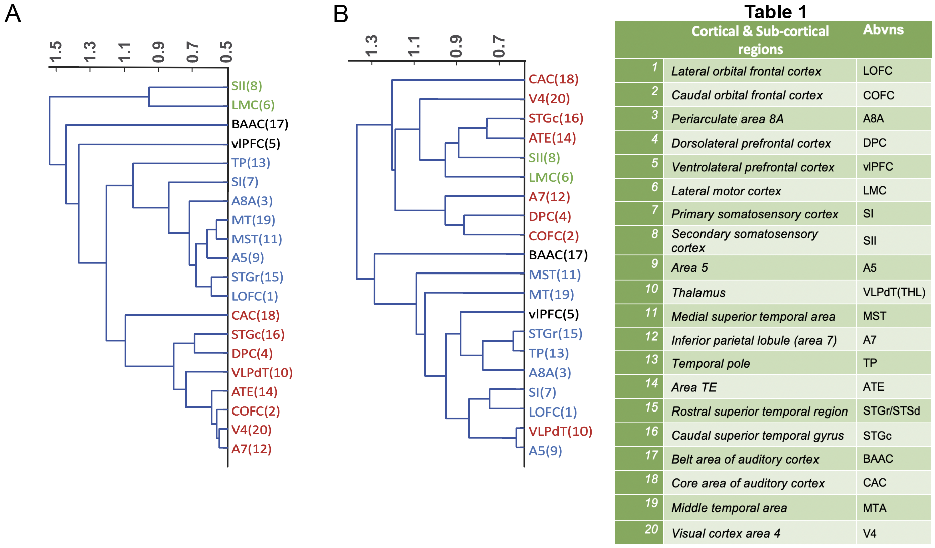
Similarity in responses in the cortical regions to the thalamic VPL seed. Hierarchical cluster organization of response similarity represented by the dendrogram plot for twenty cortical regions (Table 1) associated with heat (A) and heat + FUS (B). The horizontal axis measures cophenetic distance between objects/clusters in the parametric space using WPGMA algorithm.
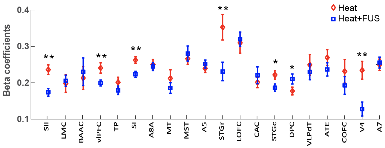
Comparison of effective connectivity measures using regression coefficients (beta value). Mean and standard error of beta coefficient using effective connectivity-based PPI regressor in the twenty subregions (red: Heat; blue: Heat + FUS). Statistical significance of difference measures using non-parametric MWW test shown (*: p < 0.05 and **: p < 0.005, FDR corrected).
DOI: https://doi.org/10.58530/2022/1830