1818
Accelerating Ultra-Low Field MRI with Compressed Sensing1ACRF Image X Institute, Faculty of Medicine and Health, The University of Sydney, Sydney, Australia, 22Department of Electrical Engineering and Computer Sciences, UC Berkeley, Berkeley, CA, United States, 3A. A. Martinos Center for Biomedical Imaging, Charlestown, MA, United States, 4Department of Physics, Harvard University, Cambridge, MA, United States, 5Harvard Medical School, Boston, MA, United States
Synopsis
Portable MRI scanners that operate at very low magnetic fields are increasingly being deployed in clinical settings. However, the intrinsic low signal-to-noise (SNR) ratio of these low-field MRI scanners often necessitates many signal averages, and therefore excessively long acquisition times. Here we propose to improve SNR through optimized k-space undersampling and Compressed Sensing reconstruction. We demonstrate this approach for 6.5 mT ultra-low-field MRI using: (1) retrospective-subsampling experiments with 2x to 4x acceleration; (2) prospectively-subsampled data acquired from a human brain phantom with a 6.5mT MRI. The results exhibit a higher SNR than the traditional averaging method, without increasing scan time.
Introduction
Recent advances in portable MRI technology have enabled the development of scanners for applications where it was previously impossible to use MR, such as at the bedside of critically ill patients in the ICU.1,2 However, the portability of these new MRI scanners is largely achieved by replacing the high-field superconducting magnet in a conventional MRI scanner with permanent magnet arrays or electromagnets operating at low (< 0.1 T) and ultra-low (< 10 mT) magnetic fields.3–6 This field reduction necessarily results in a degradation of image signal-to-noise ratio due to a drastic reduction in Boltzmann polarization. Methods that improve low-field MR image quality without compromising acquisition times are therefore highly needed for improving the diagnostic accuracy of the modality.7One approach to accelerating high-field MRI is the use of undersampled acquisitions and Compressed Sensing (CS) reconstructions.8 However, studies validating the efficacy of this approach for low-field MRI have been limited.9,10 Traditionally, low-field MRI scans apply full k-space sampling, with many averages, and reconstruction using the inverse Fourier transform. Here, we propose to improve the low-field MRI SNR through optimizing k-space undersampling and Compressed Sensing reconstruction. We demonstrate this by: (1) retrospective subsampling experiments with different subsampling schemes and accelerations, using data from a low field (6.5 mT) MRI scanner. (2) Deployment of a Poisson Disc undersampling mask11 to a 6.5 mT scanner and prospecective acquisition of undersampled data. We observe that reconstructed images exhibit higher quality than those yielded from a traditional fully-sampled acquisition of the same duration.
Methods
Imaging experiments were performed on a 6.5 mT MRI scanner with a quadrature head coil12 and a brain-shaped phantom.3 A 3D balanced-steady state free precession (bSSFP) sequence with TR/TE = 22/11 ms and matrix size 64 x 70 x 15 (Readout x Phase Encode 1 x Phase Encode 2) was used for signal acquisitions.To create a fully-sampled baseline for image quality, the phantom was imaged with a 3D fully-sampled cartesian acquisition with various numbers of signal averages (NA). Fully sampled images were reconstructed from the two-channel k-space data using a conventional root sum of squares (RSS) approach. Fully-sampled acquisitions were acquired with an NA of 128/32/16/8 over a duration 51/12.6/6.3/3.2 minutes. Images from the NA = 128 acquisition were taken as the ground truth.
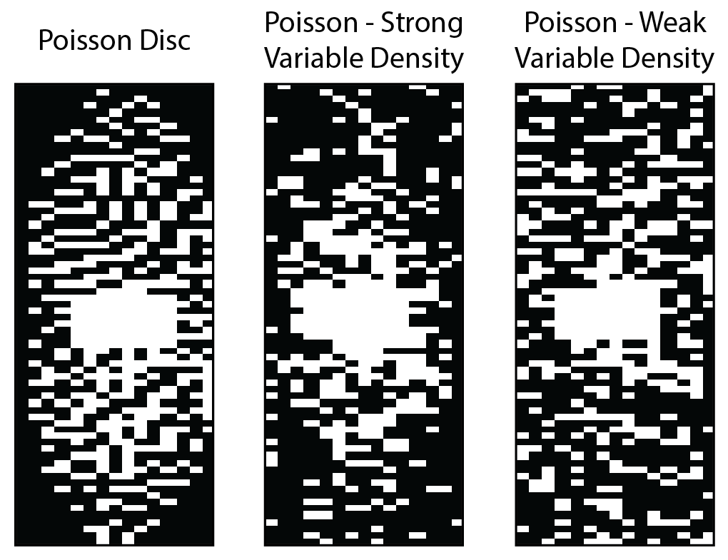
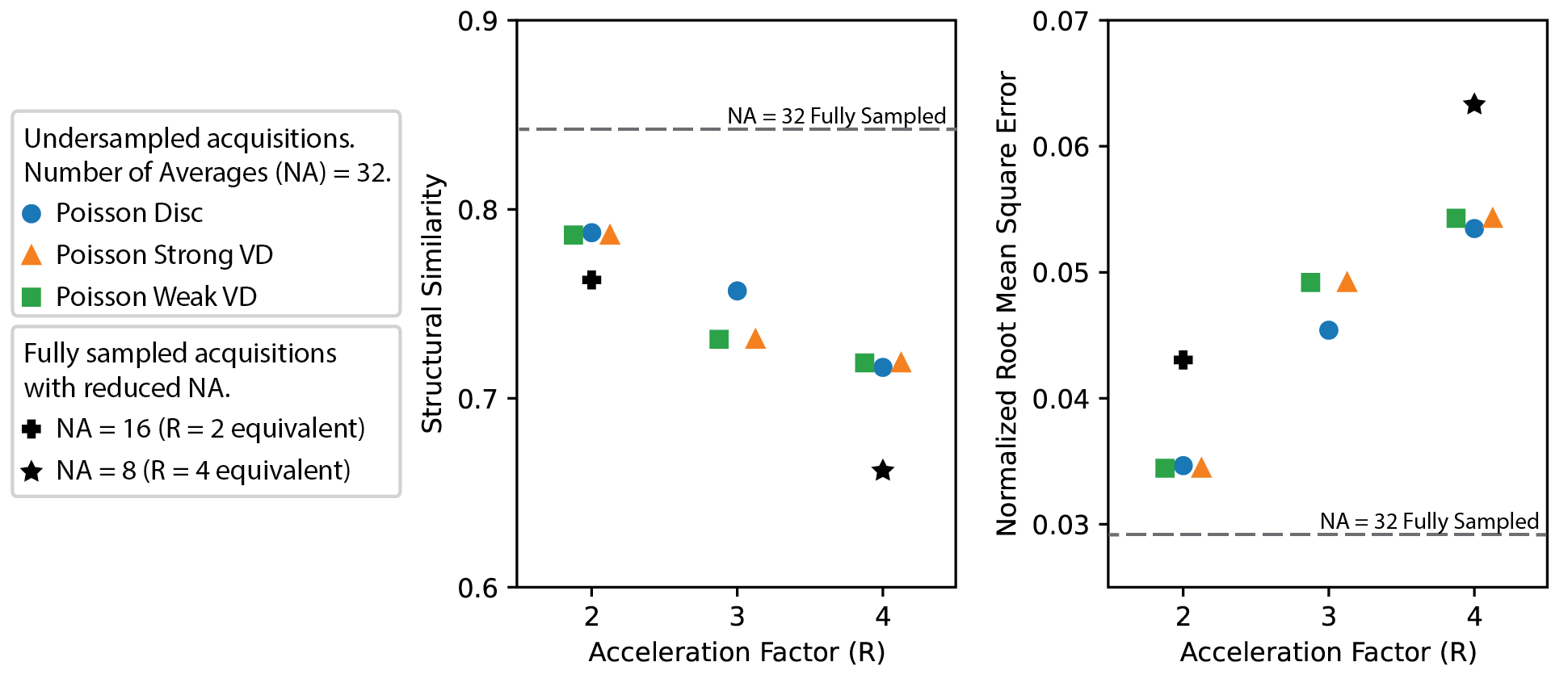
In the retrospective experiments, three types of Variable-density (VD) masks were generated and optimized: (i) Poisson-disc, (ii) weak-VD Poisson and (iii) strong-VD Poisson (see examples in Figure 1).13 Different masks were generated for acceleration factors of R = 2, 3 and 4. Sensitivity maps were computed with ESPIRiT,14 and L1-wavelet CS reconstructions were performed using SigPy.15 Normalized root-mean-square error (NRMSE) and structural similarity (SSIM)16 metrics were calculated to evaluate the quality of image reconstruction. The regularization parameter (λ) was optimized for each mask and acceleration factor separately by performing a grid search over the range of 10-6-10-1.
For prospective acquisition of undersampled data, the masks were then deployed to our 6.5 mT MRI scanner. CS reconstruction was then performed using the optimized hyperparameters found in the retrospective analysis.
Results
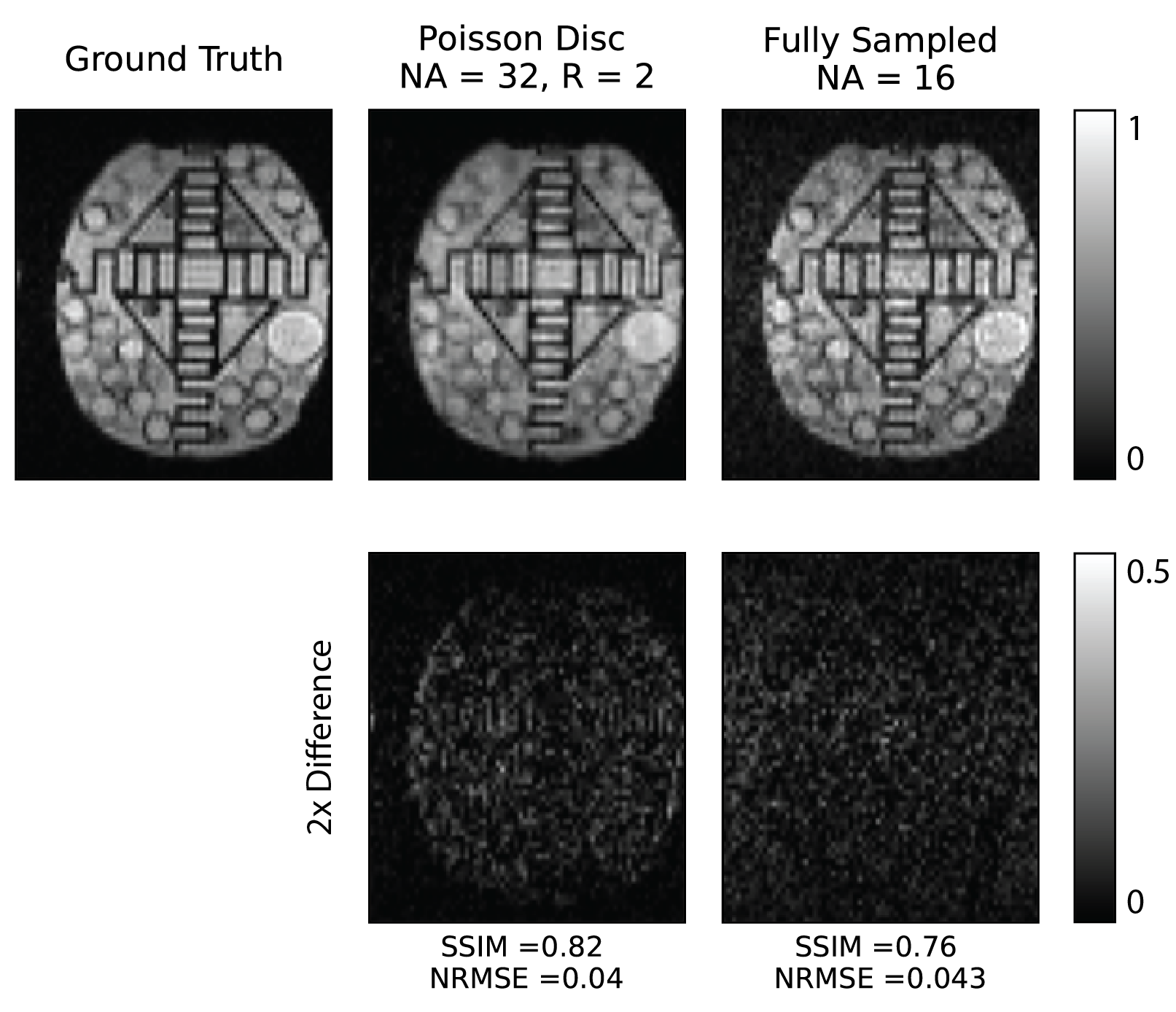
The quality of images yielded from optmized CS reconstruction in terms of NRMSE and SSIM metrics is summarized in Figure 2. Notably, our results show that all the undersampling approaches tested yield reconstructions with an NRMSE of lower than 0.05 for R=3. We found that the reconstruction quality metrics were slightly better for Poisson disc sampling in comparison to Poisson sampling with variable density. Importantly, we observed that for equivalent acquisition times, all undersampling strategies with CS reconstrucion yielded images of higher quality than fully-sampled images reconstructed with the traditional RSS approach.Noting that that metrics do not fully encapsulate the quality of reconstructed images, in Figure 3 we show an example of Poisson disc sampled (R = 2) and fully sampled images with equivalent acquisition times (6.6 min). We observe that the Poisson disc image reconstructed with CS appears sharper with less grainy features, which is also reflected in the higher quality image metrics. We also observe faint brain-shaped structures in the CS reconstructed image, indicating that λ likely cannot be increased further before the L1-wavelet regularized reconstruction begins blurring image features.
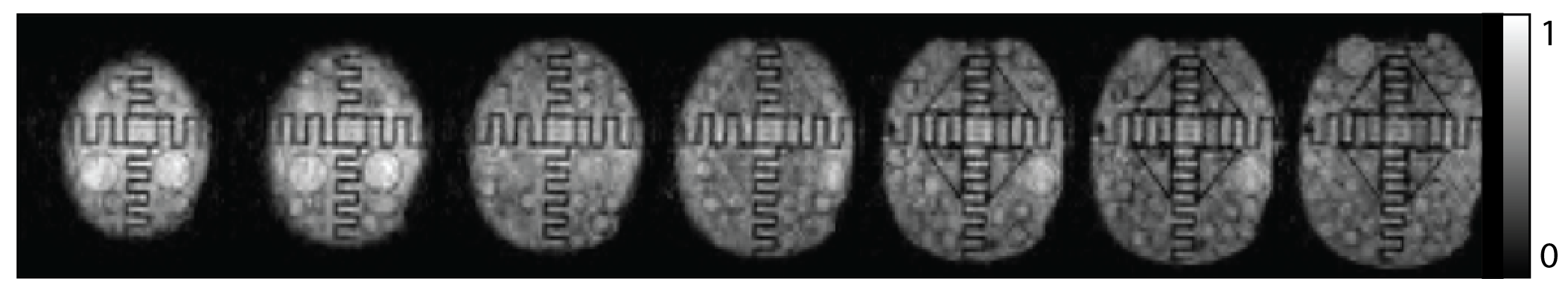
We successfully deployed a 3x accelerated Poisson disc sampling pattern to our 6.5 mT MRI scanner, prospectively acquiring data for the CS reconstruction shown in Figure 4 in 4.6 minutes.
Discussion
Low- and especially ultra-low field MRI is unusual in that images are almost always acquired with many averages of k-space. Here, we have shown that undersampling and reconstruction strategies developed for conventional high-field MRI can be applied to low-field MRI to increase the quality of images obtainable within a fixed imaging time.The development of multi-channel receiver coils for low-field MRI may also enable the application of higher acceleration factors than we have utilized here with a quadrature head coil.17 Significant scope exists to use artificial-intelligence-based reconstruction techniques to improve the image quality from low-field MRI.18
Conclusion
We have used CS reconstruction in combination with various sampling patterns to improve the SNR of low-field MRI images. These results will inform the development of faster imaging strategies for portable MRI scanners.Acknowledgements
D.E.J.W. is supported by a Cancer Institute of NSW Early Career Fellowship 2019/ECF1015. The information, data, or work presented herein was funded in part by the Advanced Research Projects Agency-Energy (ARPA-E), U.S. Department of Energy, under Award Number DE-AR0000823. The views and opinions of authors expressed herein do not necessarily state or reflect those of the United States Government or any agency thereof.
References
1. Mazurek MH, Cahn BA, Yuen MM, et al. Portable, bedside, low-field magnetic resonance imaging for evaluation of intracerebral hemorrhage. Nat Commun. 2021; 12: 1-11.2. Sheth KN, Mazurek MH, Yuen MM, et al. Assessment of Brain Injury Using Portable, Low-Field Magnetic Resonance Imaging at the Bedside of Critically Ill Patients. JAMA Neurol. 2021; 78: 41-47.
3. Sarracanie M, Lapierre CD, Salameh N, Waddington DEJ, Witzel T, Rosen MS. Low-Cost High-Performance MRI. Sci Rep. 2015; 5: 15177.
4. Cooley CZ, McDaniel PC, Stockmann JP, et al. A portable scanner for magnetic resonance imaging of the brain. Nat Biomed Eng. 2020.
5. Waddington DEJ, Boele T, Maschmeyer R, Kuncic Z, Rosen MS. High-sensitivity in vivo contrast for ultra-low field magnetic resonance imaging using superparamagnetic iron oxide nanoparticles. Sci Adv. 2020; 6: eabb0998.
6. Vogel MW, Guridi RP, Su J, Vegh V, Reutens DC. 3D-Spatial encoding with permanent magnets for ultra-low field magnetic resonance imaging. Sci Rep. 2019; 9: 1522.
7. Wald LL, Mcdaniel PC, Witzel T, Stockmann JP, Cooley CZ. Low-Cost and Portable MRI. J Magn Reson Imaging. 2019; 49: e65-e77.
8. Lustig M, Donoho D, Pauly JM. Sparse MRI: The application of compressed sensing for rapid MR imaging. Magn Reson Med. 2007; 58: 1182-1195.
9. Tamada D, Kose K. Two-dimensional compressed sensing using the cross-sampling approach for low-field MRI systems. IEEE Trans Med Imaging. 2014; 33: 1905-1912.
10. Sarracanie M, Armstrong BD, Stockmann J, Rosen MS. High speed 3D overhauser-enhanced MRI using combined b-SSFP and compressed sensing. Magn Reson Med. 2013; 71: 735-745.
11. Bridson R. Fast poisson disk sampling in arbitrary dimensions. ACM SIGGRAPH Sketches. 2007; 10.
12. Koonjoo N, Primavera B, Stockmann JP, Witzel T, Wald LL, Rosen MS. Quadrature Head Coil for Brain Imaging at 6.5 mT. In: 25th Meeting of the International Society of Magnetic Resonance in Medicine. Honolulu; 2017:2664.
13. Shimron E, Tamir JI, Wang K, Lustig M. Subtle Inverse Crimes: Naively training machine learning algorithms could lead to overly-optimistic results. arXiv. 2021: 2109.08237.
14. Uecker M, Lai P, Murphy MJ, et al. ESPIRiT - An eigenvalue approach to autocalibrating parallel MRI: Where SENSE meets GRAPPA. Magn Reson Med. 2014; 71: 990-1001.
15. Ong F, Lustig M. SigPy: a python package for high performance iterative reconstruction. In: 27th Annual Meeting of the International Society of Magnetic Resonance in Medicine. ; 2019:4819.
16. Wang Z. Image Quality Assessment: From Error Visibility to Structural Similarity. IEEE Trans Image Process. 2004; 13: 600-612.
17. Lapierre CD, Wald LL, Rosen MS. An Optimized 8-Channel Helmet Array for Head Imaging at 6.5 mT. In: 23rd Annual Meeting of the International Society of Magnetic Resonance in Medicine. ; 2015:1325.
18. Koonjoo N, Zhu B, Bagnall GC, Bhutto D, Rosen MS. Boosting the signal-to-noise of low-field MRI with deep learning image reconstruction. Sci Rep. 2021; 11: 8248.
Figures